Product: Myelin Basic Protein/MBP Antibody
Catalog: AF4085
Description: Rabbit polyclonal antibody to Myelin Basic Protein/MBP
Application: WB IHC IF/ICC
Reactivity: Human, Mouse, Rat
Prediction: Pig, Bovine, Horse, Rabbit, Dog, Chicken, Xenopus
Mol.Wt.: 14~33kd; 33kD(Calculated).
Uniprot: P02686
RRID: AB_2835364

View similar products>>

   Size Price Inventory
 50ul $250 In stock
 100ul $350 In stock
 200ul $450 In stock

Lead Time: Same day delivery

For pricing and ordering contact:
Local distributors

Product Info

Source:
Rabbit
Application:
WB 1:500-1:1000, IHC 1:50-1:200, IF/ICC 1:100-1:500
*The optimal dilutions should be determined by the end user.
*Tips:

WB: For western blot detection of denatured protein samples. IHC: For immunohistochemical detection of paraffin sections (IHC-p) or frozen sections (IHC-f) of tissue samples. IF/ICC: For immunofluorescence detection of cell samples. ELISA(peptide): For ELISA detection of antigenic peptide.

Reactivity:
Human,Mouse,Rat
Prediction:
Pig(100%), Bovine(100%), Horse(100%), Rabbit(100%), Dog(100%), Chicken(86%), Xenopus(86%)
Clonality:
Polyclonal
Specificity:
Myelin Basic Protein/MBP Antibody detects endogenous levels of total Myelin Basic Protein/MBP.
RRID:
AB_2835364
Cite Format: Affinity Biosciences Cat# AF4085, RRID:AB_2835364.
Conjugate:
Unconjugated.
Purification:
The antiserum was purified by peptide affinity chromatography using SulfoLink™ Coupling Resin (Thermo Fisher Scientific).
Storage:
Supplied at 1.0mg/mL in phosphate buffered saline (without Mg2+ and Ca2+), pH 7.4, 150mM NaCl, 0.02% sodium azide and 50% glycerol. Store at -20 °C. Stable for 12 months from date of receipt.
Alias:

Fold/Unfold

GDB; Golli MBP; Golli MBP; myelin basic protein; Hemopoietic MBP; HMBPR; HUGO; MBP; MBP_CAVPO; MBP_HUMAN; MGC99675; MLD; Myelin A1 protein; Myelin A1 Protein, basic; Myelin basic protein; Myelin Deficient; Myelin membrane encephalitogenic protein; OTTHUMP00000163776; OTTHUMP00000174387; OTTHUMP00000174388; SHI; Shiverer; SP;

Immunogens

Immunogen:
Uniprot:
Gene(ID):
Expression:
P02686 MBP_HUMAN:

MBP isoforms are found in both the central and the peripheral nervous system, whereas Golli-MBP isoforms are expressed in fetal thymus, spleen and spinal cord, as well as in cell lines derived from the immune system.

Description:
The classic group of MBP isoforms (isoform 4-isoform 14) are with PLP the most abundant protein components of the myelin membrane in the CNS. They have a role in both its formation and stabilization. The smaller isoforms might have an important role in remyelination of denuded axons in multiple sclerosis. The non-classic group of MBP isoforms (isoform 1-isoform 3/Golli-MBPs) may preferentially have a role in the early developing brain long before myelination, maybe as components of transcriptional complexes, and may also be involved in signaling pathways in T-cells and neural cells. Differential splicing events combined with optional post-translational modifications give a wide spectrum of isomers, with each of them potentially having a specialized function. Induces T-cell proliferation.
Sequence:
MGNHAGKRELNAEKASTNSETNRGESEKKRNLGELSRTTSEDNEVFGEADANQNNGTSSQDTAVTDSKRTADPKNAWQDAHPADPGSRPHLIRLFSRDAPGREDNTFKDRPSESDELQTIQEDSAATSESLDVMASQKRPSQRHGSKYLATASTMDHARHGFLPRHRDTGILDSIGRFFGGDRGAPKRGSGKDSHHPARTAHYGSLPQKSHGRTQDENPVVHFFKNIVTPRTPPPSQGKGRGLSLSRFSWGAEGQRPGFGYGGRASDYKSAHKGFKGVDAQGTLSKIFKLGGRDSRSGSPMARR

Predictions

Predictions:

Score>80(red) has high confidence and is suggested to be used for WB detection. *The prediction model is mainly based on the alignment of immunogen sequences, the results are for reference only, not as the basis of quality assurance.

Species
Results
Score
Pig
100
Horse
100
Bovine
100
Dog
100
Rabbit
100
Xenopus
86
Chicken
86
Sheep
0
Zebrafish
0
Model Confidence:
High(score>80) Medium(80>score>50) Low(score<50) No confidence

PTMs - P02686 As Substrate

Site PTM Type Enzyme
S16 Phosphorylation
S19 Phosphorylation
S26 Phosphorylation
S36 Phosphorylation
T38 Phosphorylation
T39 Phosphorylation
S40 Phosphorylation
T65 Phosphorylation
S87 Phosphorylation
S96 Phosphorylation P27361 (MAPK3)
T106 Phosphorylation
S114 Phosphorylation
S141 Phosphorylation
S146 Phosphorylation P17612 (PRKACA)
Y148 Phosphorylation
S153 Phosphorylation
T154 Phosphorylation
T169 Phosphorylation P17612 (PRKACA)
S190 Phosphorylation P17612 (PRKACA)
Y203 Phosphorylation
S205 Phosphorylation
T229 Phosphorylation P28482 (MAPK1)
T232 Phosphorylation P28482 (MAPK1) , P27361 (MAPK3)
R241 Methylation
S249 Phosphorylation P17612 (PRKACA)
Y261 Phosphorylation
S266 Phosphorylation P17612 (PRKACA)
T283 Phosphorylation
S285 Phosphorylation
S295 Phosphorylation P17612 (PRKACA)
S299 Phosphorylation Q8TAS1 (UHMK1)

Research Backgrounds

Function:

The classic group of MBP isoforms (isoform 4-isoform 14) are with PLP the most abundant protein components of the myelin membrane in the CNS. They have a role in both its formation and stabilization. The smaller isoforms might have an important role in remyelination of denuded axons in multiple sclerosis. The non-classic group of MBP isoforms (isoform 1-isoform 3/Golli-MBPs) may preferentially have a role in the early developing brain long before myelination, maybe as components of transcriptional complexes, and may also be involved in signaling pathways in T-cells and neural cells. Differential splicing events combined with optional post-translational modifications give a wide spectrum of isomers, with each of them potentially having a specialized function. Induces T-cell proliferation.

PTMs:

Several charge isomers of MBP; C1 (the most cationic, least modified, and most abundant form), C2, C3, C4, C5, C6, C7, C8-A and C8-B (the least cationic form); are produced as a result of optional PTM, such as phosphorylation, deamidation of glutamine or asparagine, arginine citrullination and methylation. C8-A and C8-B contain each two mass isoforms termed C8-A(H), C8-A(L), C8-B(H) and C8-B(L), (H) standing for higher and (L) for lower molecular weight. C3, C4 and C5 are phosphorylated. The ratio of methylated arginine residues decreases during aging, making the protein more cationic.

The N-terminal alanine is acetylated (isoform 3, isoform 4, isoform 5 and isoform 6).

Arg-241 was found to be 6% monomethylated and 60% symmetrically dimethylated.

Phosphorylated by TAOK2, VRK2, MAPK11, MAPK12, MAPK14 and MINK1.

Subcellular Location:

Plasma membrane>Myelin membrane>Peripheral membrane protein>Cytoplasmic side.
Note: Cytoplasmic side of myelin.

Nucleus.
Note: Targeted to nucleus in oligodendrocytes.

Extracellular region or secreted Cytosol Plasma membrane Cytoskeleton Lysosome Endosome Peroxisome ER Golgi apparatus Nucleus Mitochondrion Manual annotation Automatic computational assertionSubcellular location
Tissue Specificity:

MBP isoforms are found in both the central and the peripheral nervous system, whereas Golli-MBP isoforms are expressed in fetal thymus, spleen and spinal cord, as well as in cell lines derived from the immune system.

Subunit Structure:

Homodimer. Isoform 3 exists as a homodimer.

Family&Domains:

Belongs to the myelin basic protein family.

References

1). The spatial arrangement of cells in a 3D-printed biomimetic spinal cord promotes directional differentiation and repairs the motor function after spinal cord injury. Biofabrication [IF=9.0]

2). EAAT3 impedes oligodendrocyte remyelination in chronic cerebral hypoperfusion-induced white matter injury. CNS neuroscience & therapeutics (PubMed: 37803915) [IF=5.5]

Application: IF/ICC    Species: Mouse    Sample:

FIGURE 4 PDC administration increases the differentiation of OPCs. (A) Representative immunofluorescence co‐staining images of MBP (red) and Olig2 (green) in primary oligodendrocytes. (B) Quantitative analysis of the number of MBP+/ Olig2+ cells in each field of the indicated groups. n = 3–5 images from at least three independent experiments. (C) The process extensions for primary oligodendrocytes were analyzed using Sholl analysis. By counting the intersections that processes make with concentric circles that are numbered 1–3 to represent increasing distance from the cell body, branching was measured, n = 3–5 images from at least three independent experiments. (D, E) Representative immunoblotting and quantification of MBP in each group, n = 3 mice. (F–I) Representative immunofluorescence images and quantification of MBP immunofluorescent intensity in media and paramedian of CC in each field of the indicated groups, n = 3, 3–5 images per animal. There was no difference in body weight between mice in each group. The data for each group conformed to a normal distribution. p Value was determined by ANOVA with Bonferroni's post‐hoc test. *p 

Application: WB    Species: Mouse    Sample:

FIGURE 4 PDC administration increases the differentiation of OPCs. (A) Representative immunofluorescence co‐staining images of MBP (red) and Olig2 (green) in primary oligodendrocytes. (B) Quantitative analysis of the number of MBP+/ Olig2+ cells in each field of the indicated groups. n = 3–5 images from at least three independent experiments. (C) The process extensions for primary oligodendrocytes were analyzed using Sholl analysis. By counting the intersections that processes make with concentric circles that are numbered 1–3 to represent increasing distance from the cell body, branching was measured, n = 3–5 images from at least three independent experiments. (D, E) Representative immunoblotting and quantification of MBP in each group, n = 3 mice. (F–I) Representative immunofluorescence images and quantification of MBP immunofluorescent intensity in media and paramedian of CC in each field of the indicated groups, n = 3, 3–5 images per animal. There was no difference in body weight between mice in each group. The data for each group conformed to a normal distribution. p Value was determined by ANOVA with Bonferroni's post‐hoc test. *p 

3). A new therapeutic strategy targeting protein deacetylation for spinal cord injury. Neuroscience (PubMed: 33039524) [IF=3.3]

Application: WB    Species: rat    Sample: spinal cord

Fig.1 |Histopathological detection and behavior tests in injured and sham rats. (D) The acetylation levels of VIM, ALB,and MBP. The total proteins were immunoprecipitated by the corresponding antibody and then subjected to immunoblotting with anti-acetylated-Lys antibody (N=3).

4). Pterostilbene protects the optic nerves and retina in a murine model of experimental autoimmune encephalomyelitis via activation of SIRT1 signaling. NEUROSCIENCE (PubMed: 35090883) [IF=3.3]

5). Combination Therapy With Hyperbaric Oxygen and Erythropoietin Inhibits Neuronal Apoptosis and Improves Recovery in Rats With Spinal Cord Injury. Physical Therapy (PubMed: 31504911) [IF=3.2]

Application: IHC    Species: rat    Sample: spinal cord tissue

Figure 4 |Hyperbaric oxygen (HBO) 
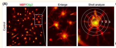
and erythropoietin (EPO) downregulate the expression of G protein–coupled receptor 17 (GPR17) and upregulate the expression of myelin basic protein (MBP), indicating improvement in the degree of demyelination.(D) Immunohistochemistry for MBP in the sham, SCI, HBO, EPO, and HBO+EPO groups at 21 days.

6). Analysis of the risk of traumatic brain injury and evaluation neurogranin and myelin basic protein as potential biomarkers of traumatic brain injury in postmortem examination. Forensic Science, Medicine and Pathology (PubMed: 35201602) [IF=1.8]

Restrictive clause


Affinity Biosciences tests all products strictly. Citations are provided as a resource for additional applications that have not been validated by Affinity Biosciences. Please choose the appropriate format for each application and consult Materials and Methods sections for additional details about the use of any product in these publications.

For Research Use Only.
Not for use in diagnostic or therapeutic procedures. Not for resale. Not for distribution without written consent. Affinity Biosciences will not be held responsible for patent infringement or other violations that may occur with the use of our products. Affinity Biosciences, Affinity Biosciences Logo and all other trademarks are the property of Affinity Biosciences LTD.