, using CDK8 Antibody at 1/1000 dilution.
5ug/NC membrane strip.
Exposure for 30s with Affinity™ ECL Kit(#KF8003).
Bands result from membrane strip incubation.")
, using CDK8 Antibody at 1/1000 dilution.
5ug/NC membrane strip.
Exposure for 30s with Affinity™ ECL Kit(#KF8003).
Bands result from membrane strip incubation.")
| Product: | CDK8 Antibody |
| Catalog: | DF3186 |
| Description: | Rabbit polyclonal antibody to CDK8 |
| Application: | WB IHC IF/ICC |
| Cited expt.: | IF/ICC |
| Reactivity: | Human, Mouse, Rat |
| Prediction: | Pig, Zebrafish, Horse, Sheep, Rabbit, Dog, Xenopus |
| Mol.Wt.: | 53 KD(Observed); 53kD(Calculated). |
| Uniprot: | P49336 |
| RRID: | AB_2835409 |
Control Products

Related Downloads
Protocols
Product Info
*The optimal dilutions should be determined by the end user. For optimal experimental results, antibody reuse is not recommended.
*Tips:
WB: For western blot detection of denatured protein samples. IHC: For immunohistochemical detection of paraffin sections (IHC-p) or frozen sections (IHC-f) of tissue samples. IF/ICC: For immunofluorescence detection of cell samples. ELISA(peptide): For ELISA detection of antigenic peptide.
Cite Format: Affinity Biosciences Cat# DF3186, RRID:AB_2835409.
Fold/Unfold
cdk8; CDK8 protein kinase; CDK8_HUMAN; Cell Division Kinase 8; Cell division protein kinase 8; Cyclin Dependent kinase 8; Cyclin-dependent kinase 8; K35; Mediator complex subunit cdk8; Mediator of RNA polymerase II transcription subunit cdk8; Protein kinase K35;
Immunogens
A synthesized peptide derived from human CDK8, corresponding to a region within N-terminal amino acids.
- P49336 CDK8_HUMAN:
- Protein BLAST With
- NCBI/
- ExPASy/
- Uniprot
MDYDFKVKLSSERERVEDLFEYEGCKVGRGTYGHVYKAKRKDGKDDKDYALKQIEGTGISMSACREIALLRELKHPNVISLQKVFLSHADRKVWLLFDYAEHDLWHIIKFHRASKANKKPVQLPRGMVKSLLYQILDGIHYLHANWVLHRDLKPANILVMGEGPERGRVKIADMGFARLFNSPLKPLADLDPVVVTFWYRAPELLLGARHYTKAIDIWAIGCIFAELLTSEPIFHCRQEDIKTSNPYHHDQLDRIFNVMGFPADKDWEDIKKMPEHSTLMKDFRRNTYTNCSLIKYMEKHKVKPDSKAFHLLQKLLTMDPIKRITSEQAMQDPYFLEDPLPTSDVFAGCQIPYPKREFLTEEEPDDKGDKKNQQQQQGNNHTNGTGHPGNQDSSHTQGPPLKKVRVVPPTTTSGGLIMTSDYQRSNPHAAYPNPGPSTSQPQSSMGYSATSQQPPQYSHQTHRY
Predictions
Score>80(red) has high confidence and is suggested to be used for WB detection. *The prediction model is mainly based on the alignment of immunogen sequences, the results are for reference only, not as the basis of quality assurance.
High(score>80) Medium(80>score>50) Low(score<50) No confidence
Research Backgrounds
Component of the Mediator complex, a coactivator involved in regulated gene transcription of nearly all RNA polymerase II-dependent genes. Mediator functions as a bridge to convey information from gene-specific regulatory proteins to the basal RNA polymerase II transcription machinery. Mediator is recruited to promoters by direct interactions with regulatory proteins and serves as a scaffold for the assembly of a functional preinitiation complex with RNA polymerase II and the general transcription factors. Phosphorylates the CTD (C-terminal domain) of the large subunit of RNA polymerase II (RNAp II), which may inhibit the formation of a transcription initiation complex. Phosphorylates CCNH leading to down-regulation of the TFIIH complex and transcriptional repression. Recruited through interaction with MAML1 to hyperphosphorylate the intracellular domain of NOTCH, leading to its degradation.
Nucleus.
Belongs to the protein kinase superfamily. CMGC Ser/Thr protein kinase family. CDC2/CDKX subfamily.
References
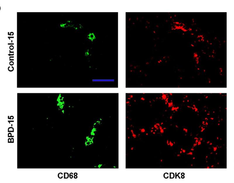
Application: IF/ICC Species: Mice Sample: lung tissues
Restrictive clause
Affinity Biosciences tests all products strictly. Citations are provided as a resource for additional applications that have not been validated by Affinity Biosciences. Please choose the appropriate format for each application and consult Materials and Methods sections for additional details about the use of any product in these publications.
For Research Use Only.
Not for use in diagnostic or therapeutic procedures. Not for resale. Not for distribution without written consent. Affinity Biosciences will not be held responsible for patent infringement or other violations that may occur with the use of our products. Affinity Biosciences, Affinity Biosciences Logo and all other trademarks are the property of Affinity Biosciences LTD.