Product: CDH11 Antibody
Catalog: DF3523
Description: Rabbit polyclonal antibody to CDH11
Application: WB IF/ICC
Reactivity: Human, Mouse
Prediction: Pig, Bovine, Horse, Sheep, Rabbit, Dog, Chicken
Mol.Wt.: 80,100kD; 88kD(Calculated).
Uniprot: P55287
RRID: AB_2835743

View similar products>>

   Size Price Inventory
 100ul $280 In stock
 200ul $350 In stock

Lead Time: Same day delivery

For pricing and ordering contact:
Local distributors

Product Info

Source:
Rabbit
Application:
WB 1:500-1:1000, IF/ICC 1:100-1:500
*The optimal dilutions should be determined by the end user.
*Tips:

WB: For western blot detection of denatured protein samples. IHC: For immunohistochemical detection of paraffin sections (IHC-p) or frozen sections (IHC-f) of tissue samples. IF/ICC: For immunofluorescence detection of cell samples. ELISA(peptide): For ELISA detection of antigenic peptide.

Reactivity:
Human,Mouse
Prediction:
Pig(100%), Bovine(100%), Horse(100%), Sheep(100%), Rabbit(100%), Dog(100%), Chicken(88%)
Clonality:
Polyclonal
Specificity:
CDH11 Antibody detects endogenous levels of total CDH11.
RRID:
AB_2835743
Cite Format: Affinity Biosciences Cat# DF3523, RRID:AB_2835743.
Conjugate:
Unconjugated.
Purification:
The antiserum was purified by peptide affinity chromatography using SulfoLink™ Coupling Resin (Thermo Fisher Scientific).
Storage:
Rabbit IgG in phosphate buffered saline , pH 7.4, 150mM NaCl, 0.02% sodium azide and 50% glycerol. Store at -20 °C. Stable for 12 months from date of receipt.
Alias:

Fold/Unfold

CAD11; CAD11_HUMAN; Cadherin 11; Cadherin 11 type 2; Cadherin 11 type 2 OB cadherin (osteoblast); Cadherin-11; Cdh11; CDHOB; OB; OB-cadherin; OBcadherin; OSF-4; OSF4; Osteoblast cadherin;

Immunogens

Immunogen:
Uniprot:
Gene(ID):
Expression:
P55287 CAD11_HUMAN:

Expressed mainly in brain but also found in other tissues. Expressed in neuroblasts.

Sequence:
MKENYCLQAALVCLGMLCHSHAFAPERRGHLRPSFHGHHEKGKEGQVLQRSKRGWVWNQFFVIEEYTGPDPVLVGRLHSDIDSGDGNIKYILSGEGAGTIFVIDDKSGNIHATKTLDREERAQYTLMAQAVDRDTNRPLEPPSEFIVKVQDINDNPPEFLHETYHANVPERSNVGTSVIQVTASDADDPTYGNSAKLVYSILEGQPYFSVEAQTGIIRTALPNMDREAKEEYHVVIQAKDMGGHMGGLSGTTKVTITLTDVNDNPPKFPQSVYQMSVSEAAVPGEEVGRVKAKDPDIGENGLVTYNIVDGDGMESFEITTDYETQEGVIKLKKPVDFETKRAYSLKVEAANVHIDPKFISNGPFKDTVTVKISVEDADEPPMFLAPSYIHEVQENAAAGTVVGRVHAKDPDAANSPIRYSIDRHTDLDRFFTINPEDGFIKTTKPLDREETAWLNITVFAAEIHNRHQEAKVPVAIRVLDVNDNAPKFAAPYEGFICESDQTKPLSNQPIVTISADDKDDTANGPRFIFSLPPEIIHNPNFTVRDNRDNTAGVYARRGGFSRQKQDLYLLPIVISDGGIPPMSSTNTLTIKVCGCDVNGALLSCNAEAYILNAGLSTGALIAILACIVILLVIVVLFVTLRRQKKEPLIVFEEEDVRENIITYDDEGGGEEDTEAFDIATLQNPDGINGFIPRKDIKPEYQYMPRPGLRPAPNSVDVDDFINTRIQEADNDPTAPPYDSIQIYGYEGRGSVAGSLSSLESATTDSDLDYDYLQNWGPRFKKLADLYGSKDTFDDDS

Predictions

Predictions:

Score>80(red) has high confidence and is suggested to be used for WB detection. *The prediction model is mainly based on the alignment of immunogen sequences, the results are for reference only, not as the basis of quality assurance.

Species
Results
Score
Pig
100
Horse
100
Bovine
100
Sheep
100
Dog
100
Rabbit
100
Chicken
88
Zebrafish
78
Xenopus
75
Model Confidence:
High(score>80) Medium(80>score>50) Low(score<50) No confidence

PTMs - P55287 As Substrate

Site PTM Type Enzyme
K43 Acetylation
S93 Phosphorylation
T99 Phosphorylation
T219 Phosphorylation
K365 Ubiquitination
Y700 Phosphorylation
Y702 Phosphorylation
S714 Phosphorylation
T723 Phosphorylation
S788 Phosphorylation
T791 Phosphorylation

Research Backgrounds

Function:

Cadherins are calcium-dependent cell adhesion proteins. They preferentially interact with themselves in a homophilic manner in connecting cells; cadherins may thus contribute to the sorting of heterogeneous cell types.

Subcellular Location:

Cell membrane>Single-pass type I membrane protein.

Extracellular region or secreted Cytosol Plasma membrane Cytoskeleton Lysosome Endosome Peroxisome ER Golgi apparatus Nucleus Mitochondrion Manual annotation Automatic computational assertionSubcellular location
Tissue Specificity:

Expressed mainly in brain but also found in other tissues. Expressed in neuroblasts.

Subunit Structure:

Interacts with PCDH8.

Family&Domains:

Three calcium ions are usually bound at the interface of each cadherin domain and rigidify the connections, imparting a strong curvature to the full-length ectodomain.

References

1). MiRNA-451a inhibits airway remodeling by targeting Cadherin 11 in an allergic asthma model of neonatal mice. International Immunopharmacology, 2020 (PubMed: 32234673) [IF=5.6]

Application: IF/ICC    Species: Mouse    Sample: lung tissues

Fig. 1. Opposite expression of miRNA-451a and CDH11 in the lung of asthma mouse pups. (A) New born mice were intraperitoneally injected with 10 μg ovalbumin (OVA) from postnatal day 5 (P5) to P10, and followed by 10-min daily challenge with 3% aerosolized OVA from P18 to P20. Twenty-four hours post the last OVA challenge (P21), the bronchoalveolar lavage fluid (BALF) was collected, and then the mice were sacrificed to collect their lung tissues. CD 4+ T cells were isolated from the control and asthmatic BALF samples. The expression levels of miRNA-451a in (B) CD 4+ T cells and (C) lung tissues were determined with real-time qPCR. The mRNA and protein levels of CDH11 were determined with (D) real-time qPCR, (E) western blot, and (F) immunofluorescent staining (bars, 50 μm). Data were expressed as mean ± S.D., and p < 0.05 was regarded statistically significant. *, **, *** indicated a p < 0.05, 0.01, 0.001.

Application: WB    Species: Mouse    Sample: lung tissues

Fig. 1. Opposite expression of miRNA-451a and CDH11 in the lung of asthma mouse pups. (A) New born mice were intraperitoneally injected with 10 μg ovalbumin (OVA) from postnatal day 5 (P5) to P10, and followed by 10-min daily challenge with 3% aerosolized OVA from P18 to P20. Twenty-four hours post the last OVA challenge (P21), the bronchoalveolar lavage fluid (BALF) was collected, and then the mice were sacrificed to collect their lung tissues. CD 4+ T cells were isolated from the control and asthmatic BALF samples. The expression levels of miRNA-451a in (B) CD 4+ T cells and (C) lung tissues were determined with real-time qPCR. The mRNA and protein levels of CDH11 were determined with (D) real-time qPCR, (E) western blot, and (F) immunofluorescent staining (bars, 50 μm). Data were expressed as mean ± S.D., and p < 0.05 was regarded statistically significant. *, **, *** indicated a p < 0.05, 0.01, 0.001.

2). BCL2L10 inhibits growth and metastasis of hepatocellular carcinoma both in vitro and in vivo. MOLECULAR CARCINOGENESIS, 2017 (PubMed: 27770580) [IF=4.6]

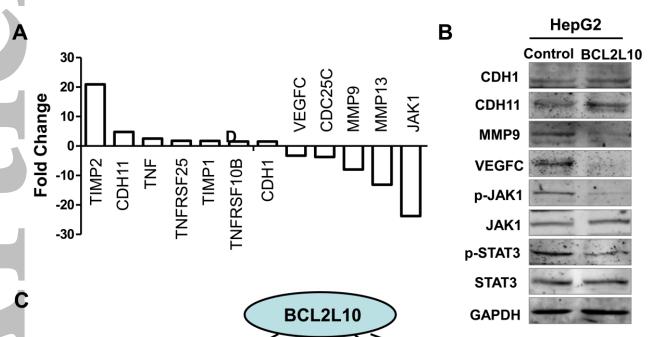
Application: WB    Species: human    Sample: HepG2

Figure 6. Effect of BCL2L10 on its downstream gene expression profiles of human cancer pathway in HepG2 cells. (A) By human cancer pathway PCR array, ectopic expression of BCL2L10 up- or down-regulated several genes related to tumor proliferation, apoptosis, metastasis and angiogenesis. (B) Western blot was performed to confirm the downstream gene expression regulated by BCL2L10 in HepG2 cells. GAPDH was used as an internal control. (C) Schematic diagram of the molecular events for BCL2L10 function as a tumor suppressor through regulating cell cycle, proliferation, apoptosis metastasis and angiogenesis effectors.

Restrictive clause


Affinity Biosciences tests all products strictly. Citations are provided as a resource for additional applications that have not been validated by Affinity Biosciences. Please choose the appropriate format for each application and consult Materials and Methods sections for additional details about the use of any product in these publications.

For Research Use Only.
Not for use in diagnostic or therapeutic procedures. Not for resale. Not for distribution without written consent. Affinity Biosciences will not be held responsible for patent infringement or other violations that may occur with the use of our products. Affinity Biosciences, Affinity Biosciences Logo and all other trademarks are the property of Affinity Biosciences LTD.