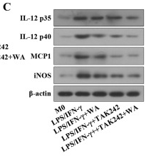
Product: IL12A Antibody
Catalog: AF5133
Description: Rabbit polyclonal antibody to IL12A
Application: WB IHC
Cited expt.: WB
Reactivity: Human, Mouse, Rat
Prediction: Pig, Bovine, Horse, Sheep, Rabbit, Dog
Mol.Wt.: 24 kDa; 25kD(Calculated).
Uniprot: P29459
RRID: AB_2837619

View similar products>>

   Size Price Inventory
 100ul $280 In stock
 200ul $350 In stock

Lead Time: Same day delivery

For pricing and ordering contact:
Local distributors

Product Info

Source:
Rabbit
Application:
WB 1:500-1:2000, IHC 1:50-1:200
*The optimal dilutions should be determined by the end user. For optimal experimental results, antibody reuse is not recommended.
*Tips:

WB: For western blot detection of denatured protein samples. IHC: For immunohistochemical detection of paraffin sections (IHC-p) or frozen sections (IHC-f) of tissue samples. IF/ICC: For immunofluorescence detection of cell samples. ELISA(peptide): For ELISA detection of antigenic peptide.

Reactivity:
Human,Mouse,Rat
Prediction:
Pig(100%), Bovine(100%), Horse(100%), Sheep(100%), Rabbit(100%), Dog(100%)
Clonality:
Polyclonal
Specificity:
IL12A Antibody detects endogenous levels of total IL12A.
RRID:
AB_2837619
Cite Format: Affinity Biosciences Cat# AF5133, RRID:AB_2837619.
Conjugate:
Unconjugated.
Purification:
The antiserum was purified by peptide affinity chromatography using SulfoLink™ Coupling Resin (Thermo Fisher Scientific).
Storage:
Rabbit IgG in phosphate buffered saline , pH 7.4, 150mM NaCl, 0.02% sodium azide and 50% glycerol. Store at -20 °C. Stable for 12 months from date of receipt.
Alias:

Fold/Unfold

CLMF; CLMF p35; Cytotoxic lymphocyte maturation factor 1 p35; Cytotoxic lymphocyte maturation factor 35 kDa subunit; IL 12 subunit p35; IL 12A; IL-12 subunit p35; IL-12A; IL12A; IL12A_HUMAN; IL35; IL35 subunit; Interleukin 12 alpha chain; Interleukin 12 p35; Interleukin 12 subunit alpha; Interleukin 12A (natural killer cell stimulatory factor 1, cytotoxic lymphocyte maturation factor 1, p35); Interleukin-12 subunit alpha; Natural killer cell stimulatory factor 1 35 kD subunit; NF cell stimulatory factor chain 1; NFSK; NK cell stimulatory factor chain 1; NKSF1; P35;

Immunogens

Immunogen:

A synthesized peptide derived from human IL12A, corresponding to a region within the internal amino acids.

Uniprot:
Gene(ID):
Description:
Cytokine that can act as a growth factor for activated T and NK cells, enhance the lytic activity of NK/lymphokine-activated Killer cells, and stimulate the production of IFN-gamma by resting PBMC.
Sequence:
MCPARSLLLVATLVLLDHLSLARNLPVATPDPGMFPCLHHSQNLLRAVSNMLQKARQTLEFYPCTSEEIDHEDITKDKTSTVEACLPLELTKNESCLNSRETSFITNGSCLASRKTSFMMALCLSSIYEDLKMYQVEFKTMNAKLLMDPKRQIFLDQNMLAVIDELMQALNFNSETVPQKSSLEEPDFYKTKIKLCILLHAFRIRAVTIDRVMSYLNAS

Predictions

Predictions:

Score>80(red) has high confidence and is suggested to be used for WB detection. *The prediction model is mainly based on the alignment of immunogen sequences, the results are for reference only, not as the basis of quality assurance.

Species
Results
Score
Pig
100
Horse
100
Bovine
100
Sheep
100
Dog
100
Rabbit
100
Chicken
67
Xenopus
0
Zebrafish
0
Model Confidence:
High(score>80) Medium(80>score>50) Low(score<50) No confidence

Research Backgrounds

Function:

Cytokine that can act as a growth factor for activated T and NK cells, enhance the lytic activity of NK/lymphokine-activated killer cells, and stimulate the production of IFN-gamma by resting PBMC.

Subcellular Location:

Secreted.

Extracellular region or secreted Cytosol Plasma membrane Cytoskeleton Lysosome Endosome Peroxisome ER Golgi apparatus Nucleus Mitochondrion Manual annotation Automatic computational assertionSubcellular location
Family&Domains:

Belongs to the IL-6 superfamily.

Research Fields

· Environmental Information Processing > Signaling molecules and interaction > Cytokine-cytokine receptor interaction.   (View pathway)

· Environmental Information Processing > Signal transduction > Jak-STAT signaling pathway.   (View pathway)

· Human Diseases > Endocrine and metabolic diseases > Type I diabetes mellitus.

· Human Diseases > Infectious diseases: Bacterial > Pertussis.

· Human Diseases > Infectious diseases: Bacterial > Legionellosis.

· Human Diseases > Infectious diseases: Parasitic > Leishmaniasis.

· Human Diseases > Infectious diseases: Parasitic > Chagas disease (American trypanosomiasis).

· Human Diseases > Infectious diseases: Parasitic > African trypanosomiasis.

· Human Diseases > Infectious diseases: Parasitic > Malaria.

· Human Diseases > Infectious diseases: Parasitic > Toxoplasmosis.

· Human Diseases > Infectious diseases: Parasitic > Amoebiasis.

· Human Diseases > Infectious diseases: Bacterial > Tuberculosis.

· Human Diseases > Infectious diseases: Viral > Measles.

· Human Diseases > Infectious diseases: Viral > Influenza A.

· Human Diseases > Infectious diseases: Viral > Herpes simplex infection.

· Human Diseases > Cancers: Overview > Pathways in cancer.   (View pathway)

· Human Diseases > Immune diseases > Inflammatory bowel disease (IBD).

· Human Diseases > Immune diseases > Allograft rejection.

· Organismal Systems > Immune system > Toll-like receptor signaling pathway.   (View pathway)

· Organismal Systems > Immune system > RIG-I-like receptor signaling pathway.   (View pathway)

· Organismal Systems > Immune system > Th1 and Th2 cell differentiation.   (View pathway)

References

1). A bioinspired polymeric membrane-enclosed insulin crystal achieves long-term, self-regulated drug release for type 1 diabetes therapy. Nature nanotechnology, 2025 (PubMed: 40011600) [IF=38.1]

2). Week-long normoglycaemia in diabetic mice and minipigs via a subcutaneous dose of a glucose-responsive insulin complex. Nature biomedical engineering, 2023 (PubMed: 38057427) [IF=28.1]

3). Long-acting IL-2 release from pressure-fused biomineral tablets promotes antitumor immune response. Nature cancer, 2025 (PubMed: 40494938) [IF=22.7]

4). RNF8 enhances the sensitivity of PD-L1 inhibitor against melanoma through ubiquitination of galectin-3 in stroma. Cell death discovery, 2023 (PubMed: 37391451) [IF=7.0]

Application: WB    Species: Mouse    Sample:

Fig. 7: Inhibition of gal-3 enhanced sensitivity of PD-L1 therapy for melanoma. PD-L1 inhibitor was dosed by intraperitoneal injection at 1 μmol per mouse. LAC was dosed by intratumor injection at a single dose of 0.1 μmol per mouse. A The 3D modeling of subcutaneous tumor-beared in RNF8+/+ host that was treated with different strategies, including PD-L1 inhibitor, LAC, and PD-L1 inhibitor plus LAC (P + L). B The representative tumors in each group were shown. C The tumor volume (left) and the ratio of tumor weight (right). D Tumor growth volume in different groups. E The Ki-67 immunohistochemical staining of tumors. F Statistical analysis of Ki-67 in (E). G HE staining of melanomas in different groups. The arrow points to lymphocytes. H The ratio of tumor area compared to stroma area in each group (left). The number of lymphocytes in groups (right). I The western blot of IL-12, IFN-γ, CXCL-9, and CXCL-10 in PD-L1 inhibitor, LacNAc, and combination group. All values are presented as the mean ± SD (n = 6).

5). A hybrid hydrogel composed of chitin and β-glucan for the effective management of wound healing and scarring. Biomaterials Science, 2022 (PubMed: 36073016) [IF=5.8]

6). TSP-1-CD47-integrin α4β1 axis drives T cell infiltration and synovial inflammation in rheumatoid arthritis. Frontiers in immunology, 2025 (PubMed: 40308591) [IF=5.7]

7). IL-12 as a Potential Prognostic Marker in Penile Cancer: Implications for Immune Dysregulation. International journal of molecular sciences, 2025 (PubMed: 41465261) [IF=5.6]

8). PAE ameliorates doxorubicin-induced cardiotoxicity via suppressing NHE1 phosphorylation and stimulating PI3K/AKT phosphorylation. International Immunopharmacology, 2022 (PubMed: 36252472) [IF=4.8]

9). Glucuronoxylomannan (GXM) modulates macrophage proliferation and apoptosis through the STAT1 signaling pathway. Cell biology international, 2025 (PubMed: 39760338) [IF=3.9]

10). Wilforlide A ameliorates the progression of rheumatoid arthritis by inhibiting M1 macrophage polarization. Journal of Pharmacological Sciences, 2022 (PubMed: 34924115) [IF=3.0]

Application: WB    Species: Mouse    Sample:

Fig. 5. Effects of Wilforlide A on M1 macrophage polarization in LPS/IFN-γ-induced macrophages. (A–B) Western blot and quantitative results of p65 in the nucleus of macrophages. §

Load more

Restrictive clause


Affinity Biosciences tests all products strictly. Citations are provided as a resource for additional applications that have not been validated by Affinity Biosciences. Please choose the appropriate format for each application and consult Materials and Methods sections for additional details about the use of any product in these publications.

For Research Use Only.
Not for use in diagnostic or therapeutic procedures. Not for resale. Not for distribution without written consent. Affinity Biosciences will not be held responsible for patent infringement or other violations that may occur with the use of our products. Affinity Biosciences, Affinity Biosciences Logo and all other trademarks are the property of Affinity Biosciences LTD.