
, using SIRT3 Antibody at 1/1000 dilution.
5ug/NC membrane strip.
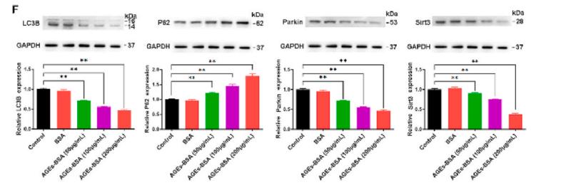
Exposure for 6s with Affinity™ ECL Kit(#KF8003).
Bands result from membrane strip incubation.")
.
Bands result from membrane strip incubation.")
.
Bands result from membrane strip incubation.")


 and mouse anti-beta tubulin Ab(T0023 1:200) for 1 hour at 37°C. An AlexaFluor594 conjugated goat anti-rabbit IgG(H+L) Ab(Red) and an AlexaFluor488 conjugated goat anti-mouse IgG(H+L) Ab(Green) were used as the secondary antibody.
The nuclear counter stain is DAPI(blue).")
 HepG2 and (B) L02 cells were incubated with different concentrations of SWNHs in 6-well plates for 48 h. The changes in the expression and the relative quantification of proteins in (C) HepG2 cells or (D) L02 cells were identified by western blotting. SW0, SW10, SW20, SW30, SW40 and SW50 correspond to the concentrations of SWNHs: 0, 0.21, 0.42, 0.64, 0.85 and 0.96 µg/cm2, respectively. Data are presented as the mean ± standard deviation (n=3). *P<0.05 compared with the SW0 group. AceCS2, acyl-CoA synthetase short chain family member 1; SCNN1A, sodium channel epithelial 1α subunit; SIRT3, sirtuin 3; SWNH, single-walled carbon nanohorn; VDAC1, voltage-dependent anion channel 1.")

 Protein expression of Sirt3 detected by western blotting; (versus the control group, * P<0.05; versus the model group, # P<0.05). Sirt3 – sirtuin; PCR – polymerase chain reaction; mRNA – messenger RNA.")
 PPI analysis and KEGG pathways demonstrated an interaction network between PARP‐1 and SIRT3 along with the related signaling pathways. Species origin: Homo sapiens. Yellow line: Textmining. Blue line: From curated databases. purple line: Experimentally determined. black line: Co‐expression. (C–E) The levels of SIRT3 in the DRGs and spinal cord were decreased from day 7 to day 21 following paclitaxel injection. One‐way ANOVA followed by Bonferroni post hoc test, *p")
 PPI analysis and KEGG pathways demonstrated an interaction network between PARP‐1 and SIRT3 along with the related signaling pathways. Species origin: Homo sapiens. Yellow line: Textmining. Blue line: From curated databases. purple line: Experimentally determined. black line: Co‐expression. (C–E) The levels of SIRT3 in the DRGs and spinal cord were decreased from day 7 to day 21 following paclitaxel injection. One‐way ANOVA followed by Bonferroni post hoc test, *p")
 is down-regulated at fibrocartilage (F) layer of rotator cuff in aged mice. A: Representative images of immunohistochemical staining for SIRT3 at fibrocartilage layer of rotator cuff among 3-, 10-, and 18-month–old mice, and quantitative analysis. The region between the dashed lines is the fibrocartilage layer. Red arrows: SIRT3+ chondrocytes. B: Immunofluorescence staining for SIRT3 in young and senescent chondrocytes and quantitative analysis. C and D: Relative mRNA expression level of SIRT3 and representative Western blot analyses for SIRT3 expression level in young and senescent chondrocytes. Data are presented as the means ± SD (A–C). ∗P < 0.05, ∗∗P < 0.01. Scale bars: 20 μm (A); 200 μm (B). B, bone; T, tendon.")
 and the statistical analysis of mitochondrial areas and the mitochondrial length/width ratio (n = 3). B Representative immunohistochemical staining of TFAM, Sirt3 and TOM20 in the mouse livers (scale bar, 50 µm). C Western blot analysis of TFAM, Sirt3 and TOM20 protein levels in the mouse liver (n = 3). D Representative TEM images from the livers of HC individuals and AIH patients. E Western blot analysis of TFAM, Sirt3 and TOM20 protein levels in the liver from HC individuals and AIH patients (n = 6).")
 and the statistical analysis of mitochondrial areas and the mitochondrial length/width ratio (n = 3). B Representative immunohistochemical staining of TFAM, Sirt3 and TOM20 in the mouse livers (scale bar, 50 µm). C Western blot analysis of TFAM, Sirt3 and TOM20 protein levels in the mouse liver (n = 3). D Representative TEM images from the livers of HC individuals and AIH patients. E Western blot analysis of TFAM, Sirt3 and TOM20 protein levels in the liver from HC individuals and AIH patients (n = 6).")
 Western blot and (B, F) statistical results of western blots of SRIT3 and AcSOD2/SOD2 (n = 5, one‐way ANOVA followed by Bonferroni's post hoc test). (C) Quantitative real‐time PCR of SIRT3 (n = 5, one‐way ANOVA followed by Bonferroni's post hoc test). (D) Immunofluorescence and (E) statistical analysis of immunofluorescence staining of SIRT3 from each group (n = 3, Scale bar = 25 μm, one‐way ANOVA followed by Bonferroni's post hoc test). (G) Statistical analysis of SOD2 activity (n = 5, one‐way ANOVA followed by Bonferroni's post hoc test). (I) ROS was measured by DCFH‐DA and (H) statistical results of fluorescence intensity (n = 5, Scale bar = 100 μm, one‐way ANOVA followed by Bonferroni's post hoc test). (J) 8‐OHdG content was detected by the 8‐OHdG assay kit (n = 5, one‐way ANOVA followed by Games–Howell post hoc test). The data represent means ± SD, *p")
 patients group (DKD-Exo) on SIRT3/AMPK signaling pathway. HKB20 cells were transfected with miR-516b-5p inhibitor or silence SIRT3, and exposed to DKD-Exo. A: the levels of miR-516b-5p were evaluated using quantitative real time-polymerase chain reaction (qRT-PCR). B: SIRT3 mRNA level and protein expression were determined using qRT-PCR and Western blot. C: the binding sites of miR-516b-5p on SIRT3 were anticipated by bioinformatics software and verified using dual-luciferase reporter gene assay. D: the expression of SIRT3, AMPK, and p-AMPK was examined using Western blot.")
 FGFR1, (B) SIRT3, and (C) DPP-4 in kidney tissues were examined by immunohistochemical staining. (D–F) Quantitative analysis of FGFR1, SIRT3 and DPP-4 expression levels in renal tissues of each group. Magnification: ×200, scale bars = 100 μm. Data are expressed as the mean ± S.D. (n = 5). *p < 0.05, **p < 0.01 vs. the control group; #p < 0.05, ##p < 0.01 vs. the DN group; and p < 0.05 vs. the DN + In group.")
| Product: | SIRT3 Antibody |
| Catalog: | AF5135 |
| Description: | Rabbit polyclonal antibody to SIRT3 |
| Application: | WB IHC IF/ICC |
| Cited expt.: | WB, IHC, IF/ICC |
| Reactivity: | Human, Mouse, Rat |
| Prediction: | Sheep, Rabbit |
| Mol.Wt.: | 29 kDa(Observed); 44kD(Calculated). |
| Uniprot: | Q9NTG7 |
| RRID: | AB_2837621 |
Control Products

Related Downloads
Protocols
Product Info
*The optimal dilutions should be determined by the end user. For optimal experimental results, antibody reuse is not recommended.
*Tips:
WB: For western blot detection of denatured protein samples. IHC: For immunohistochemical detection of paraffin sections (IHC-p) or frozen sections (IHC-f) of tissue samples. IF/ICC: For immunofluorescence detection of cell samples. ELISA(peptide): For ELISA detection of antigenic peptide.
Cite Format: Affinity Biosciences Cat# AF5135, RRID:AB_2837621.
Fold/Unfold
hSIRT 3; hSIRT3; Mitochondrial nicotinamide adenine dinucleotide dependent deacetylase; NAD dependent deacetylase sirtuin 3 mitochondrial; NAD-dependent protein deacetylase sirtuin-3, mitochondrial; Regulatory protein SIR2 homolog 3; Silent mating type information regulation 2 S.cerevisiae homolog 3; Sir 2 like 3; SIR 2 like protein 3; SIR 3; SIR2 L3; Sir2 like 3; SIR2 like protein 3; SIR2-like protein 3; SIR2L3; SIR3_HUMAN; SIRT 3; SIRT3; Sirtuin 3; Sirtuin silent mating type information regulation 2 homolog 3 (S. cerevisiae); Sirtuin type 3; Sirtuin3;
Immunogens
A synthesized peptide derived from human SIRT3, corresponding to a region within C-terminal amino acids.
- Q9NTG7 SIR3_HUMAN:
- Protein BLAST With
- NCBI/
- ExPASy/
- Uniprot
MAFWGWRAAAALRLWGRVVERVEAGGGVGPFQACGCRLVLGGRDDVSAGLRGSHGARGEPLDPARPLQRPPRPEVPRAFRRQPRAAAPSFFFSSIKGGRRSISFSVGASSVVGSGGSSDKGKLSLQDVAELIRARACQRVVVMVGAGISTPSGIPDFRSPGSGLYSNLQQYDLPYPEAIFELPFFFHNPKPFFTLAKELYPGNYKPNVTHYFLRLLHDKGLLLRLYTQNIDGLERVSGIPASKLVEAHGTFASATCTVCQRPFPGEDIRADVMADRVPRCPVCTGVVKPDIVFFGEPLPQRFLLHVVDFPMADLLLILGTSLEVEPFASLTEAVRSSVPRLLINRDLVGPLAWHPRSRDVAQLGDVVHGVESLVELLGWTEEMRDLVQRETGKLDGPDK
Predictions
Score>80(red) has high confidence and is suggested to be used for WB detection. *The prediction model is mainly based on the alignment of immunogen sequences, the results are for reference only, not as the basis of quality assurance.
High(score>80) Medium(80>score>50) Low(score<50) No confidence
Research Backgrounds
NAD-dependent protein deacetylase. Activates or deactivates mitochondrial target proteins by deacetylating key lysine residues. Known targets include ACSS1, IDH, GDH, SOD2, PDHA1, LCAD, SDHA and the ATP synthase subunit ATP5PO. Contributes to the regulation of the cellular energy metabolism. Important for regulating tissue-specific ATP levels. In response to metabolic stress, deacetylates transcription factor FOXO3 and recruits FOXO3 and mitochondrial RNA polymerase POLRMT to mtDNA to promote mtDNA transcription. Acts as a regulator of ceramide metabolism by mediating deacetylation of ceramide synthases CERS1, CERS2 and CERS6, thereby increasing their activity and promoting mitochondrial ceramide accumulation (By similarity).
Processed by mitochondrial processing peptidase (MPP) to give a 28 kDa product. Such processing is probably essential for its enzymatic activity.
Mitochondrion matrix.
Widely expressed.
Belongs to the sirtuin family. Class I subfamily.
Research Fields
· Human Diseases > Cancers: Overview > Central carbon metabolism in cancer. (View pathway)
References

Application: WB Species: human Sample: HCT116 and LoVo cells
Application: WB Species: mice Sample: bone marrow mesenchymal stem (BMSCs)

Application: IF/ICC Species: Mouse Sample:

Application: WB Species: Rat Sample: H9C2 cell
Restrictive clause
Affinity Biosciences tests all products strictly. Citations are provided as a resource for additional applications that have not been validated by Affinity Biosciences. Please choose the appropriate format for each application and consult Materials and Methods sections for additional details about the use of any product in these publications.
For Research Use Only.
Not for use in diagnostic or therapeutic procedures. Not for resale. Not for distribution without written consent. Affinity Biosciences will not be held responsible for patent infringement or other violations that may occur with the use of our products. Affinity Biosciences, Affinity Biosciences Logo and all other trademarks are the property of Affinity Biosciences LTD.