Product: ACE2 Antibody
Catalog: AF5165
Description: Rabbit polyclonal antibody to ACE2
Application: WB IHC IF/ICC
Reactivity: Human, Mouse, Rat
Prediction: Pig, Bovine, Horse, Sheep, Rabbit, Dog
Mol.Wt.: 92 kDa, 100-120kD(Glycosylated); 92kD(Calculated).
Uniprot: Q9BYF1
RRID: AB_2837651

View similar products>>

   Size Price Inventory
 100ul $280 In stock
 200ul $350 In stock

Lead Time: Same day delivery

For pricing and ordering contact:
Local distributors

Product Info

Source:
Rabbit
Application:
WB 1:500-1:2000, IF/ICC 1:100-1:500, IHC 1:50-1:200
*The optimal dilutions should be determined by the end user.
*Tips:

WB: For western blot detection of denatured protein samples. IHC: For immunohistochemical detection of paraffin sections (IHC-p) or frozen sections (IHC-f) of tissue samples. IF/ICC: For immunofluorescence detection of cell samples. ELISA(peptide): For ELISA detection of antigenic peptide.

Reactivity:
Human,Mouse,Rat
Prediction:
Pig(82%), Bovine(82%), Horse(82%), Sheep(82%), Rabbit(82%), Dog(100%)
Clonality:
Polyclonal
Specificity:
ACE2 Antibody detects endogenous levels of total ACE2.
RRID:
AB_2837651
Cite Format: Affinity Biosciences Cat# AF5165, RRID:AB_2837651.
Conjugate:
Unconjugated.
Purification:
The antiserum was purified by peptide affinity chromatography using SulfoLink™ Coupling Resin (Thermo Fisher Scientific).
Storage:
Rabbit IgG in phosphate buffered saline , pH 7.4, 150mM NaCl, 0.02% sodium azide and 50% glycerol. Store at -20 °C. Stable for 12 months from date of receipt.
Alias:

Fold/Unfold

ACE 2; ACE related carboxypeptidase; ACE-related carboxypeptidase; ACE2; ACE2_HUMAN; ACEH; Angiotensin converting enzyme 2; Angiotensin converting enzyme homolog; Angiotensin converting enzyme like protein; Angiotensin I Converting Enzyme (peptidyl dipeptidase A) 2; Angiotensin I converting enzyme 2; Angiotensin-converting enzyme homolog; DKFZP434A014; EC 3.4.17; metalloprotease MPROT 15; Metalloprotease MPROT15; OTTHUMP00000022963; Processed angiotensin-converting enzyme 2;

Immunogens

Immunogen:
Uniprot:
Gene(ID):
Expression:
Q9BYF1 ACE2_HUMAN:

Expressed in endothelial cells from small and large arteries, and in arterial smooth muscle cells (at protein level) (PubMed:15141377). Expressed in lung alveolar epithelial cells, enterocytes of the small intestine, Leydig cells and Sertoli cells (at protein level) (PubMed:15141377). Expressed in the renal proximal tubule and the small intestine (at protein level) (PubMed:18424768). Expressed in heart, kidney, testis, and gastrointestinal system (PubMed:10969042, PubMed:10924499, PubMed:15231706, PubMed:12459472, PubMed:15671045).

Description:
Carboxypeptidase which converts angiotensin I to angiotensin 1-9, a peptide of unknown function, and angiotensin II to angiotensin 1-7, a vasodilator. Also able to hydrolyze apelin-13 and dynorphin-13 with high efficiency. May be an important regulator of heart function. In case of human coronaviruses SARS and HCoV-NL63 infections, serve as functional receptor for the spike glycoprotein of both coronaviruses.
Sequence:
MSSSSWLLLSLVAVTAAQSTIEEQAKTFLDKFNHEAEDLFYQSSLASWNYNTNITEENVQNMNNAGDKWSAFLKEQSTLAQMYPLQEIQNLTVKLQLQALQQNGSSVLSEDKSKRLNTILNTMSTIYSTGKVCNPDNPQECLLLEPGLNEIMANSLDYNERLWAWESWRSEVGKQLRPLYEEYVVLKNEMARANHYEDYGDYWRGDYEVNGVDGYDYSRGQLIEDVEHTFEEIKPLYEHLHAYVRAKLMNAYPSYISPIGCLPAHLLGDMWGRFWTNLYSLTVPFGQKPNIDVTDAMVDQAWDAQRIFKEAEKFFVSVGLPNMTQGFWENSMLTDPGNVQKAVCHPTAWDLGKGDFRILMCTKVTMDDFLTAHHEMGHIQYDMAYAAQPFLLRNGANEGFHEAVGEIMSLSAATPKHLKSIGLLSPDFQEDNETEINFLLKQALTIVGTLPFTYMLEKWRWMVFKGEIPKDQWMKKWWEMKREIVGVVEPVPHDETYCDPASLFHVSNDYSFIRYYTRTLYQFQFQEALCQAAKHEGPLHKCDISNSTEAGQKLFNMLRLGKSEPWTLALENVVGAKNMNVRPLLNYFEPLFTWLKDQNKNSFVGWSTDWSPYADQSIKVRISLKSALGDKAYEWNDNEMYLFRSSVAYAMRQYFLKVKNQMILFGEEDVRVANLKPRISFNFFVTAPKNVSDIIPRTEVEKAIRMSRSRINDAFRLNDNSLEFLGIQPTLGPPNQPPVSIWLIVFGVVMGVIVVGIVILIFTGIRDRKKKNKARSGENPYASIDISKGENNPGFQNTDDVQTSF

Predictions

Predictions:

Score>80(red) has high confidence and is suggested to be used for WB detection. *The prediction model is mainly based on the alignment of immunogen sequences, the results are for reference only, not as the basis of quality assurance.

Species
Results
Score
Dog
100
Pig
82
Horse
82
Bovine
82
Sheep
82
Rabbit
82
Zebrafish
55
Chicken
55
Xenopus
0
Model Confidence:
High(score>80) Medium(80>score>50) Low(score<50) No confidence

PTMs - Q9BYF1 As Substrate

Site PTM Type Enzyme
S2 Phosphorylation
S10 Phosphorylation
N53 N-Glycosylation
N90 N-Glycosylation
N103 N-Glycosylation
S128 Phosphorylation
T129 Phosphorylation
N322 N-Glycosylation
N432 N-Glycosylation
S545 Phosphorylation
N546 N-Glycosylation
S547 Phosphorylation
T548 Phosphorylation
K625 Ubiquitination
K676 Ubiquitination
S692 Phosphorylation
K770 Acetylation
K771 Acetylation
K773 Acetylation
Y781 Phosphorylation
S783 Phosphorylation

Research Backgrounds

Function:

Carboxypeptidase which converts angiotensin I to angiotensin 1-9, a peptide of unknown function, and angiotensin II to angiotensin 1-7, a vasodilator. Also able to hydrolyze apelin-13 and dynorphin-13 with high efficiency. By cleavage of angiotensin II, may be an important regulator of heart function. By cleavage of angiotensin II, may also have a protective role in acute lung injury (By similarity). Plays an important role in amino acid transport by acting as binding partner of amino acid transporter SL6A19 in intestine, regulating trafficking, expression on the cell surface, and its catalytic activity.

(Microbial infection) Acts as a receptor for SARS coronavirus/SARS-CoV.

(Microbial infection) Acts as a receptor for Human coronavirus NL63/HCoV-NL63.

PTMs:

N-glycosylation on Asn-90 may limit SARS infectivity.

Proteolytic cleavage by ADAM17 generates a secreted form. Also cleaved by serine proteases: TMPRSS2, TMPRSS11D and HPN/TMPRSS1.

Subcellular Location:

Secreted.

Cell membrane>Single-pass type I membrane protein. Cytoplasm.
Note: Detected in both cell membrane and cytoplasm in neurons.

Extracellular region or secreted Cytosol Plasma membrane Cytoskeleton Lysosome Endosome Peroxisome ER Golgi apparatus Nucleus Mitochondrion Manual annotation Automatic computational assertionSubcellular location
Tissue Specificity:

Expressed in endothelial cells from small and large arteries, and in arterial smooth muscle cells (at protein level). Expressed in lung alveolar epithelial cells, enterocytes of the small intestine, Leydig cells and Sertoli cells (at protein level). Expressed in the renal proximal tubule and the small intestine (at protein level). Expressed in heart, kidney, testis, and gastrointestinal system.

Subunit Structure:

Interacts with ITGB1. Interacts with the catalytically active form of TMPRSS2. Interacts with SLC6A19; this interaction is essential for expression and function of SLC6A19 in intestine (By similarity).

(Microbial infection) Interacts with SARS coronavirus/SARS-CoV.

(Microbial infection) Interacts with Human coronavirus NL63/HCoV-NL63 spike glycoprotein.

Family&Domains:

Belongs to the peptidase M2 family.

Research Fields

· Organismal Systems > Endocrine system > Renin-angiotensin system.   (View pathway)

· Organismal Systems > Digestive system > Protein digestion and absorption.

References

1). Biomimetic Nanoparticle-Mediated Target Delivery of Hypoxia-Responsive Plasmid of Angiotensin-Converting Enzyme 2 to Reverse Hypoxic Pulmonary Hypertension. ACS Nano, 2023 (PubMed: 37071566) [IF=17.1]

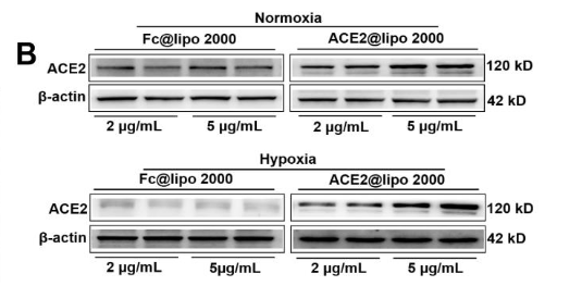
Application: WB    Species: Rat    Sample:

further compare the transfection efficiency(Data are means ± SEM., n=2independent experiments. **P< 0.01 vs. ACE2@lipo 2000 (2 μg/mL)treated PMVECsin hypoxia).(C)Representative diagram of ACE2-CS-PRT stability within 7 days in PBS(Data are means ± SEM., n=3independent experiments).(D-E) Hemolysistest for assessment the hemolytic effect of ACE2-CS-PRT@PM by using rat blood red cell(Data are means ± SEM., n=3 independent experiments. **P< 0.01 vs. ACE2-CS-PRT 1mg/mL; ##P < 0.01 vs. ACE2-CS-PRT 2mg/mL).

2). ACE2 correlated with immune infiltration serves as a novel prognostic biomarker in clear cell renal cell carcinoma: implication for COVID-19. International Journal of Biological Sciences, 2021 (PubMed: 33390830) [IF=9.2]

Application: WB    Species: Human    Sample: HK2 cell

Figure 1 Expression status of ACE2 in ccRCC tissues and cell lines. (A) Comparison of ACE2 mRNA expression in ccRCC and adjacent normal tissues (AN) based on GSE40435, GSE53757, GSE66272 and GSE126964 datasets. (B) Comparison of ACE2 protein expression in 54 pairs of matched paraffin section samples (ccRCC and adjacent normal tissue) using immunohistochemistry (C) Comparison of ACE2 protein expression in HK2 cell line and three ccRCC cell lines (786-O, OSRC2 and A498). ****p < 0.0001

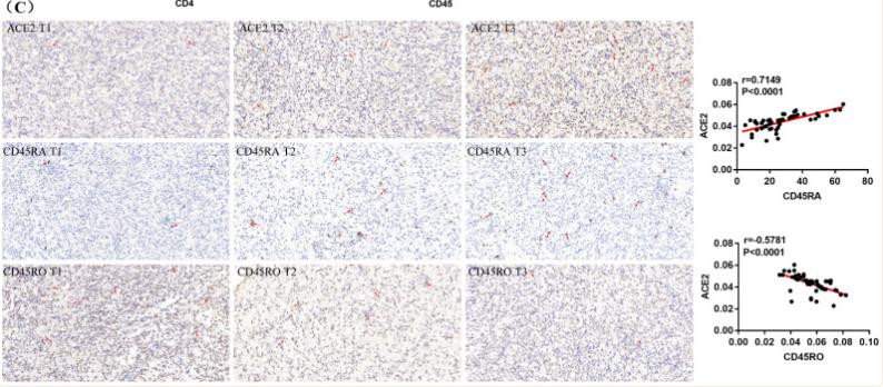
Application: IHC    Species: Human    Sample: immune cell

Figure 7 ACE2 expression was associated with CD4 Naïve and CD4 Memory infiltration. (A) Correlation between ACE2 expression and CD4 Naïve, CD4 Memory and DC infiltration levels based TCGA and TIP data. (B) Correlation between ACE2 expression and CD4 and CD45 expression based on TCGA data. (C) Correlation between ACE2 protein expression and CD45RA and CD45RO protein expression based on the immunohistochemistry staining results of 54 clinical ccRCC samples.

3). Single-cell RNA Analysis on ACE2 Expression Provides Insight into SARS-CoV-2 Blood Entry and Heart Injury. JOURNAL OF CELLULAR PHYSIOLOGY, 2020 (PubMed: 32510598) [IF=5.6]

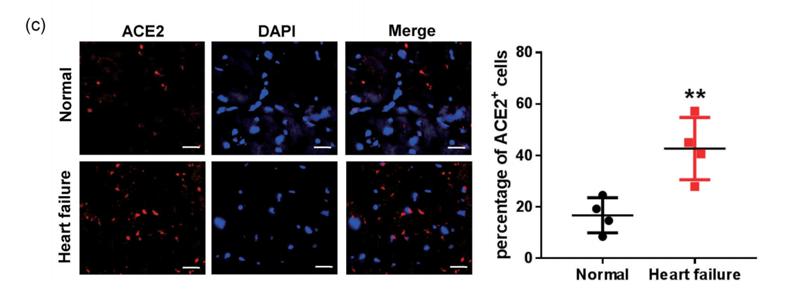
Application: IF/ICC    Species: Human    Sample: Heart

FIGURE 3 Different distribution of angiotensin converting enzyme II (ACE2)‐expressing cells in normal and failed hearts. (a,b) Feature plot showing the distribution of ACE2, GJA4, and MYH6 expression levels in normal hearts (a) and in failed hearts (b). (c) Immunofluorescence staining of ACE2 (red) expression and DAPI, 4',6‐diamidino‐2‐phenylindole (DAPI) (blue) in normal hearts, and in failed hearts. Scale bar = 20 μm (n = 4). Significance is determined with a two‐tailed unpaired Student's t test, **p < .01

4). GTS-21 attenuates ACE/ACE2 ratio and glycocalyx shedding in lipopolysaccharide-induced acute lung injury by targeting macrophage polarization derived ADAM-17. International immunopharmacology, 2024 (PubMed: 38310766) [IF=5.6]

Application: WB    Species: Mouse    Sample: lung tissue

Fig. 2. GTS-21 regulated the ACE/ACE2 ratio and glycocalyx shedding in LPS-ALI mice A-D ELISA kits were used to quantify the concentrations of major renin-angiotensin system (RAS) regulating enzymes (ACE, Ang II, ACE2, and Ang 1-7) in the lung tissue (n = 6 per group). E Protein levels of important RAS-regulating enzymes (ACE, AT1R, ACE2, and MasR) (n = 3 per group). The relative protein levels are displayed. F Levels of NF-κB phosphoproteins (p-P65 and p-IκB) (n = 3 per group). The relative protein levels are displayed. The immunofluorescence of HS, SDC-1, and surfactant protein D in the lungs is shown in G in representative images and densitometry (scar bar, 20 mm, n = 3 per group). H Evans Blue staining tested lung permeability (n = 3 for each group). Using ELISA, we assessed the BALF concentrations of HS I and HPA J; SP-D in serum K and BALF L (n = 5 for each group). M Relative protein levels of SDC-1, HPA, EXT-1, and SP-D in The lung tissues (n = 3 for each group). All data were shown as mean ± SD.

5). ACE2 correlated with immune infiltration serves as a prognostic biomarker in endometrial carcinoma and renal papillary cell carcinoma: implication for COVID-19. Aging-US, 2020 (PubMed: 32339157) [IF=5.2]

6). The Potential Effect of the Angiotensin-Converting Enzyme 2 (ACE2) Receptor of 2019-nCoV in Lung Adenocarcinoma Patients on Age. Research Square, 2020

7). ACE2/ANG-(1-7)/Mas Receptor Axis Activation Prevents Inflammation, Glial Activation And Cognitive Decline In a Rat Model of STZ Induced Impairment of Learning and Memory. Research Square, 2021

8). Categorization of lung mesenchymal cells in development and fibrosis. , 2021

9). Vitamin C is an efficient natural product for prevention of SARS-CoV-2 infection by targeting ACE2 in both cell and in vivo mouse models. bioRxiv, 2022

Restrictive clause


Affinity Biosciences tests all products strictly. Citations are provided as a resource for additional applications that have not been validated by Affinity Biosciences. Please choose the appropriate format for each application and consult Materials and Methods sections for additional details about the use of any product in these publications.

For Research Use Only.
Not for use in diagnostic or therapeutic procedures. Not for resale. Not for distribution without written consent. Affinity Biosciences will not be held responsible for patent infringement or other violations that may occur with the use of our products. Affinity Biosciences, Affinity Biosciences Logo and all other trademarks are the property of Affinity Biosciences LTD.