


 and mouse anti-beta tubulin Ab(T0023 1:200) for 1 hour at 37°C. An AlexaFluor594 conjugated goat anti-rabbit IgG(H+L) Ab(Red) and an AlexaFluor488 conjugated goat anti-mouse IgG(H+L) Ab(Green) were used as the secondary antibody.
The nuclear counter stain is DAPI(blue).")

 CCK-8 assay of hBMSCs on the indicated surfaces after 1, 3 and 7 days of culture.
* denotes p < 0.05 and ** denotes p < 0.01 compared to Ti-NTs, # denotes p < 0.05 compared to the corresponding control group without peptides. Error bars denote the
standard deviations over quadruplicate measurements with separately implants. (B) Confocal fluorescence microscopy images of hBMSCs stained with vinculin, F-actin and DAPI
after being cultured for 24 h. Scale bar, 50 μm. (C-F) qRT-PCR assay of osteogenic gene expression of (C) ALP, (D) RUNX-2, (E) COL1 and (F) OPN of hBMSCs after 7 and 14
days of culture. * denotes p < 0.05, ** denotes p < 0.01 and *** denotes p < 0.001 compared to Ti-NTs-P-A; # denotes p < 0.05, & denotes p < 0.01 and $ denotes p < 0.001
compared to the corresponding control group without peptides. All error bars denote the standard deviations over quadruplicate measurements with separately implants. (G)
Immunofluorescence staining of hBMSCs cultured on Ti-NTs-P-A for 7 days (ALP and RUNX-2) and 14 days (OPN). The images were obtained by confocal fluorescence
microscopy. Scale bar, 50 μm. (H) Western blotting of hBMSCs cultured on the substrates for 7 and 14 days. At each time point, left lane was Ti-NTs, middle lane was Ti-NTs-A
and right lane was Ti-NTs-P-A. * denotes p < 0.05, ** denotes p < 0.01 and *** denotes p < 0.001 compared to Ti-NTs-P-A. Error bars denote the standard deviations over
triplicate measurements with separately Western blotting results.")
 and VE‐Cadherin
(b) in mice defect implanted the
LV‐NC‐iPSC‐MSCs/HA scaffold or
LV‐Sema3A/HIF1α‐iPSC‐MSCs/HA
scaffold at 8 weeks after surgery (scale
bar = 100 μm). HA, hydroxyapatite; HIF1α,
hypoxia‐inducible factor‐1α; iPSC, induced
pluripotent stem cell; LV, lentivirus; MSC,
mesenchymal stem or stromal cell; RUNX2,
runt‐related transcription factor 2;
Sema3A, semaphorin 3A")
 Diabetes was mediated by STZ in rats. Subsequently, bilateral
OVX was carried out. The rats were fed for 8 weeks, and body weight was detected once a week. Then the animals were fasted
overnight, and fasting blood glucose and blood insulin contents were assessed using the commercial kits. Finally, all the animals
were sacrificed, and rat femur tissues were collected for following experiments. (d) Detection of BMD. (e) ALP staining was
performed in femur tissues. Scale bar = 100 μm. (f) Measurement of miR-340-5p expression by qRT-PCR. (g) Evaluation of OCN,
collagen-I, and RUNX2 levels with immunoblotting. β-actin was used as the internal reference. STZ, streptozotocin; OVX, ovariectomy;
BMD, bone mineral density; ALP, alkaline phosphatase; OCN, osteocalcin. Data were expressed as means ± SD (N = 6 per group). #
P < 0.05, ##P < 0.01, and ###P < 0.001 versus sham group; **P < 0.01 and ***P < 0.001 versus OVX group.")
 Western Blot showing the expression of runt‐related transcription factor‐2 (RUNX2), osteopontin (OPN), osteocalcin (OCN), and GAPDH in five groups (n = 3). G1: negative control group; G2: running group; G3: weight‐bearing group; G4: positive control group; Neonatal: neonatal bone, control.")
 The Alizarin Red S (yellow arrow head) and TRAP staining (red arrow head) of bone tissues in groups. (B) Immunohistochemical analysis of bone tissue among groups for MMP9 and RUNX2 (x 400).")
 The Alizarin Red S (yellow arrow head) and TRAP staining (red arrow head) of bone tissues in groups. (B) Immunohistochemical analysis of bone tissue among groups for MMP9 and RUNX2 (x 400). (C, D) Western blotting results of MMP, RUNX2, Cath-K, OPG and RANKL expression in bone marrow from rat femurs. The data are expressed as the means ± SD (n = 6 in each group). ***P<0.001; ****P<0.0001 vs. SHAM, and #P<0.05; ####P<0.0001 vs. OVX by one-way ANOVA and Tukey’s post hoc test")
 method, mineralized nodule area and lipid droplets formation. Data were analyzed from three independent experiments. P values were calculated by Student’s t-test. Results are presented as mean ± SD. * , ** or ***Significant difference due to the ALP activity of 2Gy (p < 0.05), 5Gy (p < 0.001) or the 10Gy(p < 0.001) group compared with 0Gy group, area of mineralized nodule of 2Gy (p < 0.01), 5Gy (p < 0.001) or the 10Gy (p < 0.001) group compared with 0Gy group and positive rate of lipid droplets staining of 5Gy (p < 0.01) or the 10Gy (p < 0.01) group compared with 0Gy group. C and D: The mRNA and protein expression of Runx2 and PPAR-γ of BMSCs under osteogenic and adipogenic differentiation treated with different doses of γ-rays were determined. Data were analyzed from three independent experiments. P values were calculated by Student’s t-test. Results are presented as mean ± SD. * or ***Significant difference due to the Runx2 expression of 2Gy (p < 0.001), 5Gy (p < 0.001) or the 10Gy (p < 0.001) group compared with 0Gy group and PPAR-γ of 2Gy, 5Gy (p < 0.001) or the 10Gy (p < 0.001) group compared with 0Gy group. ALP: alkaline phosphatase; mRNA: messenger RNA. SD:standard deviation. Scale bar: 100μm (A).")

 and western blotting
(B-C). Cells were exposed to 1000 μmol/L IS for the indicated times, and the MGP levels
were determined by qRT-PCR (D) and western blotting (E-F). Cells were stimulated with IS
ranging from 0 to 1000 μmol/L for 6 days. The expression changes of BMP2, Cbfα1, and
α-SMA was determined by qRT-PCR (G) and western blotting (H-I). J. ALP expression was
Journal Pre-proof 31 expressed relative to total cellular protein. K. After stimulation with 1000μmol/L IS for 6 days, calcium deposition was assessed in HASMCs by Alizarin Red S staining (*P < 0.05, **P <0.01).")
.")
. B:
Apoptosis was measured in the tibia of P21 mice by TUNEL assay and western blot
Journal Pre-proof
Journal Pre-proof
22
with anti-Bax and anti-Bcl-2 antibodies. The relative apoptosis was calculated by
comparing the number of apoptotic chondrocytes to total number of chondrocytes in
the hypertrophic zone. Nuclei were stained with DAPI (blue) and apoptotic cells were
labeled by Cy3 (red) (n=5:5:2). C: Immunohistochemistry of cell hypertrophy with
anti-Ihh, anti-Runx2 and anti-Col10a1 antibodies in P21 mice. The expression area of
Ihh (the area between the black lines) in mice. qRT-PCR of the cartilage from tibial
plateau of Smoc2+/+and Smoc2L359R/+ mice showed mRNA expression of Ihh, Runx2
and Col10a1 (n=5:5,
★
P<0.05,★★★P<0.001 by t-test)")
 MC3T3-E1 cells were transfected with adenoviruses. GFP marker genes were detected 48 h after transfection under both bright field and fluorescence microscopy (100×). (B) Real-time PCR analysis showing Nampt gene expression normalized to β-actin and (C) Western blot results showing the protein expression of Nampt 3 days after the transfection. On the 7th d of culture in OBM, (D) ALP staining and (E) Measurement of ALP activity showing that ALP was decreased in sh-Nampt cells and increased in ad-Nampt cells. (F) Western blot results showing the protein expression of Sirt1, Runx2, Col1a1 and OCN. Con (control) cells were not transfected; sh-con and ad-con (negative control) cells were transfected with nontargeting control viruses; sh-Nampt cells were transfected with Nampt knockdown shRNA; ad-Nampt cells were transfected with vector over expressing Nampt. The data are expressed as the mean ± SD. *, p<0.05; **, p<0.01.")
. mRNA expression level of RUNX2 (B), osterix (D), and COL1A1 (F) of the callus ares. Immunohistochemistry of RUNX2 (C), osterix (E), and type I collagen (G) within the callus area. Magnification, ×200, scale bar = 100 µm. The typical positive stained cells or regions were indicated by arrows. n=6, Mean ± SD, **p < 0.01.")
. mRNA expression level of RUNX2 (B), osterix (D), and COL1A1 (F) of the callus ares. Immunohistochemistry of RUNX2 (C), osterix (E), and type I collagen (G) within the callus area. Magnification, ×200, scale bar = 100 µm. The typical positive stained cells or regions were indicated by arrows. n=6, Mean ± SD, **p < 0.01.")
. a Calcium deposition in cultured VSMCs was detected by alizarin red S staining. Scale bar = 80 mm. b Calcified nodules in cultured VSMCs was detected by alizarin red S staining. Scale bar = 100 µm. c Osteocalcin, Runx2, and α-SMA protein levels were analyzed by Western blot and quantified by densitometry. Statistical significance was assessed using t-test. All graphs show mean + SD. d Calcium content was measured as described in methods. e ALP activity was assessed by spectrophotometry. f Osteocalcin mRNA expression was determined by qRT-PCR. g Runx2 mRNA expression was determined by qRT-PCR. Statistical significance was assessed using t-test or ANOVA. All graphs show mean + SD. *P < 0.05, **P < 0.01, ***P < 0.001 vs. Control. ##P < 0.01, ###P < 0.001 vs. β-GP for 3 days")
")
 The effect of melatonin on BMSC proliferation measured by CCK-8 assays. (B, C) Images and quantification of ALP activity after 7 days of osteogenic induction (scale bars, 200 μm). (D, E) Calcium mineralization was assessed via ARS staining and quantification (scale bars, 200 μm). (F) mRNA expression levels of osteogenesis-related markers in BMSCs following treatment with/without melatonin. (G, H) Protein expression levels of osteogenesis-related markers (OCN, RUNX2, and ALP). (I, J) Immunofluorescent images of BMSCs stained for ALP and OCN (scale bars, 100 μm). All the experiments were repeated at least 3 times independently. CCK-8, cell counting kit-8; BMSCs, bone marrow mesenchymal stem cells; ARS, alizarin red S; ALP, alkaline phosphatase; OCN, osteocalcin; RUNX2, runt-related transcription factor 2. The data are presented as means ± SEM. * p < 0.05, ** p < 0.01 vs. control group, # p < 0.05, ## p < 0.01 vs. 100 nM melatonin group.")
 of IL-18, western blot and qPCR were used to evaluate the protein and mRNA (A, B) expression levels of ALP, BMP2, RUNX2, GAPDH. hBMSCs were treated with 100 ng/ml IL-18 for 0, 3, 6, 12 and 24 h, and Western Blotting and qPCR were used to evaluate the protein expression of osteoblast-specific markers ALP, BMP2, RUNX2, GAPDH and their mRNA expression (C, D). GAPDH was used as an internal control. Data are expressed as the mean ± standard deviation (SD). Concentration groups vs. the control (0 ng) group; time-point groups vs. the control (0 h) group, *p < 0.05, **p < 0.01, ***p < 0.001.")
 Representative images of wound healing assays in the normoxic or hypoxic group. Scale bar, 1 mm. (B) Percentages of wound closure from three independent experiments are quantified. (C) Representative images of migratory cells stained with crystal violet. (D) Statistical quantification of the number of migratory cells from three independent experiments are shown. Scale bar, 100 µm. (E) Cell viability of HDPCs was detected using Cell Counting Kit-8 analysis. (F) ALP staining and (G) activity in the normoxic or hypoxic group on day 3. Scale bar, 200 µm. (H) Representative western blotting images showing the protein expression levels of RUNX2, Col I, OSX and OPN in the normoxic or hypoxic group on day 3, (I) which were quantified. Results are presented as the means ± SD from ≥ three independent experiments. **P<0.01 and ***P<0.001 vs. normoxia. Nor, normoxia; Hypo, hypoxia; OD, optical density; ALP, alkaline phosphatase; RUNX2, runt-related transcription factor 2; Col I, collagen type I; OSX, osterix; OPN, osteopontin.")
 qRT-PCR detection shows expression of lncRNA Malat1 in BMSCs after transfection with siRNA against lncRNA Malat1 (ASO) or the negative control (NC) vector for 24 h. Gapdh was used as an internal control. Data are presented as means ± SD. (B) qRT-PCR detection shows expression of miR-129-5p in BMSCs after transfection with siRNA against lncRNA Malat1 (ASO) or the negative control (NC) vector for 24 h. U6 was used as an internal control. Data are presented as means ± SD. Relative mRNA expression of Stat1 (C), Alp (D), Runx2 (E), Ocn (F) and Opn (G) was measured by qRT-PCR at day 7 of OM induction. Gapdh was used for normalization. Data are presented as means ± SD. (H) The expression of STAT1 and RUNX2 was detected by Western blot analysis when lncRNA Malat1 was knocked down in BMSCs. (I) Protein expression of STAT1 and RUNX2 in BMSCs transfected with ASO or NC vector. Asterisk indicates P < 0.05, double asterisks indicate P < 0.01 vs. Blank. (J) Images of ALP staining in BMSCs after culture in GM or OM for 7 days. Three independent experiments were performed for all of the blots and qPCR assays, and representative images are shown. (K) Histograms show ALP activity by spectrophotometry. Data are presented as means ± SD.")
 enhanced osteogenesis via Erk1/2 MAPK signaling pathway: (A) Schematic representation of the relative linear location in which the FGFR2 mutation is identified as illustrated by the large red arrow shown in the context of the protein structure. (B) CCK-8 assay was carried out to assess cell proliferation. Proliferation of MC3T3-E1 cells was more active in the MT group. (C) Relative expressions of osteogenic marker measured by qPCR. The expressions of Alp, ColIα2, Runx2, Opn and Ocn mRNA in differentiated MC3T3-e1 were remarkably increased in the MT group. Western blot analysis showed that the level of ALP, COLI and RUNX2 were also increased in the MT group. (D) ALP staining, alizarin red staining and quantitative tests. ALP staining showed increased crystal violet-staining cells in the MT group compared with the WT group. Quantitative experiment demonstrated that ALP activity is more active in the MT group. Alizarin red staining and quantitative test showed there were more mineralized nodules and mineral content in the MT group than in the WT group. (E) Western blot analysis demonstrated that the levels of p-FGFR2 and p-Erk1/2 were increased in the MT group. There was no significant change in the expression of key proteins in other downstream pathways. The western blot results of FGF/FGFR2-Erk1/2 was circled by the red frame. p values were significant at * p < 0.05, ** p < 0.01, *** p < 0.001 and **** p < 0.0001.")
 Representative western blot of HDAC2, histone H3, Ac-histone H3, RUNX2, RANKL and β-actin proteins in the SHAM, OVX model, EA and E2 treatment group; comparison of HDAC2 (B) histone H3 (C) Ac-histone H3 (D) RUNX2 (E) RANKL (F) protein expression in caput femoris of the four groups; comparison of (G) RUNX2 and (H) RANKL gene expression in caput femoris of the four groups. *P")
 ALP activity in the femoral heads. (b) mRNA levels of OCN, COL I, OPN, and Runx2 in the femoral heads. (c) Expression levels of OCN, COL I, OPN, and Runx2 in the femoral heads. (d) Representative images of OCN staining and COL I staining (scale bar, 200 μm) in the femoral heads. ∗, p < 0.05; ∗∗, p < 0.01; ns, no significant. ALP, alkaline phosphatase; OCN, osteocalcin; COL I, type I collagen; OPN, osteopontin; Runx2, Runt-related transcription factor 2.")
 ALP or (B) ARS on days 7 and 21, respectively. Scale bar, 100 μm. (C) MC3T3-E1 cells were harvested after treatment with FAC with or without ARC intervention for 48 h, before Col I and Runx2 expression levels were measured by western blotting. (D) Ratio of area of purple nodules of calcium phosphate to total area after staining. (E) Mineralized nodules were determined by calculating the ratio of mineralized area to total area after staining. Expression levels of (F) Col I and (G) Runx2 were semi-quantified by western blotting. Data are presented as the mean ± SD. ****P")
 promoted cell viability and osteogenic differentiation of rat bone marrow mesenchymal stem cells (BMSCs). (A) Morphological observation of primary cultured BMSCs at 1, 3, 5, and 7 days ( × 100, Scale bar = 100 μm). Then, BMSCs were cultured in the osteogenic induction and differentiation medium with/without IS (50, 100, and 200 mg/L) for 72 h. (B) IS (50, 100, and 200 mg/L) promoted cell viability of BMSCs, which was measured by the cell counting kit-8 analysis (n = 5). (C) IS advanced the levels of collagen type I (collagen I), alkaline phosphatase (ALP), and osteocalcin (OCN) of cultured supernatant, which was detected by the enzyme-linked immunosorbent assay (n = 6). (D) Alizarin red S (ARS) staining showed the area of ARS staining of BMSC with IS treatment was increased (n = 3). (E) They are characteristic photos of alizarin red S staining ( × 400, Scale bar = 300 μm). The expression levels of (F) bone morphogenetic protein-2 (BMP2), (G) smad1, (H) runt-related transcription factor 2 (RUNX2), (I) collagen I, (J) ALP in BMSCs were measured by Western blot (n = 3). IS promoted their expression. (K) The characteristic photos of Western blot. The experiment was repeated thrice, and all data were expressed with mean ± standard deviation. *P < 0.05, **P < 0.01, compared to the control (CN) group.")
 Cell viability of L929 cells cocultured with pure poly(acrylamide) (PAM), GEL, and GEL-30% calcium/magnesium phosphate cement (CMPC) hydrogels determined by cell counting kit-8 (CCK-8) assay. (B) In vitro cytocompatibility evaluation of PAM, GEL, and GEL-30% CMPC hydrogels using L929 cells stained by Calcein-AM and propidium iodide fluorescent dyes. In control, the cells were cultured on 2D tissue culture plates. (C) Alkaline phosphatase (ALP) expression of L929 cells cocultured with different hydrogels in the nonosteogenic differentiation medium on Days 1, 3, and 7. (D) Quantitative analysis of osteogenic gene expression in the human bone-derived mesenchymal stem cells (hBMSCs) cocultured for 7 days with different hydrogels. The error bars represent means ± SD, n = 6; (E) Representative western blot images of COL I, Runx2, and OCN proteins expression in hBMSCs cocultured with different hydrogels for 7 days.")
 Overall observation of joint cartilage in rats. (B) H&E staining of cartilage. Immunohistochemical analysis of (C) Col X and (E) Runx2 expression levels. Immunofluorescent intensity of (D) collagen X and (F) Runx2. The arrows indicate positive staining. *P")
 Col X, (A and C) Runx2, (A and D) BMPER, (A and E) BMP4 and (A and F) p-Smad1/Smad1. Immunofluorescent intensity of (G) Col X and (H) Runx2. *P")
. B 10− 6 M insulin promoted the protein expressions of COL-1, ALP, OCN, and RUNX2 in DPSCs at day 7 (n = 3). Representative western blotting (left) and quantification analysis (right). Full-length blots/gels are presented in Supplementary Fig. 1. C 10− 6 M insulin increased the secretion of extracellular bone metabolism and biochemical markers in DPSCs at day 1–7 and day 7–14 (n = 6). D 10− 6 M insulin enhanced alkaline phosphatase staining of DPSCs at day 21 (scale bars: 100 μm). E 10− 6 M insulin enhanced Alizarin red staining of DPSCs at day 21 (scale bars: 100 μm). F 10− 6 M insulin promoted the mineralized matrix formation of DPSCs at day 21 (n = 6). Data are expressed as the mean ± SD. *P")
, the immunohistochemical results of Piezo1, RUNX2, BMP2, CD31, HIF-a, and VEGF are presented. Additionally, (B–G) display the corresponding quantitative immunohistochemical results. In comparison to the model group, both the blank and WBVT groups exhibited significantly higher expression levels of Piezo1, RUNX2, BMP2, CD31, HIF-a, and VEGF. These differences were statistically significant (p < 0.05).")
. *, p < 0.05; **, p < 0.01 (n = 6). C–J, HASMCs were cultured in complete DMEM/F-12 medium (control) or calcification medium (complete DMEM/F-12 medium was added with 10 mm β-glycerophosphate disodium salt and 250 μm ascorbic acid; vehicle/CM) or plus TL1A (200 ng/ml) for 2 weeks. Calcium deposition in cells was determined by Alizarin Red S staining (C). Cellular calcium content was quantified using the calcium assay kit (D). Expression of RUNX2 protein in HASMCs was determined by immunofluorescence staining with quantitation of MFI (E). *, p < 0.05; NS, not significantly different (n = 6). Expression of RUNX2 protein in total cellular and nuclear extract (F and G) and BMP2, ALP, and OPN in total cellular extract (H and I) was determined by Western blotting with quantitative analysis of band density. Expression of BMP2, ALP, and OPN mRNA was determined by qRT-PCR (J). *, p < 0.05; NS, not significantly different (n = 3). K and L, ∼90% confluent HASMCs in serum-free medium were transfected with the plasmid DNA for empty vector or RUNX2 promoter and Renilla (as an internal control) for 24 h, followed by TL1A (200 ng/ml) treatment in serum-free normal medium or serum-free calcification medium (CM) for 24 h (K), or ∼90% confluent HASMCs in 48-well plates were precultured in serum-free normal medium or serum-free calcification medium for 24 h and then transfected with the indicated DNA for 24 h in serum-free medium, followed by 24-h TL1A (200 ng/ml) treatment in the indicated medium (L). Total cellular lysate was extracted and used to determine firefly and Renilla luciferase (luc) activity. *, p < 0.05; NS, not significantly different (n = 5). Error bars, S.D.")
 POL chemical structure; (B) BMSCs surface markers were detected by flow cytometry; (C) The result of CCK-8 assay (n = 5); (D-E) The result of CFU assay (n = 5); (F) Images of ALP staining (scale bar = 250 μm); (G) Quantification of ALP staining (n = 5); (H) Images of ARS staining (scale bar = 250 μm); (I) Quantification of ARS staining (n = 5); (J-K) The protein expression levels of OCN, RUNX2, ALP, and COL1A1 were evaluated by western blot (n = 3); (L-M) Immunofluorescence staining of OCN and ALP (scale bar = 200 μm). Data were presented as mean ± SEM. Compared with control group: * P < 0.05, **P < 0.01, ***P < 0.001. Compared with 1 μM group: # P < 0.05, ##P < 0.01, ###P < 0.001.")
 The mRNA expression of Runx2, OPN, and ALP was measured after 3 and 7 days of CTS. (D) The protein expression of Runx2, OPN, and ALP was also measured after 3 and 7 days of CTS. (E) The ALP staining and quantitative analysis was performed after 3 and 7 days of CTS. (F-G) The mRNA expression of miR-187-3p and CNR2 was measured after 3 and 7 days of CTS. (H) The protein expression of CNR2 was also measured after 3 and 7 days of CTS. (I) Immunofluorescence of CNR2 was performed after 7 days of CTS, with a scale bar of 50 μm.*p")
 qRT-PCR analysis of hPDLSCs osteogenesis (ALP, Runx-2, COL-1, OCN), cementogenesis (CEMP-1, CAP) and osteoclast (RANKL, OPG) related gene expression. (B) Western blotting analysis of hPDLSCs osteogenesis (ALP, Runx-2, COL-1, OCN), cementogenesis (CEMP-1, CAP) related protein expression. Variances were compared by one-way ANOVA. n ≥ 3. *P < 0.05, **P < 0.01, ***P < 0.001, ****P < 0.001.")
, RunX2 (B), Col-1 (C), and OCN (D). E and F: Western blotting assay used to detect osteogenic-related proteins. All statistical data are represented as mean ± SD (n = 3; ∗∗∗∗P < 0.0001).")
 hBMSCs were transfected with siLncMSTRG25-1, siLncMSTRG25-2, and the negative control and induced to differentiate into osteoblasts for 7 d. To evaluate protein expression levels, Western blotting was performed like PAX8, ALP, BMP2, RUNX2, COLL1, OPN, and OCN. (C and D) qPCR was used to detect the relative expression levels of miR-939-5p, LncMSTRG25-1, PAX8, ALP, BMP2, RUNX2, COLL1, OPN, and OCN. (E) hBMSCs were transfected with siLncMSTRG25-1, siLncMSTRG25-2, or a negative control and induced to differentiate into osteoblasts for 14 d. ARS and ALP staining methods were utilized to identify osteoblast differentiation. (F) Immunofluorescence with specific antibodies was used to detect the expression of PAX8, BMP2, and RUNX2 following 7 d of osteoblast differentiation. Data are the mean ± SD of 3 independent experiments. *P < 0.05, **P < 0.01, ***P < 0.001, and ****P < 0.0001.")
. PCR and WB were performed to measure osteogenic gene and protein expression on days 14 and 28 in different groups. (a) PCR analysis of the expression profile of ALP, RUNX2 and OCN on day 14. (b) PCR analysis of the expression profile of ALP, RUNX2 and OCN on day 28. (c) WB analysis of the expression profile of ALP, RUNX2 and OCN on day 14. (d) WB analysis of the expression profile of ALP, RUNX2 and OCN on day 28. Group A: MDSC, group B: MDSC+macrophage conditioned medium +OSM neutralising antibody, group C: MDSC+OSM, group D: MDSC+macrophage conditioned medium. * indicates p")
 Relative mRNA, (B) representative western blot bands, and (C) statistical results of knee cartilage 7-day male mice offspring in the control group versus obese group. O-WT-P7: offspring 7-day male mice from the control mothers. O-OB-P7: offspring 7-day male mice from the obese mothers. (A) n = 6 represents six mice in each group from at least three litters of different mothers. (B) n = 6, each band represents the expression of cartilage tissue proteins in two mice from at least three litters of different mothers. Results are expressed as mean ± SD. ns not significant, * p < 0.05, ** p < 0.01, and *** p < 0.001.")
 and empty AAV (AAV-vector) by femoral intramedullary cavity injection, respectively. Eight weeks after surgery, femur tissues were collected for (D) micro-CT to quantify (E) bone mineral density (BMD), bone volume/total volume (BV/TV), trabecular thickness (Tb. Th), trabecular bone number (Tb. N), and trabecular space (Tb. Sp). F Representative images of H&E staining of decalcified bone sections. Scale bar, 500 μm or 100 μm. TB, trabecular bone. G The protein expression levels of Col1A1, OCN, OPN, and Runx2 were detected by western blot. Data represented mean ± SD (n = 3 biological replicates in cell experiments, n = 6 biological replicates in animal experiments).")
 TRAP staining with the dark blue-purple stained area indicated by the red arrow. (B) Runx2 immunohistochemical staining with dark brown areas. (C) OCN immunohistochemical staining with dark brown areas. (D) CD31 immunofluorescence staining with red fluorescence area. (E–H) TRAP staining, Runx2 immunohistochemical staining, OCN immunohistochemical staining and CD31 immunofluorescence staining were used for quantitative analysis.")
; B TLR9 protein expression level in aortic valve tissue (n = 3 one-way analysis of variance); C the expression level of RUNX2 protein in aortic valve tissue (n = 3 one-way analysis of variance); D the expression level of BMP2 protein in aortic valve tissue (n = 3 one-way analysis of variance); E the expression level of TNF-α protein in aortic valve tissue (n = 3 one-way analysis of variance); F the expression level of IL-10 protein in aortic valve tissue (n = 3 one-way analysis of variance); G RT-qPCR line chart of aortic valve tissue in each group (n = 3 one-way analysis of variance); H BMP2 mRNA expression level in aortic valve tissue (n = 3 one-way analysis of variance); I RUNX2 mRNA expression level in aortic valve tissue (n = 3 one-way analysis of variance); J TLR9 mRNA expression level in aortic valve tissue (n = 3 one-way analysis of variance); K the expression level of IL-1β mRNA in aortic valve tissue (n = 3 one-way analysis of variance); L The expression level of IL-10 mRNA in aortic valve tissue (n = 3 one-way analysis of variance)")
 Osteogenic differentiation-related markers (ALP, OCN, OPN and RUNX2) were assessed by immunohistochemical staining in infection human clinical tissues and healthy human clinical tissues. (b) Osteogenic differentiation-related markers (ALP, OCN, OPN and RUNX2) were assessed by immunohistochemical staining in infected bone marrow cavity tissues and healthy tissues of rats. (c) Alkaline phosphatase (ALP) staining and alizarin red staining. (d) Semi-quantitative analysis of osteogenic differentiation-related markers (ALP, OCN, OPN and RUNX2) in the human clinical tissues. (e) Semi-quantitative analysis of osteogenic differentiation-related markers (ALP, OCN, OPN and RUNX2) in the bone marrow cavity tissue of rats. (f) Western blot of osteogenesis-related proteins (RUNX2 and ALP). (g, h) Semi-quantitative analysis of ALP staining and Alizarin red staining. n = 3 independent experiments per group,")
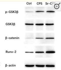
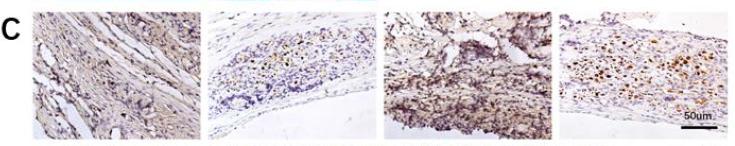
 Proper concentration of PLGA/MgO/Que increased the expression of osteogenesis associated genes including RUNX2, ALP, BMP2, COL1A1 and OPN in BMSCs; (B) in BMSCs, the expression of collagen I and Runx2 increased in Que membranes immersing medium; (C) Alizarin Red stain and ALP Stain showed Que could promote osteogenesis in 0.1% concentration; (D) cultured in membranes immersing extracts, Que increasing the expression of Collagen I and OPN, the scale is 20 μm (left) and 50 μm (right); (E) Statistical analysis of fluorescence intensity of Collagen I (left) and OPN (right); (F) and (G) 0.1% Que immersing medium increased the expression of Wnt3a and non-phospho catenin in BMSCs")
 The expression of liver typical function-related parameter (serum alaninetransaminase (ALT), Aspartate aminotransferase (AST) and kidney typical function-related parameters (blood urea nitrogen (BUN) and serum creatinine (Crea) from mice treated with PBS and 20 μg/mL RD-EVLP (n = 3). (B) H&E staining of the different groups' heart, liver, spleen, lung, and kidney section (Scale bar = 200 μm). (C) Schematic representation of the method used to research the anti-osteoporosis effect of RD-EVLP in OVX mice. (D-E) Immunohistochemical staining of RUNX2 and OCN protein expression in the femur section from different groups and osteoblasts exhibiting upregulated OCN and RUNX2 expression are explicitly highlighted with red arrows (Scale bar = 50 μm, n = 3). *P < 0.05, ***P < 0.001, and ns means not significant. (For interpretation of the references to colour in this figure legend, the reader is referred to the web version of this article.)")
 From top to bottom are the macroscopic view and microscopic view of Alizarin Red S-stained calcium nodules during osteogenic differentiation in h-BMSCs. (Scale bar = 10 μm). (B) The mRNA levels of typical osteogenic differentiation markers OCN, RUNX2 and BMP2 were measured by qRT-PCR at 0, 7, and 14 days. (C) The protein levels of typical osteogenic differentiation markers RUNX2 and BMP2 were detected by western-blotting at 0, 7, and 14 days. (D) Expression of hsa_circ_0001275/miR-422a during osteogenic differentiation of h-BMSCs at 0, 7, and 14 days. (E) Hsa_circ_0001275 was pictured under confocal microscopy by FISH in situ fluorescence hybridization technique. hsa_circ_0001275 appeared as red and DAPI staining as blue confirming that hsa_circ_0001275 was mainly located in the cytoplasm of h-BMSCs cells. *P < 0.05, **P < 0.01 and ****P < 0.0001. (For interpretation of the references to colour in this figure legend, the reader is referred to the web version of this article.)")
 and 12 (B) weeks (400 ×).")
 MIR155HG level was detected by RT-PCR assay in the BMSCs following cell transfection with OE-MIR155HG/OE-NC or infection with sh-MIR155HG/sh-NC. (B-F) The protein levels of OSX, OPN, ALP and RUNX2 in MIR155HG silenced and overexpressed BMSCs were detected by western blotting assay. (G-I) The levels of DKK1, p-β-catenin and β-catenin at translational level were tested by western blotting assay in MIR155HG silenced and overexpressed BMSCs. Following the downregulation or overexpression of MIR155HG, BMSC osteogenic differentiation ability was evaluated by (J-K) ALP activity and (L) staining with alizarin red S. (*P")
 The relative expression levels of miR-25-3p in ONFH-BMSCs and control BMSCs were examined by RT-qPCR analysis. (B) The BMSCs were transfected with miR-25-3p mimics or its control mimics NC, and the relative expression levels of miR-25-3p were determined by RT-qPCR analysis. (C) The BMSCs were treated with miR-25-3p inhibitor or its control inhibitor NC, and the relative expression levels of miR-25-3p were determined by RT-qPCR analysis. (D and E) The cell proliferation ability of BMSCs with corresponding treatment was revealed by the cell-cycle analysis, with the cell population of G1, G2, and S cell-cycle phase indicated. #p < 0.05 versus the corresponding NC group for comparing cell percentage in S phase; &p < 0.05 versus the corresponding NC group for comparing cell percentage in G1 phase; n = 3. (F and G) The protein expression levels of osteogenic marker genes in BMSCs of different groups were detected by western blot analysis. The quantitative analysis showed the protein expression levels of ALP, BMP2, RUNX2, and OCN were increased in the miRNA mimics group compared with the mimics NC group and were decreased in the miRNA inhibitor group compared with the inhibitor NC group. (H and I) The osteogenic differentiation ability of BMSCs at the 7th and 14th day after corresponding treatment was evaluated by alizarin red staining. Data were shown as mean ± SD. ∗∗p < 0.01, ∗∗∗p < 0.001, n = 3. Data between two groups were analyzed by Student's t test. Data among multiple groups were analyzed by one-way ANOVA test.")
 Surface morphology (FE-SEM) of cells adhered on different specimens after incubation for 4 h. (b) Cell proliferation ability evaluated through CCK-8 for 1, 3, and 5 days. (c and d) ALP quantitative and staining results of cells incubated on the different specimens for 7 and 14 days of osteogenic induction. (e and f) Alizarin red staining and quantitative results of the cells seeded on different specimens at 21 days of osteogenic induction. (g) mRNA levels of osteogenic differentiation-related genes (β-catenin, AXIN2 and ALP) in cells cultured on the different specimens at 14 days after seeding in osteogenic medium. (h) Western blotting assay of β-catenin, RUNX2 and OCN protein expression of MC3T3-E1 cells cultured on different specimens for 7 days, and (i) relative density quantification was normalized to β-actin.")
 Immunofluorescence staining of tibial sections. Blue: DAPI; green: MMP9; red: Runx2; magnification: ×200, scale bar: 20 μm. Statistics of (B) MMP9 and (C) Runx2 expression in the tibia. Values are expressed as mean ± standard error of the mean (n = 3–4 rats in each group). Statistical significance was evaluated by one-way ANOVA. ##P < 0.01 vs. Control; *P < 0.05, **P < 0.01 vs. CTX. Abbreviations: CTX, cyclophosphamide; AL, alendronate (7.35 mg/kg); DHG-L, deer-hide gelatin low dose (0.27 g/kg); DHG-M, deer-hide gelatin medium dose (0.54 g/kg); DHG-H, deer-hide gelatin high dose (1.08 g/kg); MMP9: matrix metalloproteinase-9; Runx2: runt-related transcription factor 2.")
 Overexpressing and knocking down tRF-23 respectively led to higher and lower levels of p-JAK2/STAT3. JAK2/STAT3 inhibitor treatment also significantly reduced levels of p-JAK2/STAT3 relative to tRF-23 overexpression in the absence of such inhibition (p-JAK2, normalized to total JAK2; p-STAT3, normalized to total STAT3). Data are presented as the mean ± SD (n = 3 independent experiments with two technical replicates per independent sample). (C) JAK2/STAT3 signaling inhibitor treatment suppressed the osteogenic differentiation of hBMSCs on day 14 relative to those in which tRF-23 was overexpressed without inhibitor treatment. Scale bars: 50 μm. Matrix mineralization and ALP activity were quantified based on absorbance. Data are presented as the mean ± SD (n = 3 independent experiments with two technical replicates per independent sample). (D and E) RUNX2, OCN, and ALP levels on day 14 of hBMSC osteogenesis were detected via qPCR and western blotting. Data are presented as the mean ± SD (n = 3 independent experiments with two technical replicates per independent sample). Data were analyzed using paired two-tailed Student’s t tests. ∗∗p < 0.01 was considered significant. OE, overexpression; shRNA, short hairpin RNA; T-JAK2, total JAK2; T-STAT3, total STAT3.")
 of rPTX3 treatment. B Cell morphology of MC3T3-E1 after treatment with different doses of rPTX3 (0, 50, 100, 200, 500, and 1000 ng/ml). Scale bar, 100 μm. C WB analysis of osteogenesis markers (ALP, Runx2 and COL1 at day 7, OCN and OPN at day 14) and apoptosis markers (Bcl-2 and Bax at 24 h) in Dex-stimulated MC3T3-E1 treated with or without rPTX3 and related quantification. Actin was used as an internal control. D, E Immunofluorescence staining and quantitative analysis of osteogenesis markers OCN (day 14), Runx2 (day 7). F Cell death/live analysis in Dex stimulated MC3T3-E1 treated with or without rPTX3 and related quantification. G Flow cytometry analysis in Dex stimulated MC3T3-E1 treated with or without rPTX3 and quantification. (Apoptotic cells: Q2 + Q3). H, I ALP staining and ARS staining in Dex stimulated MC3T3-E1 treated with or without rPTX3 and quantification. Control: Standard OIM, n = 3; Dex: Standard OIM co-cultured with dexamethasone (10 μM), n = 3; Dex+rPTX3: Standard OIM co-cultured with dexamethasone (10 μM) and rPTX3 (200 ng/mL), n = 3. Statistical analysis: Dunnett’s post-hoc tests (n = 3 independent experiments). Error bars: standard deviation, SD. The images provided in all figures represent typical examples from each experimental group.")
 Representative images of ALP staining and (B) quantification of ALP activity (n=3; ×5 magnification). (C) Representative images of ARS staining and (D) quantification of ARS staining (n=3; ×5 magnification). The mRNA expression levels of (E) Col1, (F) Osterix, (G) Runx2 and (H) Alp (n=3). (I) Representative western blot images, and semi-quantification of the protein expression levels of (J) Runx2 and (K) Osterix (n=3). Data are presented as the mean ± standard deviation. *P")
. *P")
 Construction of IR BMSCs and validations for diminished osteogenic and migrative potential of IR BMSCs. (a) Among 2, 4, and 6 Gy, notable changes were observed starting from 2 Gy, as determined by ALP activity assay. (b) CCK-8 assay revealed no significant differences in cell proliferation between BMSCs and IR BMSCs (2 Gy radiation). (c) Flow cytometry analysis of apoptosis levels showed no significant difference between IR BMSCs and its control. (d) Expression of ALP, OSX, and RUNX2. (e) The results of ALP, OSX, and RUNX2 expression. (f) Histograms showed the quantification of band intensities. (g) ALP staining results. (h) ARS staining results. (i) Would healing assay showed IR BMSCs’ decreased migrative potential. (j) Transwell assay results. Scalar bar = 100 µm. Data are represented as mean ± SEM, n = 3")
 Micro-CT 3D reconstruction images of the Achilles tendon 4 and 8 weeks after modeling. (B&C) H&E and SOFG staining images of normal Achilles tendon and Achilles tendon specimens 4 weeks and 8 weeks after modeling. (D&E) Immunohistochemical staining of RUNX2 and OCN expression in Achilles tendon specimens of mice in each group. (F) Statistical chart of quantitative analysis of Micro-CT results. (G&H) Statistics of RUNX2 and OCN positive cell rates. n = 6 in each group, data are presented as mean ± SD. Scale bar = 50 μm. *p<0.05 Vehicle group vs TF group.")
 in the Control group, marked increase in expression (black arrows) in L-MTZ, increased expression (black arrows) in S-MTZ. BMP-2: expression findings between the groups, moderate expression (black arrow) in the Control group, increase in expression (black arrows) in L-MTZ, marked expression (black arrow) in S-MTZ. Runx2: moderate expression (black arrows) in the Control group, increase in expression (black arrows) in L-MTZ, marked expression (black arrows) in S-MTZ. ALP: moderate expression (black arrow) in the Control group, marked increase in expression (black arrows) in L-MTZ, moderate expression (black arrow) in S-MTZ. OCN: moderate expression (black arrow) in the control group, markedly increased expression (black arrow) in L-MTZ, and increased expression (black arrow) in S-MTZ. RANKL: Similar RANKL expressions (black arrows) in (A) Control group, (B) L-MTZ, and (C) S-MTZ, Streptavidin biotin peroxidase method, Scale bars = 50 μm Representative histopathological images among the groups. (A) Marked fibrous tissue, moderate new bone formation (white arrow with a black border), and residual graft materials (RG) in the Control group. (B) Decreased fibrous tissue, moderate residual graft material (RG), and marked new bone formation (white arrow with a black border) in L-MTZ. (C) Decreased fibrous tissue, moderately increased new bone formation (white arrow with a black border), and residual graft materials (RG) in S-MTZ. HE, Scale bars = 200 μm")
 ALP staining (100 × ); (B) Alizarin red staining (100 × ); (C–I) Relative expression levels of mRNA and protein of ALP, Runx2 and OCN (n = 3), ∗∗∗p < 0.001, ∗∗p < 0.01, ∗p < 0.05. (For interpretation of the references to colour in this figure legend, the reader is referred to the Web version of this article.)")
 Immunohistochemical staining and quantification of average optical density for RUNX2 and OPN of the alveolar bone defect. (b) Immunohistochemical staining and quantification of average optical density for PIEZO1, ITGα5 and TNS1 of the alveolar bone defect. (c) Immunohistochemical co-staining of PIEZO1 (green) and ITGα5 (red). Yellow indicates their spatial co-localization. Right panel: Quantitative curve of the fluorescence assay analyzed by Image J software. (**P")
 Exosome uptake assay demonstrating the internalization of PKH67-labeled exosomes by BMSCs. (B) Cytotoxicity test demonstrating the appropriate exosomes dose for treatment (n = 3 per group). (C-F) ALP staining and Alizarin red staining indicating mineralization in BMSCs. Quantitative analysis illustrates the mineralization proportion, which was enhanced by L-exos and reduced by H-exos in comparison to the PBS group (n = 3 per group). (J-M) Western blotting and (G-I) qRT-PCR analyses of osteogenesis-related genes in BMSCs treated with 25 µg/ml L-exos, 25 µg/ml H-exos, or equal quantities of PBS. Protein and mRNA levels of RUNX2, BMP2, and OCN were downregulated in H-exo groups compared to L-exo group (n = 3 per group). The error bars represent the means ± SDs.")
| Product: | RUNX2 Antibody |
| Catalog: | AF5186 |
| Description: | Rabbit polyclonal antibody to RUNX2 |
| Application: | WB IHC IF/ICC |
| Cited expt.: | WB, IHC, IF/ICC |
| Reactivity: | Human, Mouse, Rat |
| Prediction: | Pig, Bovine, Horse, Sheep, Rabbit, Chicken, Xenopus |
| Mol.Wt.: | 56kDa,48kDa(Observed); 57kD(Calculated). |
| Uniprot: | Q13950 |
| RRID: | AB_2837672 |
Control Products

Related Downloads
Protocols
Product Info
*The optimal dilutions should be determined by the end user. For optimal experimental results, antibody reuse is not recommended.
*Tips:
WB: For western blot detection of denatured protein samples. IHC: For immunohistochemical detection of paraffin sections (IHC-p) or frozen sections (IHC-f) of tissue samples. IF/ICC: For immunofluorescence detection of cell samples. ELISA(peptide): For ELISA detection of antigenic peptide.
Cite Format: Affinity Biosciences Cat# AF5186, RRID:AB_2837672.
Fold/Unfold
Acute myeloid leukemia 3 protein; Alpha subunit 1; AML3; CBF alpha 1; CBF-alpha-1; CBFA1; CCD; CCD1; Cleidocranial dysplasia 1; Core binding factor; Core binding factor runt domain alpha subunit 1; Core binding factor subunit alpha 1; Core-binding factor subunit alpha-1; MGC120022; MGC120023; Oncogene AML 3; Oncogene AML-3; OSF 2; OSF-2; OSF2; Osteoblast specific transcription factor 2; Osteoblast-specific transcription factor 2; OTTHUMP00000016533; PEA2 alpha A; PEA2-alpha A; PEA2aA; PEBP2 alpha A; PEBP2-alpha A; PEBP2A1; PEBP2A2; PEBP2aA; PEBP2aA1; Polyomavirus enhancer binding protein 2 alpha A subunit; Polyomavirus enhancer-binding protein 2 alpha A subunit; Runt domain; Runt related transcription factor 2; Runt-related transcription factor 2; RUNX2; RUNX2_HUMAN; SL3 3 enhancer factor 1 alpha A subunit; SL3-3 enhancer factor 1 alpha A subunit; SL3/AKV core binding factor alpha A subunit; SL3/AKV core-binding factor alpha A subunit;
Immunogens
A synthesized peptide derived from human RUNX2, corresponding to a region within the internal amino acids.
- Q13950 RUNX2_HUMAN:
- Protein BLAST With
- NCBI/
- ExPASy/
- Uniprot
MASNSLFSTVTPCQQNFFWDPSTSRRFSPPSSSLQPGKMSDVSPVVAAQQQQQQQQQQQQQQQQQQQQQQQEAAAAAAAAAAAAAAAAAVPRLRPPHDNRTMVEIIADHPAELVRTDSPNFLCSVLPSHWRCNKTLPVAFKVVALGEVPDGTVVTVMAGNDENYSAELRNASAVMKNQVARFNDLRFVGRSGRGKSFTLTITVFTNPPQVATYHRAIKVTVDGPREPRRHRQKLDDSKPSLFSDRLSDLGRIPHPSMRVGVPPQNPRPSLNSAPSPFNPQGQSQITDPRQAQSSPPWSYDQSYPSYLSQMTSPSIHSTTPLSSTRGTGLPAITDVPRRISDDDTATSDFCLWPSTLSKKSQAGASELGPFSDPRQFPSISSLTESRFSNPRMHYPATFTYTPPVTSGMSLGMSATTHYHTYLPPPYPGSSQSQSGPFQTSSTPYLYYGTSSGSYQFPMVPGGDRSPSRMLPPCTTTSNGSTLLNPNLPNQNDGVDADGSHSSSPTVLNSSGRMDESVWRPY
Predictions
Score>80(red) has high confidence and is suggested to be used for WB detection. *The prediction model is mainly based on the alignment of immunogen sequences, the results are for reference only, not as the basis of quality assurance.
High(score>80) Medium(80>score>50) Low(score<50) No confidence
Research Backgrounds
Transcription factor involved in osteoblastic differentiation and skeletal morphogenesis. Essential for the maturation of osteoblasts and both intramembranous and endochondral ossification. CBF binds to the core site, 5'-PYGPYGGT-3', of a number of enhancers and promoters, including murine leukemia virus, polyomavirus enhancer, T-cell receptor enhancers, osteocalcin, osteopontin, bone sialoprotein, alpha 1(I) collagen, LCK, IL-3 and GM-CSF promoters. In osteoblasts, supports transcription activation: synergizes with SPEN/MINT to enhance FGFR2-mediated activation of the osteocalcin FGF-responsive element (OCFRE) (By similarity). Inhibits KAT6B-dependent transcriptional activation.
Phosphorylated; probably by MAP kinases (MAPK). Phosphorylation by HIPK3 is required for the SPEN/MINT and FGF2 transactivation during osteoblastic differentiation (By similarity). Phosphorylation at Ser-451 by CDK1 promotes endothelial cell proliferation required for tumor angiogenesis probably by facilitating cell cycle progression. Isoform 3 is phosphorylated on Ser-340.
Nucleus.
Specifically expressed in osteoblasts.
A proline/serine/threonine rich region at the C-terminus is necessary for transcriptional activation of target genes and contains the phosphorylation sites.
Research Fields
· Human Diseases > Cancers: Overview > Transcriptional misregulation in cancer.
References
Application: WB Species: Mice Sample: bone and osteoid tissues
Application: IHC Species: Mice Sample: bone and osteoid tissues

Application: IF/ICC Species: Rat Sample:
Restrictive clause
Affinity Biosciences tests all products strictly. Citations are provided as a resource for additional applications that have not been validated by Affinity Biosciences. Please choose the appropriate format for each application and consult Materials and Methods sections for additional details about the use of any product in these publications.
For Research Use Only.
Not for use in diagnostic or therapeutic procedures. Not for resale. Not for distribution without written consent. Affinity Biosciences will not be held responsible for patent infringement or other violations that may occur with the use of our products. Affinity Biosciences, Affinity Biosciences Logo and all other trademarks are the property of Affinity Biosciences LTD.