
.
Bands result from membrane strip incubation.")
, diluted 1/600 was used as secondary antibody.")
 and 1, 10, and 100 IU/mL hCG for 24 h. The protein levels of CD200 and CD200R (A), Cyp19a1, IL-1β, and TNFα (B), and of NF-κB, p-NF-κB, p38 MAPK, and p-p38 MAPK (C) were examined. HUVECs were treated with condition culture medium from KGN for 24 h. (D) The levels of vasoactive proteins VEGF and VEGFR2, and cell junction proteins of Occludin, Claudin 5, and ZO-1 in HUVECs.")

 Schematic showing the protein pull down experimental setup. Immunoblotting (left panel) and its semi-quantitative analysis (right panel)
of (b) VEC, SOD1, claudin-5 and α-tublin from whole cell lysate and pulled down by different NPs. (c) Y658 (p-VEC(Y658)), Y731 (p-VEC(Y731), VEC
and α-tublin from the whole cell lysate with/without the Src kinase inhibitor, PP1.")

 Protein expression level of Occludin and Claudin-5, 24 h after HI brain injury.")
 urea (TPPU) recovered expression of tight junction proteins down-regulated by permanent cerebral middle artery occlusion (pMCAO). (A) Brains were removed from animals at 1 or 3 days after surgery, and cortical tissue was homogenized and assayed by Western blot against Occludin, ZO-1, and Claudin-5. (B) Quantitation of relative levels of ZO-1 (n = 5). (C) Quantitation of Occludin levels (n = 5). (D) Quantitation of relative levels of Claudin-5 (n = 5). Data are the mean ± SEM. **p < 0.01, ***p < 0.001, ****p < 0.0001.")
. Effect of miR-429 on the permeability of the BSCB via HRP flux. Data represent the mean ± S.D. (n ¼ 3, each). *P < 0.05 vs. agomiR-429 NC group, #P < 0.05 vs.
antagomiR-429 NC group. (B). RT-qPCR analysis of TJ-related protein expression in the BSCB following changes in the expression of miR-429. Data represent the mean ± S.D. (n ¼ 3,
each). *P < 0.05 vs. agomiR-429 NC group, #P < 0.05 vs. antagomiR-429 NC group. (CeD). Western blotting analysis of TJ-related protein expression in the BSCB following changes in
the expression of miR-429. GAPDH was used as an endogenous control. Representative protein expression and their integrated light density values (IDVs) are shown. Data represent
the mean ± S.D. (n ¼ 3, each). *P < 0.05, vs. agomiR-429 NC group, #P < 0.05, vs. antagomiR-429 NC group. (E). Immunofluorescence staining of TJ-related proteins in the different
groups. ZO-1 (red), occludin (red), and claudin-5 (red) are labeled with their respective antibodies. Nuclei (blue) are labeled with DAPI (n ¼ 3). Scale bars represent 20 mm. (For
interpretation of the references to colour in this figure legend, the reader is referred to the Web version of this article.)")
, p-β-catenin (B), Dvl1 (C), p-GSK3β (D), nuclear β-catenin (E), ZO-1 (F), claudin-5 (G) and occludin (H). Date are expressed as mean
± SD, n = 3. *P < 0.05, **P < 0.01 vs. Sham group; ▴P < 0.05, ▴▴P < 0.01 vs. MCAO/R group.")
 Immunofluorescence assay of claudin-5. Scale bar, 50 μm.
Yellow triangle indicate the joint of claudin-5.")
 Representative brains with EB staining from each group. (B) EB content in the hippocampus of each
group after 4 weeks of EE intervention. (C) Representative Western blot analysis. (D-F) Quantification of the relative expression of MMP-9(D), ZO-1(E), and claudin-5
(F). (G) Representative microphotograph of the ultrastructure of BBB. The black arrow refers to the tight junction, and the red arrow refers to the large gaps between
the tight junctions. L: capillary cavity; EC: endothelial cell; All data are expressed as the mean ± SEM (n = 6 per group). # P < 0.05, ## P < 0.01 compared with the
Sham + SE group; * P < 0.05 compared with the 2VO + SE group.")
 Representative western blots and quantification data of TJ and AJ proteins in M, IM groups.
Columns represent mean ± SD, n = 5. (C) Claudin-5, Occludin, p120-Catenin, β-Catenin/CD31/Hoechst staining of sections from the spinal cord in M, IM groups. Red:
Claudin-5/Occludin/p120-Catenin/β-Catenin; green: CD31; blue: Hoechst. Scale bar, 20 μm. (
$
p < 0.05; $$p < 0.01; $$$p < 0.001).")
 Representative images of ICH staining for MMP-9 in the cerebral cortex vascular tissues after SAH. (B) Representative western blot bands and quantitative analysis of the expression of (C) MMP-9, (D) tight junction-related proteins, (E) claudin-5, (F) occludin, and (G) ZO-1 in the ipsilateral cortex at 24 h post-SAH. Scale bar = 50 μm; *p < 0.05 vs. sham group, # p < 0.05 vs. SAH + vehicle group; n = 6 per group.")
. B BSEP, OATP2 and NTCP1 expression in liver tissues of different groups (n = 3 for each group). C, D Liver COL1A1, α-SMA, TGF-β1 and β-catenin expression among different groups (n = 3 for each group). *p < 0.05, **p < 0.01, ***p < 0.001, ****p < 0.0001")
 Western blotting results of ZO1, occludin, and claudin-5 in each group and quantification (n = 6/group, mean ± SD). (b) Brain water content in each group after TBI (n = 6/group, mean ± SD). (c, d) Fluorescence of Evans blue and Evans blue extravasation in each group around the injury site (n = 12/group, mean ± SD) (scale bar = 50 μm, 200x) (∗P < 0.05 vs. the overexpression vector group, &P < 0.05 vs. the knockdown vector group by one-way ANOVA).")
, Occludin (B), Claudin-5 (C), and VE-Cadherin (D) were selected as the markers reflecting the integrity of the vascular endothelium. CD31 was applied explicitly for labeling the microvessels. Scale bar indicates 50 μm. (E) The qPCR analysis of ZO-1, Occludin, Claudin-5, and VE-Cadherin transcription in hBMECs treated by multiple dosages of IL-17A (0, 1, 5, 10, and 20 ng/mL). GAPDH was used as the internal reference. Data were presented as mean ± SD from three independent experiments. (F) Western blot analysis of ZO-1, Occludin, Claudin-5, and VE-Cadherin in hBMECs in response to multiple dosages of IL-17A. β-Actin was used as the loading control.")
 and claudin-5 (C) were detected by immunofluorescence staining, in which VE-cadherin and claudin-5 were labeled with green fluorescence, while the nuclear was stained by DAPI with blue fluorescence. The mean values ± SD was shown for each bar. * (P < 0.05) or ** (P < 0.01) or *** (P < 0.001) represents significance, ns represents no significance. Original magnification: × 20. BLM: Bleomycin, WYHZTL: Wenyang Huazhuo Tongluo formula")
 electrophoretic and statistical correlation histograms of occludin and claudin-5 proteins in brain tissue of rats in each group. (b,d) electrophoretic and statistical correlation histograms of MMP2 and MMP9 proteins in brain tissue of rats in each group. Data are presented as mean ± SEM; *P < 0.05, **P < 0.01 vs sham group; #P < 0.05, ##P < 0.01 vs model group.")
 PAECs were co-cultured with wild-type or resistin knockout PAMs, and the PAMs were infected by H. parasuis (100 MOI) for 2 h. mRNA levels of claudin-5, occludin, JAM-1, JAM-2, and VE-cadherin in PAECs were determined using qRT-PCR (a, b), and relative protein levels were detected using western blot analysis (c). (d, e, f) PAECs were stimulated or unstimulated with recombined resistin protein (2.5, 5, or 0 nM) for 2 h. Cells were lysed, mRNA levels of claudin-5 and occludin were analysed using qRT-PCR (d, e), and protein levels were analysed using western blot analysis (f). The mRNA level of each gene was standardized to GAPDH. The antibodies against claudin-5, occludin, JAM-1, JAM-2, and VE-cadherin were diluted at 1/1000, and the antibody against β-actin was diluted at 1/50000. β-actin served as a loading control, and the relative protein level was calculated with ImageJ software and normalized to β-actin. **p")
, with unprocessed rats serving as the control. (a) Brain tissues were collected from sacrificed rats to calculate infarction area by TTC staining. (b) HE staining was applied to evaluate histopathological conditions in rat brain (magnification ×100, scale bar: 100 μm). (c) TUNEL staining indicated neuron nuclei (brown). Red arrows indicated TUNEL-positive neurons after I/R injury (magnification ×100, scale bar: 100 μm). (d–j) Western blot was employed to detect relative protein levels of VWF, Occludin, Claudin-5, ZO-1, LC3I, LC3II, and P62 in rat brain tissues. GAPDH was used as the loading control. (k–n) Western blot was applied to detect relative protein levels of ATF5, ATG7, and Beclin1 in rat brain tissues. GAPDH was used as the loading control. ** p < 0.01, *** p < 0.001 vs Control; ++ p < 0.01, +++ p < 0.001 vs I/R, ischemia/reperfusion; DHA, dihydroartemisinin; VWF, von Willebrand factor; ATF5, transcription factor 5; ATG7, autophagy-related gene 7; ZO-1, zonula occludens 1; LC3, microtubule associated protein 1 light chain 3 alpha; GAPDH, glyceraldehyde-3-phosphate dehydrogenase; TTC, triphenyltetrazolium chloride; HE, hematoxylin and eosin.")
 in the hippocampus cortex of rats stimulated by D-gal. (a) The representative images and statistical analysis of relative expression for claudin-1, claudin-5, occludin CX43, and ZO-1 in the hippocampus. (b) The representative images and statistical analysis of relative expression for claudin-1, claudin-5, occludin CX43, and ZO-1 in the cortex. Values are mean ± SEM of six independent experiments. #p < 0.05, ##p < 0.01, ###p < 0.001 vs. the control group, ∗p < 0.05, ∗∗p < 0.01 vs. the model group.")
 or negative control (NC) siRNA (1.5 μL/10 g body weight) 48 h prior to MCAO/R surgery. Twenty-four hours after the surgery, brains were harvested for immunohistochemical and Western-blotting assays. (A) Immunohistochemical localization and quantification of ICAM-1 and VCAM-1 expression (n = 6). (B) Representative immunostaining images and quantification of neutrophiles (MPO) and macrophages (F4/80) (n = 6). (C-E) Representative Western-blotting images and quantification of tight junction proteins (Claudin-5, Occludin, and ZO-1) (n = 3). Scale bars: 40 μm. One-way ANOVA followed by the post hoc Tukey test. All data are mean ± SD, *P")
 were transfected with P-glycoprotein (P-gp) or negative control (NC) siRNA, or un-transfected, and then subjected to either oxygen glucose deprivation/reoxygenation (OGD/R) treatment or normal culture conditions. Twenty-four hours thereafter, cells were harvested for immunofluorescence staining, adhesion and transendothelial migration, real-time PCR gene expression and Western-blotting analyses. (A) Representative Western-blotting images and quantification of P-gp levels (n = 3). (B) Quantification of mRNA levels for TNF-α, IL-1β, MMP-2, and MMP-9 via quantitative real-time PCR assay (n = 4). (C) Representative immunofluorescence staining images and quantification of ICAM-1 and VCAM-1 expression (n = 3). (D) Representative images and quantification for fluorochrome-labeled leukocyte adhesion to endothelial cells and transendothelial migration (n = 3). (E) Representative immunofluorescence staining images for tight junction proteins (Claudin-5, Occludin, and ZO-1). (F) Representative immunoblots and quantification for the expressions of Claudin-5, Occludin, and ZO-1 (n = 3). Scale bars, 40 μm. One-way ANOVA followed by the post hoc Tukey test for A, E, and F. Mann-Whitney test for B, C, and D. All data are mean ± SD, *P")
 challenge, lung tissues were obtained for claudin5, occludin and ZO-1 expression. The proteins of claudin5, occludin, ZO-1 and β-actin were detected by the Western blot (A). The β-actin was used as the internal control. Quantification of claudin5, occludin and ZO-1 are shown in (B), (C) and (D) respectively. Distributions of claudin5 (E), occludin (F) and ZO-1 (G) and associated fluorescence intensity analysis of claudin5 (H), occludin (I) and ZO-1 (J) in the lungs of mice in the WT, WT + LPS, PLD2-/- and PLD2-/- + LPS groups (magnification × 200; scale bar, 50 μm). All data are presented as the mean ± SD of three independent experiments. *p < 0.05 vs the WT group. ∮p < 0.05 vs the WT + LPS group.")
 challenge, lung tissues were obtained for claudin5, occludin and ZO-1 expression. The proteins of claudin5, occludin, ZO-1 and β-actin were detected by the Western blot (A). The β-actin was used as the internal control. Quantification of claudin5, occludin and ZO-1 are shown in (B), (C) and (D) respectively. Distributions of claudin5 (E), occludin (F) and ZO-1 (G) and associated fluorescence intensity analysis of claudin5 (H), occludin (I) and ZO-1 (J) in the lungs of mice in the WT, WT + LPS, PLD2-/- and PLD2-/- + LPS groups (magnification × 200; scale bar, 50 μm). All data are presented as the mean ± SD of three independent experiments. *p < 0.05 vs the WT group. ∮p < 0.05 vs the WT + LPS group.")

 Hematoxylin and eosin staining of representative colonic tissues from the three groups. Compared with the WT and MgT groups, the APP/PS1 group showed slight mucosal epithelial cell damage, hyperchromatic nuclear pyknosis, enhanced eosinophilia in the cytoplasm (black arrows), and slight inflammatory cell infiltration in the lamina propria (red arrows). (B) Western blotting and quantification of protein levels of ZO-1, occludin, and claudin-5 in the WT, APP/PS1, and MgT groups. Results presented as means ± SEM (n = 3), analyzed by one-way analysis of variance followed by Tukey’s honestly significant difference test; *P < 0.05, **P < 0.01, and ***P < 0.001 between the APP/PS1 and MgT groups. APP/PS1: Amyloid-β precursor protein and mutant human presenilin 1; MGT: magnesium-L-threonate; WT: wild-type; ZO-1: zona occludens 1.")
 24 h after intravenous EB injection. The results showed a significant reduction in EB extravasation after VNS treatment but an increase in EB extravasation after C48/80 treatment (n = 5). C: Representative western blot showing representative ZO-1, Occludin, and Claudin-5 protein bands in the cerebral cortex. D-F: Quantitative results (n = 4). D-F: Quantitative results (n = 5). The data are presented as the mean ± SD. * P < 0.05, ** P < 0.01, *** P < 0.001 using one-way ANOVA.")
 perfusion and also measured the protein levels of tight junction in the peri-hematoma region at day 3 after ICH. A, Immunofluorescence images of mice brain perfused with FITC-dextran. we co-stained CD31 and ZO-1 as markers of blood vessels. The image on the right shows the sites taken by confocal microscopy. Scale bars = 100um, all images captured with 40 × objective. magnification = 400x. B, OLT1177 significantly reduces the extent of FITC-dextran Leakage at day 3 after ICH. n = 5–6 per group. **P < 0.01C, Western blot images show the expression of ZO-1, occludin, and claudin-5 in the brains of indicated groups of mice receiving vehicle or OLT1177. D, Data points show the protein expression levels of ZO-1, occludin, and claudin-5 in the brains of indicated groups of mice receiving vehicle or OLT1177. n = 4 mice per group. Data are presented as mean ± SD.")
 Representative immunohistochemistry images of ZO-1 and Claudin-5 in the mouse retina (magnification, ×40). (B-C) Quantification of the expression of ZO-1 (B) and Claudin-5 (C). (D) Western blotting detection of Bax, Bcl-2, and Caspase-9. (E-F) Bar charts indicate Bax/Bcl-2 (E) and active caspase-9/pro-caspase-9 (F). nsp > 0.05, ***p ≤ 0.001.")
 for 24 h. (a) The levels of vasoactive proteins VEGF and VEGFR2, and cell junction proteins of Occludin, Claudin 5 and ZO-1 were examined by western blotting. (b) The relative protein levels of A are expressed as fold changes over Ctrl (n=3). Data were presented as means ± SD, *P < 0.5, **P < 0.01, ***P < 0.001 versus Ctrl. (c) Fluorescent photomicrographs of HUVEC monolayer stained with TRITC-phalloidin, final concentration 20 mg/ml.")
 Polyacrylamide gel electrophoresis and protein expression levels of claudin-5, occludin, E-cadherin, and VE-cadherin in the upper lobe of the right lung. (B) Polyacrylamide gel electrophoresis and protein expression levels of claudin-5, occludin, E-cadherin, and VE-cadherin in the middle lobe of the right lung. (C) Polyacrylamide gel electrophoresis and protein expression levels of claudin-5, occludin, E-cadherin, and VE-cadherin in the lower lobe of the right lung. (D) Protein expression levels of claudin-5, occludin, E-cadherin, and VE-cadherin in the whole right lung. The average WB results of the upper, middle, and lower lobes were taken to compare the expression of protein in the whole lung. *p < 0.05. APRV, airway pressure release ventilation; LTV, low tidal volume ventilation.")
 The content of S100β in hippocampus on the third day following exposure to acute noise at different intensities. (b) Expression levels of blood-brain barrier-related proteins in the hippocampus of male rats on the third day following exposure to acute noise at different intensities. Quantification of ZO-1 (c), Claudin-5 (d), and Occludin (e) protein expression. (f) Expression levels of blood-brain barrier-related proteins in the hippocampus of male rats one month after exposure to acute noise at 135dB. Quantification of ZO-1 (g), Claudin-5 (h), and Occludin (i) protein expression. N = 4 rats per condition.")
 The expression level of ZO-1 and claudin-5 in lung tissue tested using immunohistochemistry (×200) and IOD of ZO-1 and claudin-5 in lung tissue of rats in all groups (n = 8), (b) ZO-1, Ang-2, SP-D and claudin-5 protein expression bands and relative protein expressions in lung tissue of rats in all groups (n = 3), (c) nuclear Nrf2, cytosolic Nrf2, HO-1, NQO1, SOD2, GCLC and GCLM protein expression bands and relative protein expressions in lung tissue of rats in all groups (n = 3). The values are expressed as mean ± SE. aP < 0.05 vs the control group, aaP < 0.01 vs the control group, bP < 0.05 vs the model group, bbP < 0.01 vs the model group.")
 The expression level of ZO-1 and claudin-5 in lung tissue tested using immunohistochemistry (×200) and IOD of ZO-1 and claudin-5 in lung tissue of rats in all groups (n = 8), (b) ZO-1, Ang-2, SP-D and claudin-5 protein expression bands and relative protein expressions in lung tissue of rats in all groups (n = 3), (c) nuclear Nrf2, cytosolic Nrf2, HO-1, NQO1, SOD2, GCLC and GCLM protein expression bands and relative protein expressions in lung tissue of rats in all groups (n = 3). The values are expressed as mean ± SE. aP < 0.05 vs the control group, aaP < 0.01 vs the control group, bP < 0.05 vs the model group, bbP < 0.01 vs the model group.")
 The penetration of FITC-dextran. n = 6. Scale bar, 2 mm. (B) Quantification of the permeability (A) of the product of FITC-dextran. (C) The presence of infiltrated IgG in each group. n = 6. Scale bar, 10 μm. (D) Quantitation of IgG (C) perivascular accumulation. (E–G) Intercellular junctions were visualized in each group. n = 6. Scale bar, 50 μm. (H) Quantification of EB extravasation. n = 5. (I) Ultrastructure of the BBB in each group was analyzed via TEM. n = 6. Scale bar, 250 nm. The data represent the means ± SD. *P < 0.05; **P < 0.01; ***P < 0.001")
, IL-6 (I), IL-4 (J), and IL-10 (K) in each group of mice after butyric acid or valeric acid treatment, n = 4. The data are expressed as the means ± SDs, with statistical significance among multiple groups determined by one-way ANOVA: *P")
 Representative fluorescence images of BTB integrity detected by the biotin tracer assay. The white asterisk (*) indicates the permeation of biotin into the seminiferous lumen. Scale bars: 100 μm. (B) Box plot illustrating the extent of BTB damage, which was calculated randomly from 50–60 seminiferous tubules for each group. (C-D) Representative images of tight junction proteins were examined by western blotting in mouse testes (n = 3). (E-H) The relative protein expression of ZO-1 (E), Occludin (F), Claudin5 (G) and Claudin11 (H) was quantified by ImageJ and normalized to vinculin or β-actin levels (n = 3). The data are presented as the mean ± SD. Statistical analyses were carried out using one-way ANOVA followed by Tukey’s post hoc test. *P")
| Product: | Claudin 5 Antibody |
| Catalog: | AF5216 |
| Description: | Rabbit polyclonal antibody to Claudin 5 |
| Application: | WB IHC IF/ICC |
| Cited expt.: | WB, IHC, IF/ICC |
| Reactivity: | Human, Mouse, Rat, Pig, Bovine, Monkey |
| Prediction: | Pig, Bovine, Rabbit, Chicken, Xenopus |
| Mol.Wt.: | 23 kDa(Observed); 23kD(Calculated). |
| Uniprot: | O00501 |
| RRID: | AB_2837702 |
Control Products

Related Downloads
Protocols
Product Info
*The optimal dilutions should be determined by the end user. For optimal experimental results, antibody reuse is not recommended.
*Tips:
WB: For western blot detection of denatured protein samples. IHC: For immunohistochemical detection of paraffin sections (IHC-p) or frozen sections (IHC-f) of tissue samples. IF/ICC: For immunofluorescence detection of cell samples. ELISA(peptide): For ELISA detection of antigenic peptide.
Cite Format: Affinity Biosciences Cat# AF5216, RRID:AB_2837702.
Fold/Unfold
Androgen withdrawal and apoptosis induced protein RVP1 like; AWAL; BEC 1; BEC1; Claudin 5 (transmembrane protein deleted in velocardiofacial syndrome); Claudin-5; Claudin5; CLD5_HUMAN; CLDN 5; Cldn5; CPETR L1; CPETRL 1; CPETRL1; TMDVCF; TMVCF; Transmembrane protein deleted in VCFS; Transmembrane protein deleted in velocardiofacial syndrome;
Immunogens
A synthesized peptide derived from human Claudin 5, corresponding to a region within C-terminal amino acids.
- O00501 CLD5_HUMAN:
- Protein BLAST With
- NCBI/
- ExPASy/
- Uniprot
MGSAALEILGLVLCLVGWGGLILACGLPMWQVTAFLDHNIVTAQTTWKGLWMSCVVQSTGHMQCKVYDSVLALSTEVQAARALTVSAVLLAFVALFVTLAGAQCTTCVAPGPAKARVALTGGVLYLFCGLLALVPLCWFANIVVREFYDPSVPVSQKYELGAALYIGWAATALLMVGGCLLCCGAWVCTGRPDLSFPVKYSAPRRPTATGDYDKKNYV
Predictions
Score>80(red) has high confidence and is suggested to be used for WB detection. *The prediction model is mainly based on the alignment of immunogen sequences, the results are for reference only, not as the basis of quality assurance.
High(score>80) Medium(80>score>50) Low(score<50) No confidence
Research Backgrounds
Plays a major role in tight junction-specific obliteration of the intercellular space.
Cell junction>Tight junction. Cell membrane>Multi-pass membrane protein.
Belongs to the claudin family.
Research Fields
· Cellular Processes > Cellular community - eukaryotes > Tight junction. (View pathway)
· Environmental Information Processing > Signaling molecules and interaction > Cell adhesion molecules (CAMs). (View pathway)
· Human Diseases > Infectious diseases: Viral > Hepatitis C.
· Organismal Systems > Immune system > Leukocyte transendothelial migration. (View pathway)
References
Application: WB Species: Mouse Sample: TM4 Sertoli cells
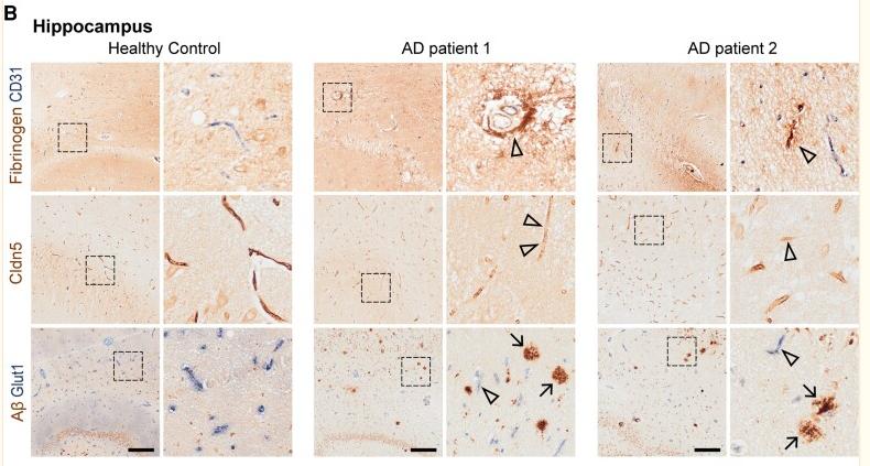
Application: IHC Species: Human Sample: hippocampus

Application: WB Species: Mouse Sample:

Application: IF/ICC Species: Mouse Sample:

Application: WB Species: human Sample:
Restrictive clause
Affinity Biosciences tests all products strictly. Citations are provided as a resource for additional applications that have not been validated by Affinity Biosciences. Please choose the appropriate format for each application and consult Materials and Methods sections for additional details about the use of any product in these publications.
For Research Use Only.
Not for use in diagnostic or therapeutic procedures. Not for resale. Not for distribution without written consent. Affinity Biosciences will not be held responsible for patent infringement or other violations that may occur with the use of our products. Affinity Biosciences, Affinity Biosciences Logo and all other trademarks are the property of Affinity Biosciences LTD.