Product: CD83 Antibody
Catalog: AF5233
Description: Rabbit polyclonal antibody to CD83
Application: WB IHC IF/ICC
Cited expt.: WB, IF/ICC
Reactivity: Human, Mouse, Rat
Prediction: Pig
Mol.Wt.: 23 kDa; 23kD(Calculated).
Uniprot: Q01151
RRID: AB_2837719

View similar products>>

   Size Price Inventory
 100ul $280 In stock
 200ul $350 In stock

Lead Time: Same day delivery

For pricing and ordering contact:
Local distributors

Product Info

Source:
Rabbit
Application:
WB 1:500-1:2000, IF/ICC 1:100-1:500, IHC 1:50-1:200
*The optimal dilutions should be determined by the end user.
*Tips:

WB: For western blot detection of denatured protein samples. IHC: For immunohistochemical detection of paraffin sections (IHC-p) or frozen sections (IHC-f) of tissue samples. IF/ICC: For immunofluorescence detection of cell samples. ELISA(peptide): For ELISA detection of antigenic peptide.

Reactivity:
Human,Mouse,Rat
Prediction:
Pig(88%)
Clonality:
Polyclonal
Specificity:
CD83 Antibody detects endogenous levels of total CD83.
RRID:
AB_2837719
Cite Format: Affinity Biosciences Cat# AF5233, RRID:AB_2837719.
Conjugate:
Unconjugated.
Purification:
The antiserum was purified by peptide affinity chromatography using SulfoLink™ Coupling Resin (Thermo Fisher Scientific).
Storage:
Rabbit IgG in phosphate buffered saline , pH 7.4, 150mM NaCl, 0.02% sodium azide and 50% glycerol. Store at -20 °C. Stable for 12 months from date of receipt.
Alias:

Fold/Unfold

B cell activation 45kDa cell surface glycoprotein Ig superfamily; B cell activation protein; B-cell activation protein; BL11; BL11 PEN; CD83; CD83 antigen (activated B lymphocytes, immunoglobulin superfamily); CD83 antigen; CD83 molecule; CD83_HUMAN; Cell surface glycoprotein; Cell surface protein HB15; HB15; hCD83;

Immunogens

Immunogen:

A synthesized peptide derived from human CD83, corresponding to a region within the internal amino acids.

Uniprot:
Gene(ID):
Expression:
Q01151 CD83_HUMAN:

Expressed by activated lymphocytes, Langerhans cells and interdigitating reticulum cells.

Description:
May play a significant role in antigen presentation or the cellular interactions that follow lymphocyte activation.
Sequence:
MSRGLQLLLLSCAYSLAPATPEVKVACSEDVDLPCTAPWDPQVPYTVSWVKLLEGGEERMETPQEDHLRGQHYHQKGQNGSFDAPNERPYSLKIRNTTSCNSGTYRCTLQDPDGQRNLSGKVILRVTGCPAQRKEETFKKYRAEIVLLLALVIFYLTLIIFTCKFARLQSIFPDFSKAGMERAFLPVTSPNKHLGLVTPHKTELV

Predictions

Predictions:

Score>80(red) has high confidence and is suggested to be used for WB detection. *The prediction model is mainly based on the alignment of immunogen sequences, the results are for reference only, not as the basis of quality assurance.

Species
Results
Score
Pig
88
Horse
67
Rabbit
63
Bovine
0
Sheep
0
Dog
0
Xenopus
0
Zebrafish
0
Chicken
0
Model Confidence:
High(score>80) Medium(80>score>50) Low(score<50) No confidence

Research Backgrounds

Function:

May play a significant role in antigen presentation or the cellular interactions that follow lymphocyte activation.

Subcellular Location:

Membrane>Single-pass type I membrane protein.

Extracellular region or secreted Cytosol Plasma membrane Cytoskeleton Lysosome Endosome Peroxisome ER Golgi apparatus Nucleus Mitochondrion Manual annotation Automatic computational assertionSubcellular location
Tissue Specificity:

Expressed by activated lymphocytes, Langerhans cells and interdigitating reticulum cells.

References

1). Targeted Covalent Nanodrugs Reinvigorate Antitumor Immunity and Kill Tumors via Improving Intratumoral Accumulation and Retention of Doxorubicin. ACS nano, 2024 (PubMed: 39760789) [IF=15.8]

2). A Multifunctional Nanoparticle for Multimodal Imaging-Guided LIFU/Immuno-Synergistic Retinoblastoma Therapy. ACS Applied Materials & Interfaces, 2020 (PubMed: 31940169) [IF=8.3]

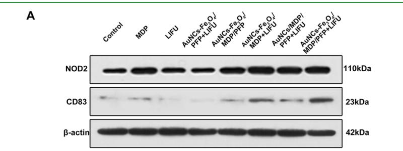
Application: WB    Species: Human    Sample: Y79 cells

Figure 12. (A,B) NOD2 and CD83 contents in Y79 tumors after different treatments. (C) IFN-γ and MCP-1 levels in Y79 tumors after different treatments. (D) H&E staining in major organs (heart, liver, spleen, lung, and kidney) of all groups after treatments. The scale bars are 50 μm. (n = 5, **p < 0.01).

3). Microglia Exhibit Distinct Heterogeneity Rather than M1/M2 Polarization within the Early Stage of Acute Ischemic Stroke. Aging and disease, 2023 (PubMed: 37199734) [IF=7.0]

Application: IF/ICC    Species: Mouse    Sample: Mic_M1L1 cells

Figure 4. Subpopulation analysis of M1-polarization-like microglia clusters. (A) Pseudotime plot (from monocle3) shows the differentiation trajectory between Mic_M1L1 and Mic_M1L2 cells. (B) Line plot of the fraction of cells in Mic_M1L1 and Mic_M1L2 subpopulations over time. (C) Expression of the ten genes with the highest Moran’s I score alongside the pseudotime trajectory. (D) Violin plot of the functional pathways revealed by gene set variation analysis (GSVA) in the Mic_M1L1 and Mic_M1L2 subpopulations. (E) Volcano plot displays the differentially expressed genes between Mic_M1L1 and Mic_M1L2 cells. Red dots represent the upregulated genes in the Mic_M1L1 cluster compared with the Mic_M1L2 cluster and blue dots represent the downregulated. (F) Relative activity of the highly expressed transcription factors (TF) in Mic_M1L2 cells compared with Mic_M1L1 cells. (G) Double immunofluorescence staining of Mic_M1L1 cells at each sampling time. Coronal brain sections are all stained with anti-Iba1(green, representing microglia), Nfkbiz (red, the marker of Mic_M1L1 cells), and DAPI (blue, representing cell nuclei) antibodies (N=3). The yellow bar represents 100μm. The arrows point to the cells simultaneously marked by Iba1 and Nfkbiz. (H) Double immunofluorescence staining of Mic_M1L2 cells at each sampling time. Coronal brain sections are all stained with anti-Iba1(green, representing microglia), Cd83 (red, the marker of Mic_M1L2 cells), and DAPI (blue, representing cell nuclei) antibodies (N=3). The yellow bar represents 100μm. The arrows point to the cells simultaneously marked by Iba1 and Cd83. (I) Bar plot of the ratio of Nfkbiz+Iba1+ cells (green plus red) compared with all DAPI+ cells in a 500μm2 area surrounding the infarction core (N=6). (J) Bar plot of the ratio of Cd83+Iba1+ cells (green plus red) compared with all DAPI+ cells in a 500μm2 area surrounding the infarction core (N=6).

4). Stromal and tumor immune microenvironment reprogramming through multifunctional cisplatin-based liposomes boosts the efficacy of anti-PD-1 immunotherapy in pancreatic cancer. Biomaterials science, 2023 (PubMed: 37921708) [IF=5.8]

5). Иммуногистохимическая характеристика биопсийных образцов десны в области беззубого альвеолярного края челюсти. Пародонтология, 2024

Restrictive clause


Affinity Biosciences tests all products strictly. Citations are provided as a resource for additional applications that have not been validated by Affinity Biosciences. Please choose the appropriate format for each application and consult Materials and Methods sections for additional details about the use of any product in these publications.

For Research Use Only.
Not for use in diagnostic or therapeutic procedures. Not for resale. Not for distribution without written consent. Affinity Biosciences will not be held responsible for patent infringement or other violations that may occur with the use of our products. Affinity Biosciences, Affinity Biosciences Logo and all other trademarks are the property of Affinity Biosciences LTD.