| Product: | LEG7 Antibody |
| Catalog: | AF5300 |
| Description: | Rabbit polyclonal antibody to LEG7 |
| Application: | WB IHC |
| Cited expt.: | WB |
| Reactivity: | Human, Mouse, Rat |
| Mol.Wt.: | 15 KD(Observed); 15kD(Calculated). |
| Uniprot: | P47929 |
| RRID: | AB_2837785 |
Control Products

Related Downloads
Protocols
Product Info
*The optimal dilutions should be determined by the end user. For optimal experimental results, antibody reuse is not recommended.
*Tips:
WB: For western blot detection of denatured protein samples. IHC: For immunohistochemical detection of paraffin sections (IHC-p) or frozen sections (IHC-f) of tissue samples. IF/ICC: For immunofluorescence detection of cell samples. ELISA(peptide): For ELISA detection of antigenic peptide.
Cite Format: Affinity Biosciences Cat# AF5300, RRID:AB_2837785.
Fold/Unfold
Gal-7; GAL7; Galectin 7B; Galectin-7; HKL-14; Human keratinocyte lectin 14; Keratinocyte lectin 14; Lectin galactoside binding soluble 7; Lectin, galactoside binding, soluble, 7B; LEG7_HUMAN; LGALS7; LGALS7A; LGALS7B; P53 induced protein 1; p53-induced gene 1 protein; Pi7; PIG1; TP53I1;
Immunogens
A synthesized peptide derived from human LEG7, corresponding to a region within the internal amino acids.
- P47929 LEG7_HUMAN:
- Protein BLAST With
- NCBI/
- ExPASy/
- Uniprot
MSNVPHKSSLPEGIRPGTVLRIRGLVPPNASRFHVNLLCGEEQGSDAALHFNPRLDTSEVVFNSKEQGSWGREERGPGVPFQRGQPFEVLIIASDDGFKAVVGDAQYHHFRHRLPLARVRLVEVGGDVQLDSVRIF
Research Backgrounds
Could be involved in cell-cell and/or cell-matrix interactions necessary for normal growth control. Pro-apoptotic protein that functions intracellularly upstream of JNK activation and cytochrome c release.
Cytoplasm. Nucleus. Secreted.
Note: May be secreted by a non-classical secretory pathway.
Mainly expressed in stratified squamous epithelium.
References
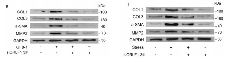
Application: WB Species: human Sample: LF cells

Application: WB Species: human Sample: LF cells
Restrictive clause
Affinity Biosciences tests all products strictly. Citations are provided as a resource for additional applications that have not been validated by Affinity Biosciences. Please choose the appropriate format for each application and consult Materials and Methods sections for additional details about the use of any product in these publications.
For Research Use Only.
Not for use in diagnostic or therapeutic procedures. Not for resale. Not for distribution without written consent. Affinity Biosciences will not be held responsible for patent infringement or other violations that may occur with the use of our products. Affinity Biosciences, Affinity Biosciences Logo and all other trademarks are the property of Affinity Biosciences LTD.