Product: MMP13 Antibody
Catalog: AF5355
Description: Rabbit polyclonal antibody to MMP13
Application: WB IHC IF/ICC
Reactivity: Human, Mouse, Rat
Mol.Wt.: 54 kDa; 54kD(Calculated).
Uniprot: P45452
RRID: AB_2837840

View similar products>>

   Size Price Inventory
 100ul $280 In stock
 200ul $350 In stock

Lead Time: Same day delivery

For pricing and ordering contact:
Local distributors

Product Info

Source:
Rabbit
Application:
WB 1:500-1:2000, IHC 1:50-1:200, IF/ICC 1:100-1:500
*The optimal dilutions should be determined by the end user.
*Tips:

WB: For western blot detection of denatured protein samples. IHC: For immunohistochemical detection of paraffin sections (IHC-p) or frozen sections (IHC-f) of tissue samples. IF/ICC: For immunofluorescence detection of cell samples. ELISA(peptide): For ELISA detection of antigenic peptide.

Reactivity:
Human,Mouse,Rat
Clonality:
Polyclonal
Specificity:
MMP13 Antibody detects endogenous levels of total MMP13.
RRID:
AB_2837840
Cite Format: Affinity Biosciences Cat# AF5355, RRID:AB_2837840.
Conjugate:
Unconjugated.
Purification:
The antiserum was purified by peptide affinity chromatography using SulfoLink™ Coupling Resin (Thermo Fisher Scientific).
Storage:
Rabbit IgG in phosphate buffered saline , pH 7.4, 150mM NaCl, 0.02% sodium azide and 50% glycerol. Store at -20 °C. Stable for 12 months from date of receipt.
Alias:

Fold/Unfold

CLG 3; CLG3; Collagenase 3; Collagenase3; MANDP1; Matrix metallopeptidase 13 (collagenase 3); Matrix Metalloproteinase 13; Matrix metalloproteinase-13; MMP 13; MMP-13; Mmp13; MMP13_HUMAN;

Immunogens

Immunogen:
Uniprot:
Gene(ID):
Expression:
P45452 MMP13_HUMAN:

Detected in fetal cartilage and calvaria, in chondrocytes of hypertrophic cartilage in vertebrae and in the dorsal end of ribs undergoing ossification, as well as in osteoblasts and periosteal cells below the inner periosteal region of ossified ribs. Detected in chondrocytes from in joint cartilage that have been treated with TNF and IL1B, but not in untreated chondrocytes. Detected in T lymphocytes. Detected in breast carcinoma tissue.

Description:
Defects in MMP13 are the cause of spondyloepimetaphyseal dysplasia Missouri type (SEMD-MO) [MIM:602111]. A bone disease characterized by moderate to severe metaphyseal changes, mild epiphyseal involvement, rhizomelic shortening of the lower limbs with bowing of the femora and/or tibiae, coxa vara, genu varum and pear-shaped vertebrae in childhood. Epimetaphyseal changes improve with age.
Sequence:
MHPGVLAAFLFLSWTHCRALPLPSGGDEDDLSEEDLQFAERYLRSYYHPTNLAGILKENAASSMTERLREMQSFFGLEVTGKLDDNTLDVMKKPRCGVPDVGEYNVFPRTLKWSKMNLTYRIVNYTPDMTHSEVEKAFKKAFKVWSDVTPLNFTRLHDGIADIMISFGIKEHGDFYPFDGPSGLLAHAFPPGPNYGGDAHFDDDETWTSSSKGYNLFLVAAHEFGHSLGLDHSKDPGALMFPIYTYTGKSHFMLPDDDVQGIQSLYGPGDEDPNPKHPKTPDKCDPSLSLDAITSLRGETMIFKDRFFWRLHPQQVDAELFLTKSFWPELPNRIDAAYEHPSHDLIFIFRGRKFWALNGYDILEGYPKKISELGLPKEVKKISAAVHFEDTGKTLLFSGNQVWRYDDTNHIMDKDYPRLIEEDFPGIGDKVDAVYEKNGYIYFFNGPIQFEYSIWSNRIVRVMPANSILWC

PTMs - P45452 As Substrate

Site PTM Type Enzyme
N117 N-Glycosylation
Y360 Phosphorylation
Y366 Phosphorylation

Research Backgrounds

Function:

Plays a role in the degradation of extracellular matrix proteins including fibrillar collagen, fibronectin, TNC and ACAN. Cleaves triple helical collagens, including type I, type II and type III collagen, but has the highest activity with soluble type II collagen. Can also degrade collagen type IV, type XIV and type X. May also function by activating or degrading key regulatory proteins, such as TGFB1 and CCN2. Plays a role in wound healing, tissue remodeling, cartilage degradation, bone development, bone mineralization and ossification. Required for normal embryonic bone development and ossification. Plays a role in the healing of bone fractures via endochondral ossification. Plays a role in wound healing, probably by a mechanism that involves proteolytic activation of TGFB1 and degradation of CCN2. Plays a role in keratinocyte migration during wound healing. May play a role in cell migration and in tumor cell invasion.

PTMs:

The proenzyme is activated by removal of the propeptide; this cleavage can be effected by other matrix metalloproteinases, such as MMP2, MMP3 and MMP14 and may involve several cleavage steps. Cleavage can also be autocatalytic, after partial maturation by another protease or after treatment with 4-aminophenylmercuric acetate (APMA) (in vitro).

N-glycosylated.

Tyrosine phosphorylated by PKDCC/VLK.

Subcellular Location:

Secreted>Extracellular space>Extracellular matrix. Secreted.

Extracellular region or secreted Cytosol Plasma membrane Cytoskeleton Lysosome Endosome Peroxisome ER Golgi apparatus Nucleus Mitochondrion Manual annotation Automatic computational assertionSubcellular location
Tissue Specificity:

Detected in fetal cartilage and calvaria, in chondrocytes of hypertrophic cartilage in vertebrae and in the dorsal end of ribs undergoing ossification, as well as in osteoblasts and periosteal cells below the inner periosteal region of ossified ribs. Detected in chondrocytes from in joint cartilage that have been treated with TNF and IL1B, but not in untreated chondrocytes. Detected in T lymphocytes. Detected in breast carcinoma tissue.

Subunit Structure:

Monomer. Interacts with TIMP1, TIMP2 and TIMP3. Binds (via the C-terminal region) to collagen.

Family&Domains:

The conserved cysteine present in the cysteine-switch motif binds the catalytic zinc ion, thus inhibiting the enzyme. The dissociation of the cysteine from the zinc ion upon the activation-peptide release activates the enzyme (By similarity).

The C-terminal region binds to collagen.

Belongs to the peptidase M10A family.

Research Fields

· Organismal Systems > Immune system > IL-17 signaling pathway.   (View pathway)

· Organismal Systems > Endocrine system > Relaxin signaling pathway.

References

1). Pentraxin 3 regulated by miR-224-5p modulates macrophage reprogramming and exacerbates osteoarthritis associated synovitis by targeting CD32. Cell Death & Disease (PubMed: 35739102) [IF=9.0]

Application: IF/ICC    Species: Mouse    Sample:

Fig. 2 PTX3 accelerates synovitis, cartilage erosion, and exacerbates OA development in mice. A, B Safranin O/fast green staining and Osteoarthritis Research Society International (OARSI) grades of knee cartilage from DMM mice treated with vehicle, recombinant mouse PTX3 (rmPTX3), or PTX3 neutralizing antibody (PTX3-NAb) for 4 weeks and 8 weeks. Scale bar: 100 µm. C–F IF staining and quantification of ADATMS5 and MMP13 in knee cartilage of controls and DMM mice treated with vehicle, rmPTX3, or PTX3-NAb for 8 weeks. Scale bar: 50 µm. G IF staining and quantification of COL2 in knee cartilage of controls and DMM mice treated with rmPTX3 or PTX3-NAb for 8 weeks. Scale bar: 50 µm. H, I H&E and synovitis score of synovial tissues from DMM mice treated with vehicle, rmPTX3, or PTX3-NAb for 4 weeks and 8 weeks. Scale bar: 100 µm. *P 

2). Naringenin protects against iron overload-induced osteoarthritis by suppressing oxidative stress. PHYTOMEDICINE (PubMed: 35905566) [IF=7.9]

3). Guanxinning injection ameliorates cardiac remodeling in HF mouse and 3D heart spheroid models via p38/FOS/MMP1-mediated inhibition of myocardial hypertrophy and fibrosis. Biomedicine & Pharmacotherapy (PubMed: 37027988) [IF=7.5]

4). A lithium-containing biomaterial promotes chondrogenic differentiation of induced pluripotent stem cells with reducing hypertrophy. Stem Cell Research & Therapy (PubMed: 32085810) [IF=7.5]

Application: IF/ICC    Species: Human    Sample: iPSCs

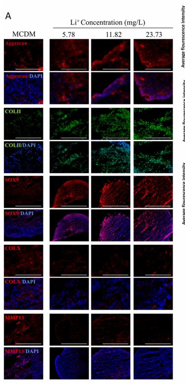
Fig. 7 The chondrogenic differentiation of iPSCs with different Li+ ions concentration in 14 days. a Immunofluorescence of chondrocytes specified proteins (COL II, Aggrecan, and SOX9) and hypertrophic specified protein (COL X and MMP13). b The average fluorescence intensity of COL II, Aggrecan, SOX9, MMP13, and COL X proteins in each group, n = 5. c The relative genes expression of chondrogenic differentiation cultured with Li+ ions, n = 3. d The relative genes expression of hypertrophic differentiation cultured with Li+ ions, n = 3. Data presented as mean ± SEM. Scale bar = 200 μm. CTR: MCDM without any Li+ ions. *p < 0.05, **p < 0.01, ***p < 0.001

5). An osteoarthritis subtype characterized by synovial lipid metabolism disorder and fibroblast-like synoviocyte dysfunction. Journal of Orthopaedic Translation (PubMed: 35330945) [IF=6.6]

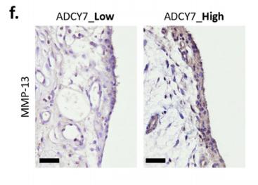
Application: IHC    Species: human    Sample: OA synovium

FIGURE 2 | Synovial inflammation assessed by inflammatory cytokines and chemokines expression (e, n ¼ 8), MMP-13 production (f) and M1 macrophage infiltration (g, n ¼ 8, bar ¼ 100 μm).

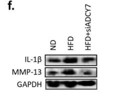
Application: WB    Species: rat    Sample: cartilage

Fig. 4.| ADCY7 plays a key role in an HFD-induced OA model.Safranin O staining and OARSI scoring of articular cartilage (n ¼ 5, bar ¼ 800 μm) (e–f). MMP-13 and IL-1β expression in cartilage assessed by immunohistochemistry (e, n ¼ 5, bar ¼ 100 μm) and western blot (f) (g–h). Lipid accumulation and lipolysis in synovium assessed by toluidine blue staining (g, bar ¼100 μm) and levels of lipolytic products (h, n ¼ 5) (i–k).

6). L-Glutamine alleviates osteoarthritis by regulating lncRNA-NKILA expression through the TGF-β1/SMAD2/3 signalling pathway. Clinical Science (PubMed: 35730575) [IF=6.0]

7). PDLIM2 protects articular chondrocytes from lipopolysaccharide-induced apoptosis, degeneration and inflammatory injury through down-regulation of nuclear factor (NF)-κB signaling. International Immunopharmacology (PubMed: 32805696) [IF=5.6]

Application: WB    Species: Mouse    Sample: chondrocytes

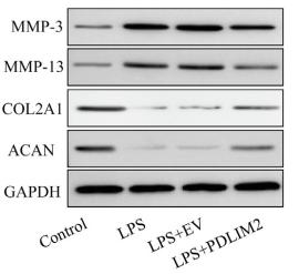
Fig. 2. Up-regulation of PDLIM2 attenuates LPS-induced injury in chondrocytes. Chondrocytes were transfected with empty vector (EV) or PDLIM2 expression vector for 48 h and then stimulated with 10 μg/ml of LPS for 6 h. (A) The relative mRNA expression of PDLIM2 was determined by RT-qPCR. (B, C) Protein expression of PDLIM2 was examined by Western blotting. (D) The effect of PDLIM2 overexpression on cell viability was detected by MTT assay. The effect of PDLIM2 overexpression on cell apoptosis was assessed by (E, F) TUNENL and (G, H) Annexin V-FITC/PI apoptosis assays. (I) The effect of PDLIM2 overexpression on MMP-3, MMP-13, COL2A1 and ACAN was evaluated by Western blotting. Quantitative analysis of protein expression of (J) MMP-3, (K) MMP-13, (L) COL2A1 and (M) ACAN. N = 3, **p < 0.01 and ***p < 0.001.

8). Fexofenadine Protects Against Intervertebral Disc Degeneration Through TNF Signaling. Frontiers in Cell and Developmental Biology (PubMed: 34504840) [IF=5.5]

Application: WB    Species: Human    Sample: discs

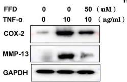
FIGURE 1 Fexofenadine (FFD) protected against tumor necrosis factor-α (TNF-α)-induced intervertebral disc degeneration. (A) Safranin O staining of mouse disc. The discs were cultured and treated with TNF-α (10 ng/ml) in the presence or absence of FFD (50 μM) for 7 days. (B) Histological score analysis based on the Safranin O staining (n = 6). Scale bar = 200 μm. (C) The mouse disc were cultured in presence of FFD with various concentration conditions (10, 50, 100, and 200 μM), the effect of the drug was determined by Safranin O staining (n = 6). (D) Histological score based on the Safranin O staining. Scale bar = 200 μm. (E) H&E staining of mouse disc. The mouse discs were cultured in medium supplemented with 10 ng/ml TNF-α in the absence or presence of FFD (50 uM) for 7 days. (F) Histological score of mouse discs in each indicated group (n = 6). Scale bar = 200 μm. (G) Protein was extracted from mouse discs, Western Blotting analysis of COX-2 and MMP-13 was determined. (H,I) Transcriptional level of Aggrecan and Col II from mouse discs in each group. Each experiment was performed three times. (J) Immunohistochemistry staining of Aggrecan in human disc organ. The human discs were treated without or with TNF-α (10 ng/mL) in the absence or presence of FFD (50 μM) for 7 days (n = 6). Brown signal indicates positive. Scale bar = 200 μm. (K) Statistical analysis of the percentage of the positive cells based on immunohistochemistry staining. The values shown represent the mean ± standard deviation *p < 0.05 and **p < 0.01 vs. control group.

Application: IHC    Species: Human    Sample: discs

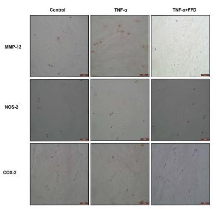
FIGURE 4 Fexofenadine (FFD) inhibited TNF-α induced inflammation in disc degeneration. (A) The protein expression of mouse COX-2 and MMP-13 were detected by Western blotting assay. The mouse NP cells were treated with TNF-α (10 ng/ml) with or without additional treatment with FFD (1 μM, 10 μM) for 48 h, followed by protein examination. (B,C) Quantification of the band density for mouse COX-2 and MMP-13 was based on the Western blotting assay. (D) The protein expression of human COX-2 and MMP-13 were detected by Western blotting assay. The human NP cells were treated with TNF-α (10 ng/ml) with or without additional treatment with FFD (1 μM, 10 μM) for 48 h, followed by protein examination. (E,F) Quantification of the band’s density for human COX-2 and MMP-13 was based on Western blotting assay. (G) Immunohistochemistry staining of MMP-13, NOS-2, and COX-2 in human discs. The human discs were treated without or with TNF-α (10 ng/mL) in the absence or presence of FFD (50 μM) for 7 days (n = 6). Scale bar = 200 μm. Brown signal indicated positive. (H–J) Quantification of the percentage of the positive cells was based on immunohistochemistry staining. (K–M) The expression of IL-1β, IL-6, and IL-17 in the culture media of each group, as detected by ELISA assay (n = 3). The human NP cells were treated without or with TNF-α (10 ng/mL) in the absence or presence of FFD (1 μM, 10 μM) for 48 h. Then the supernatant were collected for ELISA. The values shown represent the mean ± standard deviation. *p < 0.05, **p < 0.01, and ***p < 0.001 vs. control group.

9). Tiao-bu-fei-shen formula promotes downregulation of the caveolin 1-p38 mapk signaling pathway in COPD - Associated tracheobronchomalacia cell model. JOURNAL OF ETHNOPHARMACOLOGY (PubMed: 35367574) [IF=5.4]

Application: WB    Species: rat    Sample:

Fig. 10.| Western blot analysis of the expression of key proteins in the caveolin 1-p38 MAPK signaling pathway. The levels of (A) caveolin 1, (B) p-p38 MAPK, (C)MMP-13, (D) IL-1β, (E) TNF-α, (F) Bax, and (G) Bcl-2. *P < 0.05 indicates statistical differences between each treatment group and the model group.

10). Asiatic acid attenuates hypertrophic and fibrotic differentiation of articular chondrocytes via AMPK/PI3K/AKT signaling pathway. ARTHRITIS RESEARCH & THERAPY (PubMed: 32398124) [IF=4.9]

Load more

Restrictive clause


Affinity Biosciences tests all products strictly. Citations are provided as a resource for additional applications that have not been validated by Affinity Biosciences. Please choose the appropriate format for each application and consult Materials and Methods sections for additional details about the use of any product in these publications.

For Research Use Only.
Not for use in diagnostic or therapeutic procedures. Not for resale. Not for distribution without written consent. Affinity Biosciences will not be held responsible for patent infringement or other violations that may occur with the use of our products. Affinity Biosciences, Affinity Biosciences Logo and all other trademarks are the property of Affinity Biosciences LTD.