 and mouse anti-beta tubulin Ab(T0023) for 1 hour at 37°C. An AlexaFluor594 conjugated goat anti-rabbit IgG(H+L) Ab(Red) and an AlexaFluor488 conjugated goat anti-mouse IgG(H+L) Ab(Green) were used as the secondary antibody.
The nuclear counter stain is DAPI (blue).")
. The total and phosphorylated expression of p65,IKKα and IKKβ after the above treatment was examined by WB and demonstrated by histogram. (a and b).The levels of inflammatory cytokines and chemokines were then measured by ELISA. (c and d) The expression of adhesion molecules (ICAM‐1 and VCAM‐1) was detected by WB (e). *p < 0.05, **p < 0.01")
 Representative western blot results for AMPK/NF-κB p65/NLRP3 signaling pathway proteins in AGEs/salidroside treated-HUVECs with or without compound C.GAPDH was used as a loading control. (B) Histograms analysis of the western blot results. The data are representative of one experiment performed in triplicate and are expressed as the mean ± S.D.")
, ticagrelor (20 μM), clopidogrel (20 μM), DMSO plus LPS (10 ng/mL) and CD14 (1 μg/mL), ticagrelor (20 μM) plus LPS (10 ng/mL) and CD14 (1 μg/mL), and clopidogrel (20 μM) plus LPS (10 ng/mL) and CD14 (1 μg/mL), separately, for 16 h. Then, the cell lysates were immunoblotted with the indicated antibodies")

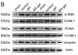
. The upper panel presents immunohistochemistry results, and the lower panel shows the quantified relative expression levels.*P<0.05 vs. control; #P<0.05 vs. model; ^P<0.05 vs. model + DEX. DEX, dexmedetomidine; 4‑PBA, 4‑phenylbutyric acid; ICAM‑1, intercellular adhesion molecule‑1; VCAM‑1, vascular adhesion molecule‑1.")

. The upper panel presents immunohistochemistry results, and the lower panel shows the quantified relative expression levels. *P<0.05 vs. control; #P<0.05 vs. model; ^P<0.05 vs. model + DEX. DEX, dexmedetomidine; 4-PBA, 4-phenylbutyric acid; ICAM-1, intercellular adhesion molecule-1; VCAM-1, vascular adhesion molecule-1.")
. The expression level of chemokines was measured by the RT-qPCR assay, (b) the production of chemokines was evaluated by the ELISA assay, (c) the expression level of CAMs was checked by the RT-qPCR assay, (d) the expression level of CAMs was measured by the Western blotting assay (p < 0.05 and p < 0.01), (e) the adhesion between hNECs and U937 monocytes was inhibited by febuxostat. The attached U937 monocytes were detected using the calcein-AM staining assay (p < 0.05 and p < 0.01), (f) the expression level of KLF6 was evaluated by the RT-qPCR assay, and (g) the expression level of KLF6 was determined by the Western blotting assay (p < 0.05 and p < 0.01). Immunofluorescence magnification was 10×.")
, SELE (B), ICAM-1 (C) and VCAM-1 (D) was observed by immunofluorescence staining. compared with the PBS group, bleomycin can significantly upregulate the expression levels of vWF, SELE, ICAM-1 and VCAM-1 in endothelial cells, while Wenyang Huazhuo Tongluo Formula and KC7F2 can significantly reverse the bleomycin-induced upregulation of vWF, SELE, ICAM-1 and VCAM-1. The mean values ± SD was shown for each bar. * (P < 0.05) or ** (P < 0.01) or *** (P < 0.001) represents significance, ns represents no significance. Original magnification: × 20. BLM: Bleomycin, WYHZTL: Wenyang Huazhuo Tongluo formula")
 The mRNA levels of ICAM-1 and VCAM-1 were measured by quantitative real-time PCR. (C-J) The level of p-MAPK/MAPK, p-ERK/ERK, ICAM, VCAM-1, Aβ1–42, and p-Tau/Tau were measured by Western blot. The data are expressed as mean ± standard deviation, n = 3. **p")
 WB analysis of VLA4, VCAM-1, RAC1, p-PYK2 and p-VE-cad. The protein expression levels of VLA4, VCAM-1, RAC1, p-PYK2 and p-VE-cad were significantly higher in the RHD group than in the control group. (C) RT‒qPCR results for VLA4, VCAM-1 and RAC1. The mRNA expression levels of VLA4, VCAM-1 and RAC1 were significantly higher in the RHD group than in the control group. (D,E) Immunohistochemical results for VLA4, VCAM-1, p-PYK2 and p-VE-cad in heart valves. Magnification, 400 × ; scale bar: 50 µm; arrows point to positive staining. The expression levels of VLA4, VCAM-1, p-PYK2 and p-VE-cad were significantly higher in the RHD group than in the control group. The data are presented as the mean ± SD; *p < 0.05 vs. the control group. VLA4, very late antigen4; VCAM-1, vascular cell adhesion molecule-1; RAC1, RAS-related C3 botulinum substrate 1; p-PYK2, phosphorylated proline-rich tyrosine kinase 2; p-VE-cad, phosphorylated VE cadherin; RHD, rheumatic heart disease.")
 WB analysis of VLA4, VCAM-1, RAC1, p-PYK2 and p-VE-cad. The protein expression levels of VLA4, VCAM-1, RAC1, p-PYK2 and p-VE-cad were significantly higher in the RHD group than in the control group. (C) RT‒qPCR results for VLA4, VCAM-1 and RAC1. The mRNA expression levels of VLA4, VCAM-1 and RAC1 were significantly higher in the RHD group than in the control group. (D,E) Immunohistochemical results for VLA4, VCAM-1, p-PYK2 and p-VE-cad in heart valves. Magnification, 400 × ; scale bar: 50 µm; arrows point to positive staining. The expression levels of VLA4, VCAM-1, p-PYK2 and p-VE-cad were significantly higher in the RHD group than in the control group. The data are presented as the mean ± SD; *p < 0.05 vs. the control group. VLA4, very late antigen4; VCAM-1, vascular cell adhesion molecule-1; RAC1, RAS-related C3 botulinum substrate 1; p-PYK2, phosphorylated proline-rich tyrosine kinase 2; p-VE-cad, phosphorylated VE cadherin; RHD, rheumatic heart disease.")
 Representative immunoblot and relative quantification of TLR4 and MyD88 in RAW264.7 cells. (D,E) Representative immunofluorescence images of TLR4-positive and MyD88-positive in RAW264.7 cells. Scale bars: 25 μm. (F–H) Representative immunoblot and relative quantification of p-IκBα (S32/S36), IκBα, p-p65 (S536), and p65 in RAW264.7 cells. (I) Representative immunofluorescence images of NF-κB p65 in RAW264.7 cells. Scale bars: 100 μm. (J–N) Representative immunoblot and relative quantification of CD68, MCP-1, ICAM1, and VCAM1 in RAW264.7 cells. (O–R) mRNA levels of Ccl2, Ccl3, Ccl4, and Cxcl10 in RAW264.7 cells. Data are presented as mean ± SEM (n = 3). * p < 0.05, ** p < 0.01, *** p < 0.001, and **** p < 0.0001 vs. the Con group. # p < 0.05, ## p < 0.01, ### p < 0.001, and #### p < 0.0001 vs. the LPS group.")
 AGS and (B) HGC-27 cells. Western blotting analysis the protein expression levels of VCAM1, ICAM1, PTGS2, IL6 and CCL2 (C) AGS and (D) HGC-27 cells. In order to save and recycle antibodies and save time, membranes were cut in horizontal strips at molecular weight ranges for target proteins and the information was shown in the Supplementary Fig. 1 and Fig. 2. Differences between two groups were assessed by Students t test, and differences among three groups were assessed by one-way analysis of variance (ANOVA).")
| Product: | VCAM1 Antibody |
| Catalog: | DF6082 |
| Description: | Rabbit polyclonal antibody to VCAM1 |
| Application: | WB IHC IF/ICC |
| Cited expt.: | WB, IHC, IF/ICC |
| Reactivity: | Human, Mouse, Rat |
| Prediction: | Pig, Bovine, Horse, Rabbit, Dog |
| Mol.Wt.: | 80~120kDa(Observed); 81kD(Calculated). |
| Uniprot: | P19320 |
| RRID: | AB_2838050 |
Control Products

Related Downloads
Protocols
Product Info
*The optimal dilutions should be determined by the end user. For optimal experimental results, antibody reuse is not recommended.
*Tips:
WB: For western blot detection of denatured protein samples. IHC: For immunohistochemical detection of paraffin sections (IHC-p) or frozen sections (IHC-f) of tissue samples. IF/ICC: For immunofluorescence detection of cell samples. ELISA(peptide): For ELISA detection of antigenic peptide.
Cite Format: Affinity Biosciences Cat# DF6082, RRID:AB_2838050.
Fold/Unfold
CD106; CD106 Antigen; INCAM 100; INCAM-100; L1CAM; MGC99561; V-CAM 1; Vascular Cell Adhesion Molecule 1; Vascular cell adhesion protein 1; VCAM 1; VCAM-1; VCAM1; VCAM1_HUMAN;
Immunogens
A synthesized peptide derived from human VCAM1, corresponding to a region within the internal amino acids.
Expressed on inflamed vascular endothelium, as well as on macrophage-like and dendritic cell types in both normal and inflamed tissue.
- P19320 VCAM1_HUMAN:
- Protein BLAST With
- NCBI/
- ExPASy/
- Uniprot
MPGKMVVILGASNILWIMFAASQAFKIETTPESRYLAQIGDSVSLTCSTTGCESPFFSWRTQIDSPLNGKVTNEGTTSTLTMNPVSFGNEHSYLCTATCESRKLEKGIQVEIYSFPKDPEIHLSGPLEAGKPITVKCSVADVYPFDRLEIDLLKGDHLMKSQEFLEDADRKSLETKSLEVTFTPVIEDIGKVLVCRAKLHIDEMDSVPTVRQAVKELQVYISPKNTVISVNPSTKLQEGGSVTMTCSSEGLPAPEIFWSKKLDNGNLQHLSGNATLTLIAMRMEDSGIYVCEGVNLIGKNRKEVELIVQEKPFTVEISPGPRIAAQIGDSVMLTCSVMGCESPSFSWRTQIDSPLSGKVRSEGTNSTLTLSPVSFENEHSYLCTVTCGHKKLEKGIQVELYSFPRDPEIEMSGGLVNGSSVTVSCKVPSVYPLDRLEIELLKGETILENIEFLEDTDMKSLENKSLEMTFIPTIEDTGKALVCQAKLHIDDMEFEPKQRQSTQTLYVNVAPRDTTVLVSPSSILEEGSSVNMTCLSQGFPAPKILWSRQLPNGELQPLSENATLTLISTKMEDSGVYLCEGINQAGRSRKEVELIIQVTPKDIKLTAFPSESVKEGDTVIISCTCGNVPETWIILKKKAETGDTVLKSIDGAYTIRKAQLKDAGVYECESKNKVGSQLRSLTLDVQGRENNKDYFSPELLVLYFASSLIIPAIGMIIYFARKANMKGSYSLVEAQKSKV
Predictions
Score>80(red) has high confidence and is suggested to be used for WB detection. *The prediction model is mainly based on the alignment of immunogen sequences, the results are for reference only, not as the basis of quality assurance.
High(score>80) Medium(80>score>50) Low(score<50) No confidence
Research Backgrounds
Important in cell-cell recognition. Appears to function in leukocyte-endothelial cell adhesion. Interacts with integrin alpha-4/beta-1 (ITGA4/ITGB1) on leukocytes, and mediates both adhesion and signal transduction. The VCAM1/ITGA4/ITGB1 interaction may play a pathophysiologic role both in immune responses and in leukocyte emigration to sites of inflammation.
Sialoglycoprotein.
Membrane>Single-pass type I membrane protein.
Expressed on inflamed vascular endothelium, as well as on macrophage-like and dendritic cell types in both normal and inflamed tissue.
Either the first or the fourth Ig-like C2-type domain is required for VLA4-dependent cell adhesion.
Research Fields
· Environmental Information Processing > Signal transduction > NF-kappa B signaling pathway. (View pathway)
· Environmental Information Processing > Signaling molecules and interaction > Cell adhesion molecules (CAMs). (View pathway)
· Environmental Information Processing > Signal transduction > TNF signaling pathway. (View pathway)
· Human Diseases > Infectious diseases: Parasitic > African trypanosomiasis.
· Human Diseases > Infectious diseases: Parasitic > Malaria.
· Human Diseases > Infectious diseases: Viral > HTLV-I infection.
· Organismal Systems > Immune system > Leukocyte transendothelial migration. (View pathway)
References
Application: WB Species: Rat Sample:

Application: WB Species: Mouse Sample:
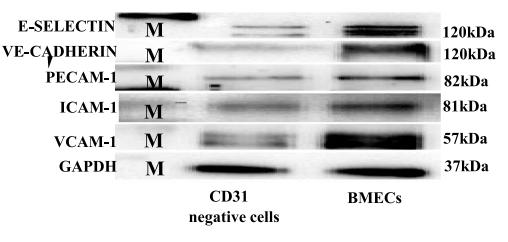
Application: WB Species: mouse Sample: primary bone marrow endothelial cells
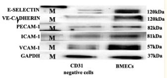
Application: WB Species: Mice Sample: bMECs

Application: WB Species: Rat Sample:
Restrictive clause
Affinity Biosciences tests all products strictly. Citations are provided as a resource for additional applications that have not been validated by Affinity Biosciences. Please choose the appropriate format for each application and consult Materials and Methods sections for additional details about the use of any product in these publications.
For Research Use Only.
Not for use in diagnostic or therapeutic procedures. Not for resale. Not for distribution without written consent. Affinity Biosciences will not be held responsible for patent infringement or other violations that may occur with the use of our products. Affinity Biosciences, Affinity Biosciences Logo and all other trademarks are the property of Affinity Biosciences LTD.