
.
Bands result from membrane strip incubation.")
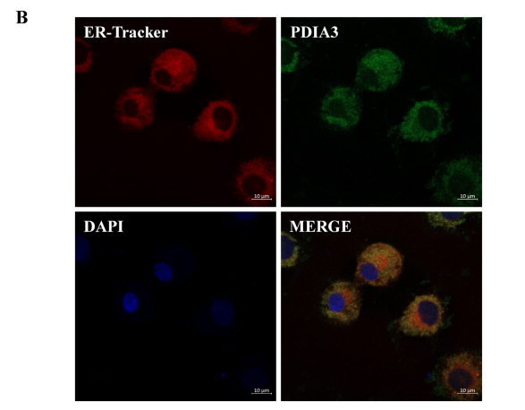
 and mouse anti-beta tubulin Ab(T0023 1:200) for 1 hour at 37°C. An AlexaFluor594 conjugated goat anti-rabbit IgG(H+L) Ab(Red) and an AlexaFluor488 conjugated goat anti-mouse IgG(H+L) Ab(Green) were used as the secondary antibody.
The nuclear counter stain is DAPI(blue).")
| Product: | PDIA3 Antibody |
| Catalog: | DF6230 |
| Description: | Rabbit polyclonal antibody to PDIA3 |
| Application: | WB IHC IF/ICC |
| Cited expt.: | WB, IF/ICC |
| Reactivity: | Human, Mouse, Rat |
| Prediction: | Pig, Bovine, Horse, Sheep, Rabbit, Dog, Chicken |
| Mol.Wt.: | 57kDa(Observed); 57kD(Calculated). |
| Uniprot: | P30101 |
| RRID: | AB_2838196 |
Control Products

Related Downloads
Protocols
Product Info
*The optimal dilutions should be determined by the end user. For optimal experimental results, antibody reuse is not recommended.
*Tips:
WB: For western blot detection of denatured protein samples. IHC: For immunohistochemical detection of paraffin sections (IHC-p) or frozen sections (IHC-f) of tissue samples. IF/ICC: For immunofluorescence detection of cell samples. ELISA(peptide): For ELISA detection of antigenic peptide.
Cite Format: Affinity Biosciences Cat# DF6230, RRID:AB_2838196.
Fold/Unfold
58 kDa glucose regulated protein; 58 kDa glucose-regulated protein; 58 kDa microsomal protein; Disulfide isomerase ER 60; Disulfide isomerase ER-60; Endoplasmic reticulum resident protein 57; Endoplasmic reticulum resident protein 60; ER p57; ER protein 57; ER protein 60; ERp 57; ERp57; ERp60; ERp61; Glucose Regulated Protein 58 Kd; GRP 57; GRP 58; GRP57; HsT17083; p58; PDIA 3; PDIA3; PDIA3_HUMAN; Phospholipase C alpha; PI PLC; Protein disulfide isomerase A3; Protein disulfide isomerase family A member 3; Protein disulfide-isomerase A3;
Immunogens
A synthesized peptide derived from human PDIA3, corresponding to a region within the internal amino acids.
- P30101 PDIA3_HUMAN:
- Protein BLAST With
- NCBI/
- ExPASy/
- Uniprot
MRLRRLALFPGVALLLAAARLAAASDVLELTDDNFESRISDTGSAGLMLVEFFAPWCGHCKRLAPEYEAAATRLKGIVPLAKVDCTANTNTCNKYGVSGYPTLKIFRDGEEAGAYDGPRTADGIVSHLKKQAGPASVPLRTEEEFKKFISDKDASIVGFFDDSFSEAHSEFLKAASNLRDNYRFAHTNVESLVNEYDDNGEGIILFRPSHLTNKFEDKTVAYTEQKMTSGKIKKFIQENIFGICPHMTEDNKDLIQGKDLLIAYYDVDYEKNAKGSNYWRNRVMMVAKKFLDAGHKLNFAVASRKTFSHELSDFGLESTAGEIPVVAIRTAKGEKFVMQEEFSRDGKALERFLQDYFDGNLKRYLKSEPIPESNDGPVKVVVAENFDEIVNNENKDVLIEFYAPWCGHCKNLEPKYKELGEKLSKDPNIVIAKMDATANDVPSPYEVRGFPTIYFSPANKKLNPKKYEGGRELSDFISYLQREATNPPVIQEEKPKKKKKAQEDL
Predictions
Score>80(red) has high confidence and is suggested to be used for WB detection. *The prediction model is mainly based on the alignment of immunogen sequences, the results are for reference only, not as the basis of quality assurance.
High(score>80) Medium(80>score>50) Low(score<50) No confidence
Research Backgrounds
Endoplasmic reticulum. Endoplasmic reticulum lumen. Melanosome.
Note: Identified by mass spectrometry in melanosome fractions from stage I to stage IV (PubMed:12643545).
Detected in the flagellum and head region of spermatozoa (at protein level).
Belongs to the protein disulfide isomerase family.
Research Fields
· Genetic Information Processing > Folding, sorting and degradation > Protein processing in endoplasmic reticulum. (View pathway)
· Organismal Systems > Immune system > Antigen processing and presentation. (View pathway)
References

Application: WB Species: human Sample:
Application: IF/ICC Species: cow Sample:
Application: WB Species: cow Sample:
Restrictive clause
Affinity Biosciences tests all products strictly. Citations are provided as a resource for additional applications that have not been validated by Affinity Biosciences. Please choose the appropriate format for each application and consult Materials and Methods sections for additional details about the use of any product in these publications.
For Research Use Only.
Not for use in diagnostic or therapeutic procedures. Not for resale. Not for distribution without written consent. Affinity Biosciences will not be held responsible for patent infringement or other violations that may occur with the use of our products. Affinity Biosciences, Affinity Biosciences Logo and all other trademarks are the property of Affinity Biosciences LTD.