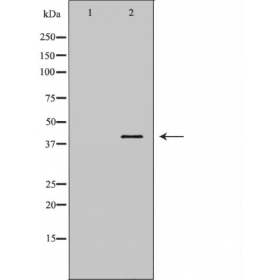
Annexin A1 Antibody - #DF6254
| Product: | Annexin A1 Antibody |
| Catalog: | DF6254 |
| Description: | Rabbit polyclonal antibody to Annexin A1 |
| Application: | WB |
| Reactivity: | Human, Mouse, Rat |
| Mol.Wt.: | 39kDa; 39kD(Calculated). |
| Uniprot: | P04083 |
| RRID: | AB_2838220 |
Product Info
*The optimal dilutions should be determined by the end user. For optimal experimental results, antibody reuse is not recommended.
*Tips:
WB: For western blot detection of denatured protein samples. IHC: For immunohistochemical detection of paraffin sections (IHC-p) or frozen sections (IHC-f) of tissue samples. IF/ICC: For immunofluorescence detection of cell samples. ELISA(peptide): For ELISA detection of antigenic peptide.
Cite Format: Affinity Biosciences Cat# DF6254, RRID:AB_2838220.
Fold/Unfold
Annexin 1; Annexin A1; Annexin I (lipocortin I); Annexin I; Annexin-1; AnnexinA1; AnnexinI; ANX 1; ANX A1; ANX1; ANXA 1; ANXA1; ANXA1 protein; ANXA1_HUMAN; Calpactin 2; Calpactin II; Calpactin-2; CalpactinII; Chromobindin 9; Chromobindin-9; Chromobindin9; HGNC:533; Lipocortin 1; Lipocortin I; Lipocortin1; LipocortinI; LPC 1; LPC1; p35; Phospholipase A2 inhibitory protein;
Immunogens
A synthesized peptide derived from human Annexin A1, corresponding to a region within N-terminal amino acids.
Detected in resting neutrophils (PubMed:10772777). Detected in peripheral blood T-cells (PubMed:17008549). Detected in extracellular vesicles in blood serum from patients with inflammatory bowel disease, but not in serum from healthy donors (PubMed:25664854). Detected in placenta (at protein level) (PubMed:2532504). Detected in liver.
- P04083 ANXA1_HUMAN:
- Protein BLAST With
- NCBI/
- ExPASy/
- Uniprot
MAMVSEFLKQAWFIENEEQEYVQTVKSSKGGPGSAVSPYPTFNPSSDVAALHKAIMVKGVDEATIIDILTKRNNAQRQQIKAAYLQETGKPLDETLKKALTGHLEEVVLALLKTPAQFDADELRAAMKGLGTDEDTLIEILASRTNKEIRDINRVYREELKRDLAKDITSDTSGDFRNALLSLAKGDRSEDFGVNEDLADSDARALYEAGERRKGTDVNVFNTILTTRSYPQLRRVFQKYTKYSKHDMNKVLDLELKGDIEKCLTAIVKCATSKPAFFAEKLHQAMKGVGTRHKALIRIMVSRSEIDMNDIKAFYQKMYGISLCQAILDETKGDYEKILVALCGGN
Research Backgrounds
Plays important roles in the innate immune response as effector of glucocorticoid-mediated responses and regulator of the inflammatory process. Has anti-inflammatory activity. Plays a role in glucocorticoid-mediated down-regulation of the early phase of the inflammatory response (By similarity). Promotes resolution of inflammation and wound healing. Functions at least in part by activating the formyl peptide receptors and downstream signaling cascades. Promotes chemotaxis of granulocytes and monocytes via activation of the formyl peptide receptors. Contributes to the adaptive immune response by enhancing signaling cascades that are triggered by T-cell activation, regulates differentiation and proliferation of activated T-cells. Promotes the differentiation of T-cells into Th1 cells and negatively regulates differentiation into Th2 cells. Has no effect on unstimulated T cells. Promotes rearrangement of the actin cytoskeleton, cell polarization and cell migration. Negatively regulates hormone exocytosis via activation of the formyl peptide receptors and reorganization of the actin cytoskeleton. Has high affinity for Ca(2+) and can bind up to eight Ca(2+) ions (By similarity). Displays Ca(2+)-dependent binding to phospholipid membranes. Plays a role in the formation of phagocytic cups and phagosomes. Plays a role in phagocytosis by mediating the Ca(2+)-dependent interaction between phagosomes and the actin cytoskeleton (By similarity).
Phosphorylated by protein kinase C, EGFR and TRPM7. Phosphorylated in response to EGF treatment.
Sumoylated.
Nucleus. Cytoplasm. Cell projection>Cilium. Cell membrane. Membrane>Peripheral membrane protein. Endosome membrane>Peripheral membrane protein. Basolateral cell membrane. Apical cell membrane. Lateral cell membrane. Secreted. Secreted>Extracellular space. Cell membrane>Peripheral membrane protein>Extracellular side. Secreted>Extracellular exosome. Cytoplasmic vesicle>Secretory vesicle lumen. Cell projection>Phagocytic cup. Early endosome. Cytoplasmic vesicle membrane>Peripheral membrane protein.
Note: Secreted, at least in part via exosomes and other secretory vesicles. Detected in exosomes and other extracellular vesicles (PubMed:25664854). Detected in gelatinase granules in resting neutrophils (PubMed:10772777). Secretion is increased in response to wounding and inflammation (PubMed:25664854). Secretion is increased upon T-cell activation (PubMed:17008549). Neutrophil adhesion to endothelial cells stimulates secretion via gelatinase granules, but foreign particle phagocytosis has no effect (PubMed:10772777). Colocalizes with actin fibers at phagocytic cups (By similarity). Displays calcium-dependent binding to phospholipid membranes (PubMed:2532504, PubMed:8557678).
Detected in resting neutrophils. Detected in peripheral blood T-cells. Detected in extracellular vesicles in blood serum from patients with inflammatory bowel disease, but not in serum from healthy donors. Detected in placenta (at protein level). Detected in liver.
The full-length protein can bind eight Ca(2+) ions via the annexin repeats. Calcium binding causes a major conformation change that modifies dimer contacts and leads to surface exposure of the N-terminal phosphorylation sites; in the absence of Ca(2+), these sites are buried in the interior of the protein core. The N-terminal region becomes disordered in response to calcium-binding.
The N-terminal 26 amino acids are sufficient for its extracellular functions in the regulation of inflammation and wound healing (PubMed:25664854). Acylated peptides that contain the first 26 amino acids of the mature protein can activate signaling via the formyl peptide receptors (PubMed:15187149, PubMed:25664854).
Belongs to the annexin family.
Restrictive clause
Affinity Biosciences tests all products strictly. Citations are provided as a resource for additional applications that have not been validated by Affinity Biosciences. Please choose the appropriate format for each application and consult Materials and Methods sections for additional details about the use of any product in these publications.
For Research Use Only.
Not for use in diagnostic or therapeutic procedures. Not for resale. Not for distribution without written consent. Affinity Biosciences will not be held responsible for patent infringement or other violations that may occur with the use of our products. Affinity Biosciences, Affinity Biosciences Logo and all other trademarks are the property of Affinity Biosciences LTD.
