

.
Bands result from membrane strip incubation.")


 by IF/ICC. The samples were fixed with PFA and permeabilized in 0.1% Triton X-100,then blocked in 10% serum for 45 minutes at 25°C. Samples were then incubated with primary Ab(DF6304 1:200) and mouse anti-beta tubulin Ab(T0023 1:200) for 1 hour at 37°C. An AlexaFluor594 conjugated goat anti-rabbit IgG(H+L) Ab(Red) and an AlexaFluor488 conjugated goat anti-mouse IgG(H+L) Ab(Green) were used as the secondary antibody.
The nuclear counter stain is DAPI(blue).")
 and mouse anti-beta tubulin Ab(T0023 1:200) for 1 hour at 37°C. An AlexaFluor594 conjugated goat anti-rabbit IgG(H+L) Ab(Red) and an AlexaFluor488 conjugated goat anti-mouse IgG(H+L) Ab(Green) were used as the secondary antibody.")
.")
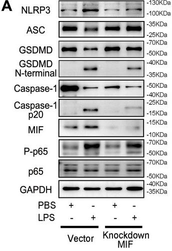
 and AZA (9.0 mg/kg) on the protein levels of NLRP3, ASC, caspase-1 p10 and pro-caspase-1 in colons of TNBS-induced colitis in rats.")
Representative immunoblot bands for the NLRP3, ASC, Pro-Caspase-1, Caspase-1, and GSDMD proteins. β-actin was used as a loading control")
,
p-p65 (b), p65, p-IκBα (c), IκBα, NLRP3 (d), ASC (e), Caspase-1 (e), and β-actin in mesenteric arteries from SO group, CLP group, CLP + HKL group, and
CLP + 3-TYP group were tested by western blot. n=5, *P<0.05 compared with SO group, #P<0.05 compared with CLP group")
, ASC (B), and Caspase-1 (C). Data are shown as mean ± SD, n = 8. #p < 0.05 and ##p < 0.01 versus the Control group. ⁎p < 0.05 and ⁎⁎p < 0.01 versus the Model group.")
 The liver levels of IL-18 and IL-1β. (B) The hepatic
mRNA expression of NLRP3. (C) Representative bands of TXNIP, NLRP3, ASC and cleaved-caspase 1. (D) Quantitative results of Western blot bands densities of
TXNIP, NLRP3, ASC and Cleaved-caspase1. Data are presented as the mean ± SD (n = 3 ~ 8). ## P < 0.01 vs. NC group; **P < 0.01 vs. Model group.")
 The
protein expression of IκB- α , NF-κBp65, and
p65 in the cytosol; p65 in the nucleus;
β-Actin, and lamin B obtained from HUV-EC-
C cells of all experimental groups. B) The
effects of PAVA and RSV on the ox-LDL-
induced inflammasome pathway in HUV-
EC-C cells. Cells were pretreated with JSH-
23 inhibitor or PAVA or RSV and their
combination for 2 h before incubation in the
presence or absence of ox-LDL for 24 h. The
protein expression of NLRP3, ASC, cleaved
caspase-1, IL-1β, CRP, and β-actin obtained
from the HUV-EC-C cells of all experimental
groups by Western blot analysis. Data are
shown as mean ± SD. Statistical analysis was
performed with one-way ANOVA by Tukey’s
multiple comparison test for comparison
between groups, (n = 3). Significant differ-
ences are shown by **, ***p < 0.01 and
0.001, respectively, vs control; #, ##, ### p <
0.05, 0.01, and 0.001, respectively, vs ox-
LDL alone. $, $$ p < 0.05 and 0.01, respec-
tively, vs the combination of PAVA + RSV.
NS indicates no significance.")
Representative Western blot analysis of NLRP3 inflammasome in Sham, SCI + vehicle and SCI + zinc group(n = 6).")
. DMSO, dimethyl sulfoxide; EAP,
experimental autoimmune prostatitis")
 The level of IL-1β in LPS/Aβ-treated BV-2 cells with different concentrations of milrinone (0, 10, 25, 50 μmol/L) was measured by ELISA analysis. (B) The level of IL-6 in LPS/Aβ-treated BV-2 cells with different concentrations of milrinone (0, 10, 25, 50 μmol/L) was measured by ELISA analysis. (C) The level of TNF-α in LPS/Aβ-treated BV-2 cells with different concentrations of milrinone (0, 10, 25, 50 μmol/L) was measured by ELISA analysis. (D) The Western blot assay images for the levels of ASC, NLRP3, active caspase 1, IL-1β, and IL-18 in LPS/Aβ-treated BV-2 cells. (E) The protein level of ASC in LPS/Aβ-treated BV-2 cells was measured by Western blot assay. (F) The protein level of NLRP3 in LPS/Aβ-treated BV-2 cells was measured by Western blot assay. (G) The protein level of active caspase 1 in LPS/Aβ-treated BV-2 cells was measured by Western blot assay. (H) The protein level of IL-1β in LPS/Aβ-treated BV-2 cells was measured by Western blot assay. (I) The protein level of IL-18 in LPS/Aβ-treated BV-2 cells was measured by Western blot assay. **P < 0.01 vs the control group; #P < 0.05, ##P < 0.01, && P < 0.01 vs the LPS/Aβ group. ns: not significant.")
 Western blotting images of IL-17, p-NF-κB, NF-κB, NLRP3, ASC, cleaved-Caspase1, pro-Caspase 1, pro-IL-1β, cleaved-IL-1β, and IL-18: Lane 1, control group; lane 2, model group; lane 3, YH 5 g crude drug/kG group; lane 4, YH 15 g crude drug/kG group; lane 5, selenious group. Bar graphs indicate the relative ratio of IL-17 over tubulin (B), p-NF-κB over NF-κB (C), NLRP3 over tubulin (D), ASC over tubulin (E), cleaved-Caspase-1 over pro-Caspase-1 (F), cleaved-IL-1β over pro-IL-1β (G), and IL-18 over tubulin (H) in thyroid. All values are expressed as mean ± SEM. *p < 0.05, **p < 0.01 vs Model.")
 Representative examples of Western blot analysis demonstrating the expression levels of NLRP3, ASC, procaspase-1, and caspase-1; quantification of the expression levels of (B) NLRP3, (C) ASC, and (D) procaspase-1 and (E) caspase-1; (F) representative examples of Western blot analysis demonstrating the expression levels of TLR4, MyD88, p-NF-κB, and NF-κB p65; quantification of the expression levels of (G) TLR4, (H) MyD88, (I) p-NF-κB, and NF-κB p65. Data presented are the means ± SD. ##p < 0.01 compared with Sham; *p < 0.05, **p < 0.01 compared with MI/R.")
 Representative Western blotting images of NLRP3, ASC, Caspase-1and Caspase-1 p10 (B–E) The protein expression levels of NLRP3, ASC, Caspase-1 and Caspase-1 p10 in the NLRP3 inflammasome. Data are expressed as the means ± SD (n = 3). # p < 0.05, ## p < 0.01 vs. control group; *p < 0.05, **p < 0.01 vs. DSS group; & p < 0.05, && p < 0.01 vs. COP group.")
 Representative Western blot bands; The protein expression of (B) NLRP3, (C) caspase-1, (D) ASC, (E) IL-1β; the gene expression of (F) NLRP3, (G) caspase-1, (H) ASC, (I) IL-1β; Protein levels were determined by Western blotting and the band optical densities were quantitated by Image J software. The renal protein levels were normalized to GAPDH. Relative gene levels were determined by RT-qPCR. The results are expressed as mean ± SD (n = 3–5). # p < 0.05, ## p < 0.01, compared with NC group; * p < 0.05, ** p < 0.01, compared with HUA group.")
 NLRP3 and ASC, (B) cleaved-caspase-1 and caspase-1, (C) iNOS, (D) p-P65, P65, p-IκBα, and IκBα. The images (in gray) were analyzedusing Image J software. Data were expressed as mean ± SEM (n = 6).")
. *, P<0.05 vs. Control; #, P<0.05 LPS + EB1089 vs. LPS. ASC,apoptosis-associated speck-like protein containing a CARD; VDR, vitamin D receptor; LPS, lipopolysaccharide.")
 on NF-κB/NLRP3 and Nrf2 signaling pathways in LPS plus MSU-activated THP-1 cells. (A) Representative Western blotting images of p-p65, p65, p-IKBα, IKBα and β-actin. (B) Representative Western blotting images of NLRP3, ASC, IL-1β, Caspase-1 and β-actin. (C) Representative Western blotting images of Nrf2, HO-1 and β-actin. Changes in the relative protein expression levels of p-p65/p65 (D), p-IKBα/IKBα (E), NLRP3 (F), ASC (G), IL-1β (H), Caspase-1 (I), Nrf2 (J) and HO-1 (K). Data are shown as mean ± SD (n = 3); ##P < 0.01 vs Control group; *P < 0.05, **P < 0.01 vs Model group.")
 Representative western blot analysis detects the NLRP3, ASC, caspase-1 and IL-1 levels of sham, subarachnoid hemorrhage (SAH)+vehicle and SAH+NBTI. (b) Quantitative analyses of NLRP3. (c) Quantitative analyses of ASC. (d) Quantitative analyses of caspase-1. (e) Quantitative analyses of IL-1. n = 4 per group. The bars represent the mean ± SD. *P < 0.05 versus sham. # P < 0.05 versus SAH+vehicle. ASC, apoptosis associated speck like protein containing CARD; NBTI, nitrobenzylthioinosine.")
 pyroptosis by upregulating the expression of nucleotide-binding oligomerization segment-like receptor family 3 (NLRP3) inflammasome in FLS and the effect of monomeric derivatives of paeoniflorin (MDP). (a, b) shHIF-1α was transfected into FLS and exposed to hypoxia for 2 h. The expression of hypoxia-inducible factor (HIF)-1α, NLRP3, speck-like protein containing CARD (ASC), caspase-1, and cleaved-caspase-1 in FLS was detected by Western blot (n = 3). (c, d) NLRP3 short hairpin (shNLRP3) construct was transfected into FLS and exposed to hypoxia for 2 h. The expression of HIF-1α, NLRP3, and N-terminal domain of human gasdermin D (GSDMD-N) in FLS after exposure to hypoxia for 2 h was detected by Western blot (n = 3). (e, f) FLS were treated with or without MDP and exposed to hypoxia for 2 h. The expression of HIF-1α, NLRP3, ASC, caspase-1, cleaved-caspase-1, and GSDMD-N in FLS was detected by Western blot (n = 3). Data are expressed as mean ± SD. #p < 0.05, ##p < 0.01 vs. the control group; ∗p < 0.05, ∗∗p < 0.01 vs. the hypoxia group. (g) Analysis of expression levels of HIF-1α in the nucleus. (h) Quantitative PCR analysis and quantification of the mRNA levels of NLRP3, ASC, and caspase-1. Data are expressed as mean SD. #p < 0.05 vs. the control group; ∗p < 0.05 vs. the hypoxia group.")
 Western blot analysis of NLRP3, ASC, caspase-1, and cleaved-IL-1β protein expression in the cardiac tissue. (B) Immunochemical staining of NLRP3, ASC, and cleaved-IL-1β. Tubulin or GAPDH was detected as the loading control. NLRP3: nod-like receptor pyrin domain 3, ASC: apoptosis-associated speck-like protein, and IL-1β: interleukin 1β. The data are the mean ± standard deviation (x¯±s) (n = 3 or 4 per group) *p < 0.05, **p < 0.01, and ***p < 0.001.")
 mRNA levels of miRNA-33, NLRP3, ASC and caspase-1. The effects of EMS on the expression of NLRP3 inflammasome-associated protein in the PMs of AA rats. (B) The protein levels of NLRP3, ASC, caspase-1, pro-caspase-1 were determined by western blotting. Densitometric analyses of the NLRP3 inflammasome-associated protein normalized to histone β-actin. (C) ASC, (D) caspase-1, (E) NLRP3 and (F) pro-caspase-1. ##p < 0.01, vs. the normal group; **p < 0.01 vs. the model group (n = 3).")
 Western blotting results of NLRP3, ASC, cleaved caspase-1, IL-1β, and GSDMD in the sham and OA synovitis rats. (c) mRNA levels of NLRP3, ASC, caspase-1, IL-1β, and GSDMD expressed in TMJ synovium between the sham and OA rats. (d) Immunohistochemistry of TMJ slices from the sham or OA rats using anti-NLRP3, anti-caspase-1, anti-GSDMD, and anti-IL-1β antibodies, scale bar = 50 μm. Positive cells are mainly around synovial membrane and vessels (black arrow). (e) Percentage of positive cells. Data in this figure are analyzed by independent samples t-test and presented as mean ± SD. ∗P < 0.05, ∗∗P < 0.01, and ∗∗∗P < 0.001.")
 Protein levels of NLRP3-related molecules shown by western blot. β-Tubulin was an internal reference control. n = 3 in each group. (b) Relative mRNA levels of Nlrp3-relaterd genes shown by real-time RT-PCR. Tubb3 served as a reference gene. n = 3 in each group. (c) Ratio of caspase-1 p12/procaspase-1. n = 3 in each group. (d) Caspase-1 activity in heart tissues. n = 6 in each group. Immunohistochemical staining of (e) NLRP3, (f) ASC, (g) GSDMD, (h) IL-1β, (i) IL-18, and (j) NEK7. Scale bar, 50 μm. (k) Double-labeling immunofluorescence of NLRP3 and CD68. Scale bar, 50 μm. Data are presented as mean ± SD. ∗∗P < 0.01 CON vs. SHAM; ##P < 0.01 SG vs. SHAM. n = 3 in each group.")
 Protein levels of NLRP3-related molecules shown by western blot. β-Tubulin was an internal reference control. n = 3 in each group. (b) Relative mRNA levels of Nlrp3-relaterd genes shown by real-time RT-PCR. Tubb3 served as a reference gene. n = 3 in each group. (c) Ratio of caspase-1 p12/procaspase-1. n = 3 in each group. (d) Caspase-1 activity in heart tissues. n = 6 in each group. Immunohistochemical staining of (e) NLRP3, (f) ASC, (g) GSDMD, (h) IL-1β, (i) IL-18, and (j) NEK7. Scale bar, 50 μm. (k) Double-labeling immunofluorescence of NLRP3 and CD68. Scale bar, 50 μm. Data are presented as mean ± SD. ∗∗P < 0.01 CON vs. SHAM; ##P < 0.01 SG vs. SHAM. n = 3 in each group.")
 Immunofluorescence staining for F4/80 (macrophages) and chemerin of placenta tissue in normal and GDM pregnant women. (B) Content of IL-18 and IL-1β in placenta tissue of normal and GDM pregnant women was measured by ELISA (n = 3). (C) Content of IL-18 and IL-1β in placenta tissue of Ctrl, CHM+sh-NC, and CHM+sh-chemR23 mice was measured by ELISA (n = 3). (D) Protein levels of NOD-like receptor family pyrin domain containing 3 (NRLP3), apoptosis-associated speck-like protein containing CARD (Asc), pro caspase 3, cleaved caspase 3, pro caspase 9, cleaved caspase 9, pro caspase 1, cleaved caspase 1, pro-IL-1β, and pro-IL-18 in placental macrophages were measured by Western blotting. Protein levels of cleaved caspase 1, IL-1β, and IL-18 in the culture supernatants of placental macrophages were measured by Western blotting as well. Glyceraldehyde-3-phosphate dehydrogenase (GAPDH) served as the internal control (n = 3). (E) Trophoblast cells were co-incubated with control (Ctrl), the supernatant from macrophage, macrophage+CHM+sh-NC, and macrophage+CHM+sh-chemR23 for 24 h, then relative exosomal miR-140-3p and miR-574-3p expression was measured by qRT-PCR (n = 3). (F) Trophoblast cells were treated with Control, recombinant protein IL-18, recombinant protein IL-1β, and recombinant protein IL-18+IL-1β. Relative exosomal miR-140-3p and miR-574-3p expression was measured by qRT-PCR (n = 3). (G) Changed protein levels of IL-18 and IL-1β in macrophage after treatment with chemerin and siRNA NC (CHM+si-NC), IL-1β siRNA (CHM+si-IL-1β), IL-18 siRNA (CHM+si-IL-18) and IL-1β siRNA and IL-18 siRNA (CHM+si-IL-1β+si-IL-18) were measured by Western blotting (n = 3). (H) Trophoblast cells were co-incubated with the supernatant of macrophage+CHM+si-NC, macrophage+CHM+si-IL-1β, macrophage+CHM+si-IL-18, and macrophage+CHM+si-IL-1β+si-IL-18, then relative exosomal miR-140-3p and miR-574-3p expression was measured by qRT-PCR (n = 3).")
 Relative mRNA levels of NLRP3, ASC, and caspase-1 were measured via RT-qPCR. (d–f) Relative expression levels of NLRP3, ASC, and caspase-1 were measured via western blot assay. (NO, normoxia group; NA, normoxia + artesunate group; HO, hyperoxia group; HA, hyperoxia + artesunate group. P value was calculated by one-way ANOVA and SNK test.")

 Protein expression of NLRP3, CASP1, and ASC. (B,C,D) The analysis of protein expression of NLRP3, CASP1, and ASC. (## p < 0.01, vs. sham group;")
 inhibits the expression of inflammatory proteins. (A,B) Western blot analyses of the expression of NLRP3, caspase-1, ASC, IL-1β, NFκB p65, and NFκB p50 in cardiac tissue. The widths of the images have been compressed at a ratio of 1:0.6. n = 3 or 4 per group. Data are represented as mean ± standard deviation (x¯ ± s); *P < 0.05, **P < 0.01.")
")


 with or without plasmon-activated water (PAW) against protein expression of (A) NLRP3 inflammasomes and (B) PYCARD in Caco-2 cells. Data are presented as mean ± SEM (n = 5 per group). The bars not sharing the same letter indicate statistical differences between the groups at p < 0.05.")
, and incubation was continued for 24 h. Next, 3 mM ATP was added to the medium and incubated for 45 min to activate the expression of NLRP3, and then, indices reflecting oxidative stress (A: ROS, B: MDA, and C: SOD) were assessed. (D) GSDMD-N immunofluorescence staining was performed to detect cellular pyroptosis; (E) Western immunoblotting assay was performed to evaluate the level of expression of GSDMD-N, NLRP3, ASC, and C-caspase-1 proteins. (F) Protein quantitative analysis was performed. (G) Caspase-1 activity was determined using the Caspase 1 Activity Assay Kit ; ELISA was performed to determine the level of expression of IL-1β (H) and IL-18 (I) to evaluate the inhibitory effect of irisin on HG-induced oxidative stress and pyroptosis in Min6 cells. Scale bar = 130 μm")
. Values are presented as mean ± SD. *P")
 NLRP3 (A), ASC (B), GSDMD (C), and Caspase-1 (D) mRNA expression levels were measured by quantitative reverse transcription-polymerase chain reaction. (E) Western blot analysis of inflammation- and pyroptosis-associated protein expression levels. β-Actin was used as an internal reference for NLRP3, ASC, GADMD, IL-18, and IL-1β. GAPDH was used as an internal reference for TLR4, P65, p-P65, IκBα, and p-IκBα. (F–M) NLRP3 (F), ASC (G), GSDMD (H), IL-18 (I), IL-1β (J), TLR4 (K), p-P65/P65 (L), and p-IκBα/IκBα (M) protein expression levels were measured by Western blot assay. All western blot data were normalized to the sham group. Data are expressed as mean ± SD (n = 3). *P < 0.05, **P < 0.01, vs. sham group; #P < 0.05, ##P < 0.01, vs. model group (one-way analysis of variance followed by Tukey's post hoc test). ASC: Apoptosis-associated speck-like protein containing CARD; BA: Biochanin A; GAPDH: glyceraldehyde-3-phosphate dehydrogenase; GSDMD: gasdermin D; IL-18: interleukin-18; IL-1β: interleukin-1β; NLRP3: NOD-like receptor thermal protein domain associated protein 3; PC: positive control; p-P65: phospho-NF-κB P65; p-IκBα: phospho-NF-kappa-B inhibitor alpha; SCI: spinal cord injury; TLR4: Toll-like receptor 4.")
 The expressions of NLRP3, ASC, caspase-1, and IL-1β mRNA were tested by RT-PCR. (E–J) The expressions of NLRP3, ASC, caspase-1, IL-1β, and IL-18 protein were examined by western blotting. Data are showed as means ± SEM. **P < 0.01 vs control group, #P < 0.05, ##P < 0.01 vs DSS group.")
 The transfection efficiency of SREBP1 was measured by Western blot analysis. (B) Quantification of knockdown efficiency of SREBP1 by siRNA. (C) Cell viability was detected using Calcein/PI staining in HT22 cells. (D) The expression of pyroptosis-related proteins was captured by Western blot analysis. (E) Quantification of the results of D. (F) IF analysis of NLRP1 and NRP3 expression in HT22 cells. Scale bar, 50 μm. (G) NLRP1 and NRP3 fluorescence intensity quantification in the control and Glu groups treated with siSREBP1 or siNC. The values were showed as the mean ± SEM (n = 3). *p < 0.05, **p < 0.01, ***p < 0.001 compared with siNC + PBS group and #p < 0.05, ##p < 0.01 compared with siNC + Glu group. NS, no significance. siSREBP1, SREBP1 target siRNA; siNC, control siRNA; Cle-cas1, Cleaved caspase-1; IL-18, interleukin-18.")
 exposure on NLRP3 and AIM2 inflammasomes activation in lung tissues(n=3). (A)The represented protein bands of NLRP3, AIM2, ASC, IL-18 and IL-1β in the lung of rats. (B-F) The relative gray values of NLRP3/GAPDH, AIM2/GAPDH, ASC/GAPDH, IL-18/GAPDH, and IL-1β/GAPDH in lung of rat. Compared with the control group in the same group, *P")
 The mRNA expression of AIM2, ASC, caspase-1, IL-1β and IL-18 in liver tissue was analyzed by qRT-PCR (n = 5 mice/group). (B) Serum levels of IL-1β and IL-18 in mice (n = 6 mice/group). (C) The protein expression of AIM2, ASC, Cleaved caspase-1, caspase-1 and IL-1β in mouse livers were determined by Western Blotting (n = 3 mice/group). (D) NLRP3 protein and mRNA expression in mouse livers were determined by Western Blotting (n = 3 mice/group) and qRT-PCR (n = 5 mice/group). Data are shown as mean ± SEM of the independent experiments, statistical analysis was performed using One-way ANOVA, * P < 0.05, ** P < 0.01, *** P < 0.001 vs LV-control mice; # p < 0.05, ## P < 0.01, ### P < 0.001 vs LV-control HFD-fed mice.")
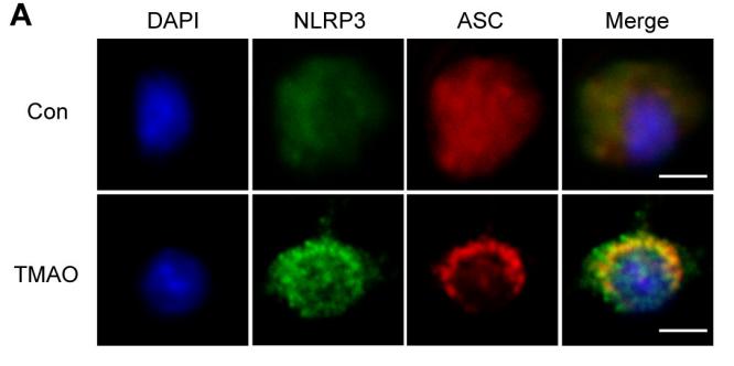
 Co-localization of NLRP3 and ASC was examined by immunofluorescence staining. (B) Relative expression levels of NLRP3 and ASC. Data are showed as means ± SEM. **P < 0.01 vs control group, ##P < 0.01 vs DSS group.")
. (n = 4). Data are presented as mean ± SEM; *P")
 Protein expressions of NLRP3, C-Caspase-1, GSDMD, IL-18, ASC, IL-1β, and GAPDH were evaluated by Western blot. GAPDH was used as an internal control for sample loading. (B-G) Quantitative analyses of the expression levels of NLRP3, C-Caspase-1, GSDMD, IL-18, ASC, and IL-1β. (n = 3 per group). Values are presented as the mean ± SD. *p")
 Cardiomyocyte viability after treatment of different concentrations of Dox measured with CCK-8 assay. (B) RT-qPCR and western blotting to detect FTO expression in cardiomyocytes after Dox treatment. (C) RT-qPCR and western blotting to test FTO expression in cardiomyocytes after upregulation or knockdown of FTO. (D) Flow cytometry to examine apoptosis. (E) Detection of LDH expression. (F) Expression of inflammatory factors (IL-18, IL-1β, TNF-α, and IL-6) determined with ELISA. (G) Expression of oxidative stress-related indicators (MDA and SOD). (H) Western blotting analysis of pyroptosis-related proteins (NLRP3, ASC, cleaved caspase-1, and GSDMD-N). (I) Immunofluorescence staining to measure the pyroptosis protein NLRP3. Data were expressed as mean ± standard deviation. Cell experiments were performed three times. Normally distributed data between two groups were compared with the unpaired t-test and among multiple groups were compared with one-way analysis of variance. Tukey’s test was used for post hoc analysis. *, P")
 In vivo experimental workflow. (B) HE (20× magnification) and Safranin O-fast green staining (5× magnification) of rabbit cartilage from OA and control groups, with damaged areas indicated by arrows (n = 6). (C) HE staining of synovial tissue from OA and control groups (20× magnification), with damaged areas indicated by arrows (n = 6). (D) OARSI cartilage scores for each group. (E) OARSI synovial scores for each group. (F) RT-PCR analysis of NLRP3, ASC, MMP13, COL2, and IRF1 mRNA expression in rabbit cartilage from OA and control groups (n = 3). (G-H) Western Blot analysis of NLRP3, ASC, cleaved Caspase-1, MMP13, COL2, and IRF1 protein expression in rabbit cartilage from OA and control groups (n = 3). (I-J) Immunohistochemical detection of the positive cell rates of NLRP3, ASC, cleaved Caspase-1, MMP13, COL2, and IRF1 in rabbit cartilage from OA and control groups (n = 3). (K) Elisa analysis of IL-1β and IL-18 levels in rabbit serum (n = 6). All data are expressed as the mean ± S.D. ⁎⁎P < 0.01, ⁎P < 0.05. (For interpretation of the references to colour in this figure legend, the reader is referred to the web version of this article.)")
 In vivo experimental workflow. (B) HE (20× magnification) and Safranin O-fast green staining (5× magnification) of rabbit cartilage from OA and control groups, with damaged areas indicated by arrows (n = 6). (C) HE staining of synovial tissue from OA and control groups (20× magnification), with damaged areas indicated by arrows (n = 6). (D) OARSI cartilage scores for each group. (E) OARSI synovial scores for each group. (F) RT-PCR analysis of NLRP3, ASC, MMP13, COL2, and IRF1 mRNA expression in rabbit cartilage from OA and control groups (n = 3). (G-H) Western Blot analysis of NLRP3, ASC, cleaved Caspase-1, MMP13, COL2, and IRF1 protein expression in rabbit cartilage from OA and control groups (n = 3). (I-J) Immunohistochemical detection of the positive cell rates of NLRP3, ASC, cleaved Caspase-1, MMP13, COL2, and IRF1 in rabbit cartilage from OA and control groups (n = 3). (K) Elisa analysis of IL-1β and IL-18 levels in rabbit serum (n = 6). All data are expressed as the mean ± S.D. ⁎⁎P < 0.01, ⁎P < 0.05. (For interpretation of the references to colour in this figure legend, the reader is referred to the web version of this article.)")
 In Naringin-treated experimental autoimmune prostatitis mice, immunofluorescence data showed reduced M1 macrophage infiltration. (C) Western blotting of ASC, procaspase-1, NLRP3, cleaved-caspase-1, NF-κB, and cleaved-IL-1β in prostate tissues (n = 3). NF-κB (D), NLRP3 (E), ASC (F), procaspase-1 (G), cleaved caspase-1 (H), and cleaved-IL-1β (I) expression levels measured by Western blotting assay. One-way ANOVA analysis was used to evaluate the data displayed as the mean ± SD. Bonferroni post-hoc test was used to determine the differences between the groups.")
 Representative TUNEL staining images of the left-ventricular samples from each group of mice. Bar = 50 μm. (B) Quantification of TUNEL-positive cells in (A); n = 100 cells per group. (C) Representative western blot images of NLRP3, caspase-1, and GSDMD expression in the left-ventricular samples from each group of mice. (D) Representative western blot images of caspase-3, caspase-7, and caspase-8 expression in the left-ventricular samples from each group of mice. (E) Representative western blot images of p-RIPK1, p-RIPK3, and p-MLKL expression in the left-ventricular samples from each group of mice. (F)–(I) Quantification of the protein expression of NLRP3, cleaved caspase-1, and GSDMD-N in (C). (J)–(L) Quantification of the protein expression of cleaved caspase-3, cleaved caspase-7, and cleaved caspase-8 in (D). (M)–(O) Quantification of the protein expression of p-RIPK1, p-RIPK3, and p-MLKL in (E); n = 6 mice per group for panels A–O. The normality of data distribution was tested using the Shapiro–Wilk method. One-way ANOVA was applied in (B) and in (F)–(O). *p")
 on nod-like receptor family protein 3 (NLRP3) inflammasome activation in HT22 cells. After treatment of HT22 cells with Hcy (1.25, 2.5, and 5 mM) for 24 h, the expressions of NLRP3 (a), ASC (b), and cleaved-caspase-1 (c) were determined by Western blot. Data were shown as the mean ± standard error of the mean. Statistical significance was determined by one-way analysis of variance with least-significant difference post hoc test. (n = 3).")
, with comparisons made among the S. copri OMVs or LPS-treated group and the untreated control group. n = 3.")
 Immunofluorescence images of ASC (red), caspase 8 (green), and RIPK3 (rose red) co-localization with nuclear DAPI staining (blue). (B–D) Average fluorescence intensity of ASC, caspase 8, and RIPK3. (Mean ± SEM, n = 3, * p < 0.05 and ** p < 0.01 vs. model group). Dashed box shows the zoomed area.")
 differential expression of USP7 in ccRCC tissues compared with that in normal tissues in TCGA cohort. ****p")
 Effect of DEP exposure on the viability of Nthy-Ori 3–1 cells (n = 3–4). (B) Protein expression of AHR and quantitative analysis (n = 3). (C) mRNA expression of NLRP3, CASP1 and PYCARD (n = 3). (D) Protein expression of NLRP3, cleaved-caspase-1, ASC, cleaved-IL-1β and GSDMD-N, and respective quantitative analysis (n = 3). (E) ELISA detection of IL-1β and IL-18 in cell supernatant (n = 3). Control: non-DEP group; 20 μg/mL: low-dose DEP exposure group; 40 μg/mL: high-dose DEP exposure group. AHR, aryl hydrocarbon receptor; NLRP3, NOD-like receptor pyrin domain-containing 3; cleaved caspase-1, cleaved cysteine-aspartic acid protease-1; ASC, apoptosis-associated speck-like protein containing a CARD.")
 The expression of NLRP3 in H9c2 cells was detected by immunofluorescence assay; (B) The activation of Caspase-1 in H9c2 cells was detected by immunofluorescence assay; (C) The expression of EGFP-NLRP3 in H9c2 cells was detected by immunofluorescence assay; (D) The protein expressions of NLRP3, ASC, Caspase1, Cleaved Caspase-1, IL-1β and Cleaved-IL-1β in H9c2 cells were detected by Western blot; scale bar = 20 μm (n = 3). Compared with control group, ###P < 0.001; Compared with LPS + ATP group")
, ASC (C,D), caspase-1, and caspase-1-p20 (E,F),")
 Levels of IL-1β and IL-18. B) Protein level of GSDMD. C) Inflammasome complex protein levels determined by Western blotting. ***p < 0.001, compared with si-NC group. Three replicate experiments were performed. Data are presented as mean ± standard deviation")
| Product: | TMS1/ASC Antibody |
| Catalog: | DF6304 |
| Description: | Rabbit polyclonal antibody to TMS1/ASC |
| Application: | WB IHC IF/ICC |
| Cited expt.: | WB, IHC, IF/ICC |
| Reactivity: | Human, Mouse, Rat |
| Prediction: | Bovine, Horse, Sheep, Rabbit |
| Mol.Wt.: | 15~25kDa(Observed); 22kD(Calculated). |
| Uniprot: | Q9ULZ3 |
| RRID: | AB_2838270 |
Control Products

Related Downloads
Protocols
Product Info
*The optimal dilutions should be determined by the end user. For optimal experimental results, antibody reuse is not recommended.
*Tips:
WB: For western blot detection of denatured protein samples. IHC: For immunohistochemical detection of paraffin sections (IHC-p) or frozen sections (IHC-f) of tissue samples. IF/ICC: For immunofluorescence detection of cell samples. ELISA(peptide): For ELISA detection of antigenic peptide.
Cite Format: Affinity Biosciences Cat# DF6304, RRID:AB_2838270.
Fold/Unfold
Apoptosis associated speck like protein containing a CARD; Apoptosis-associated speck-like protein containing a CARD; ASC; ASC_HUMAN; CARD 5; CARD5; Caspase recruitment domain containing protein 5; Caspase recruitment domain protein 5; Caspase recruitment domain-containing protein 5; hASC; MGC10332; PYCARD; PYD and CARD domain containing; PYD and CARD domain containing protein; PYD and CARD domain-containing protein; Target of methylation induced silencing 1; Target of methylation-induced silencing 1; TMS 1; TMS; TMS1;
Immunogens
A synthesized peptide derived from human TMS1/ASC, corresponding to a region within N-terminal amino acids.
Widely expressed at low levels. Detected in peripheral blood leukocytes, lung, small intestine, spleen, thymus, colon and at lower levels in placenta, liver and kidney. Very low expression in skeletal muscle, heart and brain. Expressed in lung epithelial cells (at protein level) (PubMed:23229815). Detected in the leukemia cell lines HL-60 and U-937, but not in Jurkat T-cell lymphoma and Daudi Burkitt's lymphoma. Detected in the melanoma cell line WM35, but not in WM793. Not detected in HeLa cervical carcinoma cells and MOLT-4 lymphocytic leukemia cells.
- Q9ULZ3 ASC_HUMAN:
- Protein BLAST With
- NCBI/
- ExPASy/
- Uniprot
MGRARDAILDALENLTAEELKKFKLKLLSVPLREGYGRIPRGALLSMDALDLTDKLVSFYLETYGAELTANVLRDMGLQEMAGQLQAATHQGSGAAPAGIQAPPQSAAKPGLHFIDQHRAALIARVTNVEWLLDALYGKVLTDEQYQAVRAEPTNPSKMRKLFSFTPAWNWTCKDLLLQALRESQSYLVEDLERS
Predictions
Score>80(red) has high confidence and is suggested to be used for WB detection. *The prediction model is mainly based on the alignment of immunogen sequences, the results are for reference only, not as the basis of quality assurance.
High(score>80) Medium(80>score>50) Low(score<50) No confidence
Research Backgrounds
Functions as key mediator in apoptosis and inflammation. Promotes caspase-mediated apoptosis involving predominantly caspase-8 and also caspase-9 in a probable cell type-specific manner. Involved in activation of the mitochondrial apoptotic pathway, promotes caspase-8-dependent proteolytic maturation of BID independently of FADD in certain cell types and also mediates mitochondrial translocation of BAX and activates BAX-dependent apoptosis coupled to activation of caspase-9, -2 and -3. Involved in macrophage pyroptosis, a caspase-1-dependent inflammatory form of cell death and is the major constituent of the ASC pyroptosome which forms upon potassium depletion and rapidly recruits and activates caspase-1. In innate immune response believed to act as an integral adapter in the assembly of the inflammasome which activates caspase-1 leading to processing and secretion of proinflammatory cytokines. The function as activating adapter in different types of inflammasomes is mediated by the pyrin and CARD domains and their homotypic interactions. Required for recruitment of caspase-1 to inflammasomes containing certain pattern recognition receptors, such as NLRP2, NLRP3, AIM2 and probably IFI16. In the NLRP1 and NLRC4 inflammasomes seems not be required but facilitates the processing of procaspase-1. In cooperation with NOD2 involved in an inflammasome activated by bacterial muramyl dipeptide leading to caspase-1 activation. May be involved in DDX58-triggered proinflammatory responses and inflammasome activation. Isoform 2 may have a regulating effect on the function as inflammasome adapter. Isoform 3 seems to inhibit inflammasome-mediated maturation of interleukin-1 beta. In collaboration with AIM2 which detects cytosolic double-stranded DNA may also be involved in a caspase-1-independent cell death that involves caspase-8. In adaptive immunity may be involved in maturation of dendritic cells to stimulate T-cell immunity and in cytoskeletal rearrangements coupled to chemotaxis and antigen uptake may be involved in post-transcriptional regulation of the guanine nucleotide exchange factor DOCK2; the latter function is proposed to involve the nuclear form. Also involved in transcriptional activation of cytokines and chemokines independent of the inflammasome; this function may involve AP-1, NF-kappa-B, MAPK and caspase-8 signaling pathways. For regulation of NF-kappa-B activating and inhibiting functions have been reported. Modulates NF-kappa-B induction at the level of the IKK complex by inhibiting kinase activity of CHUK and IKBK. Proposed to compete with RIPK2 for association with CASP1 thereby down-regulating CASP1-mediated RIPK2-dependent NF-kappa-B activation and activating interleukin-1 beta processing. Modulates host resistance to DNA virus infection, probably by inducing the cleavage of and inactivating CGAS in presence of cytoplasmic double-stranded DNA.
Phosphorylated.
Cytoplasm. Endoplasmic reticulum. Mitochondrion. Nucleus.
Note: Upstream of caspase activation, a redistribution from the cytoplasm to the aggregates occurs. These appear as hollow, perinuclear spherical, ball-like structures. Upon NLRP3 inflammasome activation redistributes to the perinuclear space localizing to endoplasmic reticulum and mitochondria. Localized primarily to the nucleus in resting monocytes/macrophages and rapidly redistributed to the cytoplasm upon pathogen infection. Localized to large cytoplasmic aggregate appearing as a speck containing AIM2, PYCARD, CASP8 and bacterial DNA after infection with Francisella tularensis (By similarity).
Golgi apparatus membrane.
Note: (Microbial infection) Upon HRSV infection, the protein is mainly located in lipid rafts in the Golgi membrane.
Widely expressed at low levels. Detected in peripheral blood leukocytes, lung, small intestine, spleen, thymus, colon and at lower levels in placenta, liver and kidney. Very low expression in skeletal muscle, heart and brain. Expressed in lung epithelial cells (at protein level). Detected in the leukemia cell lines HL-60 and U-937, but not in Jurkat T-cell lymphoma and Daudi Burkitt's lymphoma. Detected in the melanoma cell line WM35, but not in WM793. Not detected in HeLa cervical carcinoma cells and MOLT-4 lymphocytic leukemia cells.
The CARD domain mediates interaction with CASP1 and NLRC4 (PubMed:14634131 and PubMed:11967258).
The pyrin domain mediates homotypic interactions with pyrin domains of proteins such as of NLRP3, PYDC1, PYDC2 and AIM2.
Research Fields
· Cellular Processes > Cell growth and death > Necroptosis. (View pathway)
· Human Diseases > Infectious diseases: Bacterial > Salmonella infection.
· Human Diseases > Infectious diseases: Bacterial > Pertussis.
· Human Diseases > Infectious diseases: Bacterial > Legionellosis.
· Human Diseases > Infectious diseases: Viral > Influenza A.
· Organismal Systems > Immune system > NOD-like receptor signaling pathway. (View pathway)
· Organismal Systems > Immune system > Cytosolic DNA-sensing pathway. (View pathway)
References
Application: IF/ICC Species: mouse Sample: BMDMs

Application: WB Species: Rat Sample:

Application: WB Species: Mouse Sample:
Application: WB Species: Human Sample: HK-2 cells
Restrictive clause
Affinity Biosciences tests all products strictly. Citations are provided as a resource for additional applications that have not been validated by Affinity Biosciences. Please choose the appropriate format for each application and consult Materials and Methods sections for additional details about the use of any product in these publications.
For Research Use Only.
Not for use in diagnostic or therapeutic procedures. Not for resale. Not for distribution without written consent. Affinity Biosciences will not be held responsible for patent infringement or other violations that may occur with the use of our products. Affinity Biosciences, Affinity Biosciences Logo and all other trademarks are the property of Affinity Biosciences LTD.