
, using Claudin 1 Antibody at 1/1000 dilution.")
, using Claudin 1 Antibody at 1/1000 dilution.")
, using Claudin 1 Antibody at 1/1000 dilution.
5ug/NC membrane strip.
Exposure for 30s with Affinity™ ECL Kit(#KF8003).
Bands result from membrane strip incubation.")
.
Bands result from membrane strip incubation.")


 Relative mRNA expression of MLCK, ZO-1, occludin and claudin-1 (n = 4); (e–i) Expressions of MLC,p-MLC, ZO-1, occludin and claudin-1proteins (n = 3). Values were represented the mean ± SEM. **P < 0.01, *P < 0.05 versus 5-FU group and ##P < 0.01 versus normal group.")
 Protein expression levels of claudin‑1, claudin‑2, occludin and ZO‑1 in each group were measured using western blot analysis.")
 in colon tissue. The bar graph of the relative intensities of ZO-1 (b), occludin (c), claudin-1 (d), claudin-2 (e), and E-cadherin (f) Western Blotting bands. The mRNA expression of occludin (g), ZO-1 (h), and claudin-1 (i) and claudin-2 (j) in colon tissue. All values are presented as the mean ± SEM. ##P < 0.01 and #P < 0.05 versus normal group; ∗P < 0.05, ∗∗P < 0.01, and ∗∗∗P < 0.001 versus DSS group.")
 Detection of relative protein expression levels of ZO-1, Claudin-1, Claudin-2 by Western blot; (B) Statistics of the ZO-1, Claudin-1, Claudin-2 Western blot results. Experiments were repeated three times and data were presented as the mean ± SEM (n = 4). * P < 0.05 vs. EpH4-Ev; # P < 0.05 vs. LPS; $ P < 0.05 vs. MSC; & P < 0.05 vs. MSC-GFP.")
 The expressions of occludin, claudin-1,2, and ZO1 protein were verified by western blot.")
 Representative the occludin, claudin-1 and ZO-1 protein repressive at day 8. CK group administrate with 0.9% saline; PE group administrate with L. paracasei; NC, CIP and TE groups administrate with E. coli. (B,D,F) Representative the occludin, claudin-1 and ZO-1 protein repressive at day 15. CK and NC group administrate with 0.9% saline; CIP group administrate with ciprofloxacin; PE group administrate with E. coli, and TE group administrate with L. paracasei. *Representative compared with CK group. *P < 0.05; **P < 0.01. ▲Representative compared with NC group. ▲▲P < 0.01, ▲▲▲P < 0.001, ▲▲▲▲P < 0.0001 as determined by Student’s t-test.")
 and 15 (B). “M” representative Protein Marker; *Compared with CK group. *P < 0.05; **P < 0.01; ▲Compared with NC group. ▲P < 0.05; ▲▲P < 0.01 as determined by Student’s t-test.")
 Serum DAO activity; (B) serum D-lactate concentration; (C,D) the mRNA abundance of jejunal and ileal OCLN, ZO-1, and CLDN-1; (E) the protein levels of jejunal OCLN, ZO-1, and CLDN-1; (F) the protein levels of ileal OCLN, ZO-1, and CLDN-1. Values were presented as mean with standard error represented by vertical bars. a,bMeans with different letters differ significantly among the groups (P < 0.05). OCLN, occludin; ZO-1, zonula occludens-1; CLDN-1, claudin-1.")
 H&E staining of jejunum; (B) TG content in jejunum; (C) Protein expressions of Zo-1, Claudin-1 and Occludin in jejunum; (D) Densitometry analysis of Zo-1, Claudin-1 and Occludin; (E) mRNA levels of Zo-1, Claudin-1 and Occludin in jejunum; (F) Protein expressions of p-JNK, JNK, p-p38 and p38 in jejunum; (G) Densitometry analysis of p-JNK, JNK, p-p38 and p38; (H) mRNA levels of Inos, Tnf-α and Il-1β in jejunum; (I) mRNA levels of yGcs, Mnsod, Nqo-1, Gpx1 and Ho-1 in jejunum. Data are presented as mean ± SEM, (A-B, n = 3; C-I, n = 6). *p < 0.05, **p < 0.01, vs. control group, #p < 0.05, ##p < 0.01 vs. HFD group.")
 TJ morphology (magnification, ×8000): (a) CTR group; (b) MOD group; (c) AZT group; and (d) GIN group. Arrows, TJ; arrow heads, desmosome. (B) Representative images of immunofluorescence of ZO-1 in colon sections. (C) Western blot analysis of claudin-1 and ZO-1 expression in colon tissue. (D) and (E) Relative levels of claudin-1 and ZO-1 (n = 4).")
 HE staining results of colon tissue; (B) AB staining results; (C) PAS staining results; (D) IHC results for Muc2 protein; (E) mRNA expression of ZO-1, occludin and claudin 1; (F) Western blotting results for ZO-1, occludin and claudin 1; (G) normalized analysis results for the ZO-1, occludin and claudin 1 proteins. Data are presented as mean ± SD (A–D,G) (n = 3–5 per group).")
 Serum DAO activity; (B) serum D-lactate concentration; (C and D) the mRNA abundance of jejunal and ileal OCLN, ZO-1, and CLDN-1; (E and F) the relative protein abundance of jejunal and ileal OCLN, ZO-1, and CLDN-1. Values were expressed as mean with standard error represented by vertical bars. a−c Means with different letters differ significantly among the groups (P < 0.05). OCLN, occludin; ZO-1, zonula occludens-1; CLDN-1, claudin-1.")
. E: Ultrastructure comparison among the sham group, MCAO/r group, VNS treatment group, VNS + Vehicle group, and VNS + C48/80 group by transmission electron microscopy (TEM). The MCAO/r group exhibited sparse epithelial microvilli, damaged epithelial cells, a reduced amount of electron-dense material, a sparse structure, and widened intercellular gaps. VNS treatment ameliorated tight junction disruption and epithelial cell injury, while the VNS + C48/80 group howed a disrupted epithelial microvilli arrangement and structural damage. F: ZO-1 immunofluorescence staining (green) and WGA cell membrane staining (red) showing that VNS treatment alleviated colonic barrier damage. The data are presented as the mean ± SD. * P < 0.05, ** P < 0.01, *** P < 0.001 using one-way ANOVA. (For interpretation of the references to colour in this figure legend, the reader is referred to the web version of this article.)")
 The protein expression levels of TJP1, CLDN1 and OCLN in different groups. (B) The ratio of TJP1, CLDN1, or OCLN grayscale values to β-actin grayscale values in different groups (n=3). (C) Bacterial load in the colonic contents of different groups (n=5). *P")
 Representative Western blotting images of the tight junction proteins ZO-1, Occludin and Claudin-1. (B) Relative expression of the ZO-1 protein. (C) Relative expression of the occludin protein. (D) Relative expression of the claudin-1 protein. (E) AB-PAS staining of the colon samples from different groups.")
; semiquantitative analysis of fluorescence intensity (B). Scale bar = 20 μm (magnification: ×400). The number of samples was 6. One‐way ANOVA and Tukey tests were applied for multigroup comparisons. # p")
 Expression of intestinal barrier markers (claudin-1, occludin, and ZO-l) and TLR4/NF-κB/NLRP3/IL1-β (IHC staining ×200); (B) mRNA expression of IL-6, TNF-α, IL-17A, ICAM-1, MCP-1, and MIP-2 in the colon tissue of rat in each treatment group (nsp > .05 vs. sham group; **p < .01 vs. sham group; ##p < .01 vs. AKI group).")
 Effect on the DAO concentration of the colonic homogenate. (B) Evaluation of TJ permeability by FITC-dextran. (C) The expressions of the tight junction proteins ZO1, Claudin and occludin in colon tissues were evaluated by Western blotting. (D) Quantitative analysis of the Western blot results normalized to that of β-actin. (E) Expression of MUC2 in the colon tissues of UC mice was detected by IHC (original magnification, ×200; scale bar = 100 μm). (F) Quantitative analysis of the IHC results (n = 3). The results are expressed as the mean ± SEM. ###p < 0.001, ##p < 0.01, #p < 0.05 vs. the control group; ***p < 0.001, *p < 0.05 vs. the DSS group.")
 Alcian staining of cuprocytes in colon tissues (magnification, 100×; scale bar, 200 μm); (B) Observation of intercellular connections between colon tissues in each group under transmission electron microscope(TEM), the red arrows refer to the state of intestinal epithelial intercellular junctions, which can be seen as tight intercellular junctions in the control group, disrupted intercellular junctions with breaks and clefts in the DSS and DSS+RSL-3 groups, and protected from disruption of intercellular junctions in the DSS+Fer-1, DSS+CSO and DSS+RSL-3+CSO groups (magnification, 10000×; scale bar, 2 μm). (C) Expression of tight junction protein (E-cadherin, ZO-1 and Claudin) in colonic tissues of different groups by immunohistochemical assay.")
 Intestinal permeability (n = 6). Expression of protein and mRNA of (B) claudin-1, (C) claudin-2, (D) ZO-1, and (E) Occludin. Three biological repeated immunoblots have been performed. Data were represented as the mean ± SEM. * P < 0.05 and ** P < 0.01.")
, TNF-α (B), IL-1β (C) and IL-10 (D) in intestinal tissues (n = 10). E: H&E and AB staining; F: IHC of ZO-1 and Claudin-1. G–H: Immunohistochemical scores for ZO-1 and Claudin-1 (n = 10). I: The number of goblet cells (n = 10). J: Representative electron microscopic images of intestinal tissues, cellular tight junctions were marked by red arrows, and intestinal villi were marked by blue arrows (n = 3). The data were presented as means ± SEM. (For interpretation of the references to colour in this figure legend, the reader is referred to the Web version of this article.)")
 HE staining of ileum. (B) Villus width, villus height and crypt depth were analyzed. (C-E) ZO-1, claudin-1, and occludin were observed in the ileum by immunochemistry. The nuclei were stained blue by hematoxylin, and the positive expression of DAB was brownish-yellow. (F) The expression of ZO-1, claudin-1, and occludin proteins in the ileum. Data are shown as means ± SD (n = 6 mice/group). (G) The expression of ZO-1, claudin-1, and occludin mRNA in the ileum. #P")
, LPS, LPS+IPA (LPS co-cultured with varying concentrations of IPA), and LPS+QF (LPS combined with fecal diluent from quercetin-treated mice). (a) The TEER value. (b) The permeability of cell monolayers. (c–e) The protein expressions of ZO-1 and Claudin-1 in Caco-2 cells. (f, g) The protein expression of AhR in Caco-2 cells. (h) IL-22 level in Caco-2 cells. Data were mean ± SEM. Pound indicated significant differences vs. the control group (##p < 0.01; ###p < 0.001). The asterisk indicated significant differences vs. the LPS group (*p < 0.05; **p < 0.01; ***p < 0.001).")
| Product: | Claudin 1 Antibody |
| Catalog: | AF0127 |
| Description: | Rabbit polyclonal antibody to Claudin 1 |
| Application: | WB IHC |
| Cited expt.: | WB, IHC |
| Reactivity: | Human, Mouse, Rat |
| Prediction: | Pig, Bovine, Horse, Sheep, Rabbit, Chicken |
| Mol.Wt.: | 22kDa(Observed); 23kD(Calculated). |
| Uniprot: | O95832 |
| RRID: | AB_2833311 |
Control Products

Related Downloads
Protocols
Product Info
*The optimal dilutions should be determined by the end user. For optimal experimental results, antibody reuse is not recommended.
*Tips:
WB: For western blot detection of denatured protein samples. IHC: For immunohistochemical detection of paraffin sections (IHC-p) or frozen sections (IHC-f) of tissue samples. IF/ICC: For immunofluorescence detection of cell samples. ELISA(peptide): For ELISA detection of antigenic peptide.
Cite Format: Affinity Biosciences Cat# AF0127, RRID:AB_2833311.
Fold/Unfold
Claudin-1; Claudin1; CLD 1; CLD1; CLD1_HUMAN; CLDN 1; Cldn1; ILVASC; SEMP 1; SEMP1; Senescence associated epithelial membrane protein 1; Senescence associated epithelial membrane protein; Senescence-associated epithelial membrane protein;
Immunogens
A synthesized peptide derived from human Claudin 1, corresponding to a region within C-terminal amino acids.
Strongly expressed in liver and kidney. Expressed in heart, brain, spleen, lung and testis.
- O95832 CLD1_HUMAN:
- Protein BLAST With
- NCBI/
- ExPASy/
- Uniprot
MANAGLQLLGFILAFLGWIGAIVSTALPQWRIYSYAGDNIVTAQAMYEGLWMSCVSQSTGQIQCKVFDSLLNLSSTLQATRALMVVGILLGVIAIFVATVGMKCMKCLEDDEVQKMRMAVIGGAIFLLAGLAILVATAWYGNRIVQEFYDPMTPVNARYEFGQALFTGWAAASLCLLGGALLCCSCPRKTTSYPTPRPYPKPAPSSGKDYV
Predictions
Score>80(red) has high confidence and is suggested to be used for WB detection. *The prediction model is mainly based on the alignment of immunogen sequences, the results are for reference only, not as the basis of quality assurance.
High(score>80) Medium(80>score>50) Low(score<50) No confidence
Research Backgrounds
Claudins function as major constituents of the tight junction complexes that regulate the permeability of epithelia. While some claudin family members play essential roles in the formation of impermeable barriers, others mediate the permeability to ions and small molecules. Often, several claudin family members are coexpressed and interact with each other, and this determines the overall permeability. CLDN1 is required to prevent the paracellular diffusion of small molecules through tight junctions in the epidermis and is required for the normal barrier function of the skin. Required for normal water homeostasis and to prevent excessive water loss through the skin, probably via an indirect effect on the expression levels of other proteins, since CLDN1 itself seems to be dispensable for water barrier formation in keratinocyte tight junctions.
(Microbial infection) Acts as a receptor for hepatitis C virus (HCV) in hepatocytes. Associates with CD81 and the CLDN1-CD81 receptor complex is essential for HCV entry into host cell. Acts as a receptor for dengue virus.
Cell junction>Tight junction. Cell membrane>Multi-pass membrane protein. Basolateral cell membrane.
Note: Associates with CD81 and the CLDN1-CD81 complex localizes to the basolateral cell membrane.
Strongly expressed in liver and kidney. Expressed in heart, brain, spleen, lung and testis.
Belongs to the claudin family.
Research Fields
· Cellular Processes > Cellular community - eukaryotes > Tight junction. (View pathway)
· Environmental Information Processing > Signaling molecules and interaction > Cell adhesion molecules (CAMs). (View pathway)
· Human Diseases > Infectious diseases: Bacterial > Pathogenic Escherichia coli infection.
· Human Diseases > Infectious diseases: Viral > Hepatitis C.
· Organismal Systems > Immune system > Leukocyte transendothelial migration. (View pathway)
References

Application: WB Species: Mouse Sample:
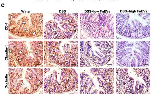
Application: IHC Species: mouse Sample: colon

Application: WB Species: Mouse Sample:
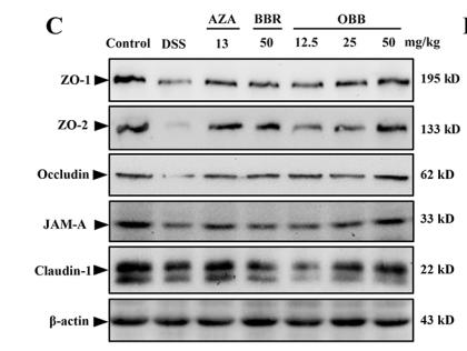
Application: WB Species: Mice Sample: colonic tissues
Application: IF/ICC Species: Mouse Sample:

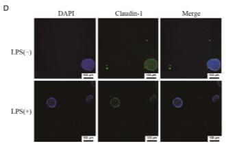
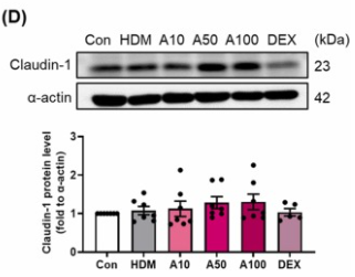
Application: WB Species: human Sample: Caco2 cells
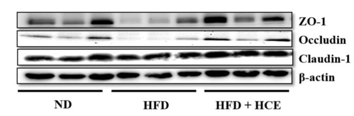
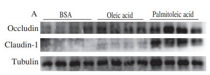
Application: WB Species: Mice Sample: epididymal white adipose tissue
Application: WB Species: Mouse Sample:
Application: WB Species: Mouse Sample: lung
Restrictive clause
Affinity Biosciences tests all products strictly. Citations are provided as a resource for additional applications that have not been validated by Affinity Biosciences. Please choose the appropriate format for each application and consult Materials and Methods sections for additional details about the use of any product in these publications.
For Research Use Only.
Not for use in diagnostic or therapeutic procedures. Not for resale. Not for distribution without written consent. Affinity Biosciences will not be held responsible for patent infringement or other violations that may occur with the use of our products. Affinity Biosciences, Affinity Biosciences Logo and all other trademarks are the property of Affinity Biosciences LTD.