
. Knockdown of SUMO1 inhibited the secretion of IL-6,
IL-8 and TNF-α in HTEC cells treated under hypoxic condition for 24 h. ***p < 0.001 vs control, !!p < 0.01 vs siNC. (B). SUMO1 silencing promoted the TEER value in
HTEC cells treated under hypoxic condition for 24 h. ***p < 0.001 vs control, !!!p < 0.001 vs siNC. (C). SUMO-1 silencing reduced the permeability of HTEC cells
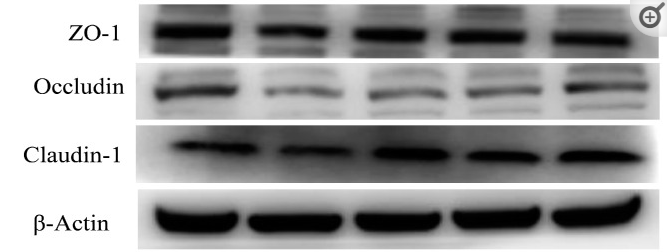
treated under hypoxic condition for 24 h. *p < 0.05 vs control, ***p < 0.001 vs siNC; !!!p < 0.001 vs siNC. (D). Western blotting was used to examine the protein
levels of SUMO1, HIF-1α, VEGF, Occluding, Claudin-1 and ZO-1 in cells as indicated. (E). Immunohistochemistry staining assay was performed to determine the
levels of ZO-1, Occluding and Claudin-1 in cells as indicated above.")
. Knockdown of SUMO1 inhibited the secretion of IL-6,
IL-8 and TNF-α in HTEC cells treated under hypoxic condition for 24 h. ***p < 0.001 vs control, !!p < 0.01 vs siNC. (B). SUMO1 silencing promoted the TEER value in
HTEC cells treated under hypoxic condition for 24 h. ***p < 0.001 vs control, !!!p < 0.001 vs siNC. (C). SUMO-1 silencing reduced the permeability of HTEC cells
treated under hypoxic condition for 24 h. *p < 0.05 vs control, ***p < 0.001 vs siNC; !!!p < 0.001 vs siNC. (D). Western blotting was used to examine the protein
levels of SUMO1, HIF-1α, VEGF, Occluding, Claudin-1 and ZO-1 in cells as indicated. (E). Immunohistochemistry staining assay was performed to determine the
levels of ZO-1, Occluding and Claudin-1 in cells as indicated above.")
. Panel G represents Western blot analysis shown in Fig. 8 , panel H–J represents data shown in Fig. 8 . A indicates the normal group, B indicates the model group, C indicates the Bifendate group, D indicates the YJSB 0.39 g/kg dose group, E indicates the YJSB 1.17 g/kg dose group, F indicates the YJSB 3.51 g/kg dose group, H indicates the expression of claudin1 in the ileum, I indicates the expression of occludin in the ileum, J indicates the expression of ZO-1 in the ileum. YJSB pretreatment upregulated the expression of claudin1, occludin, and ZO-1 in the ileal mucosa; ##p < 0.01 compared to the normal group; **p < 0.01 and *p < 0.05 compared to the model group.")
| Product: | Claudin 1 Antibody |
| Catalog: | DF6919 |
| Description: | Rabbit polyclonal antibody to Claudin 1 |
| Application: | WB IHC IF/ICC |
| Cited expt.: | WB, IHC, IF/ICC |
| Reactivity: | Human, Mouse, Rat |
| Prediction: | Pig, Bovine, Horse, Sheep, Rabbit, Chicken |
| Mol.Wt.: | 23kDa(Observed); 23kD(Calculated). |
| Uniprot: | O95832 |
| RRID: | AB_2838878 |
Control Products

Related Downloads
Protocols
Product Info
*The optimal dilutions should be determined by the end user. For optimal experimental results, antibody reuse is not recommended.
*Tips:
WB: For western blot detection of denatured protein samples. IHC: For immunohistochemical detection of paraffin sections (IHC-p) or frozen sections (IHC-f) of tissue samples. IF/ICC: For immunofluorescence detection of cell samples. ELISA(peptide): For ELISA detection of antigenic peptide.
Cite Format: Affinity Biosciences Cat# DF6919, RRID:AB_2838878.
Fold/Unfold
Claudin-1; Claudin1; CLD 1; CLD1; CLD1_HUMAN; CLDN 1; Cldn1; ILVASC; SEMP 1; SEMP1; Senescence associated epithelial membrane protein 1; Senescence associated epithelial membrane protein; Senescence-associated epithelial membrane protein;
Immunogens
A synthesized peptide derived from human Claudin 1, corresponding to a region within C-terminal amino acids.
Strongly expressed in liver and kidney. Expressed in heart, brain, spleen, lung and testis.
- O95832 CLD1_HUMAN:
- Protein BLAST With
- NCBI/
- ExPASy/
- Uniprot
MANAGLQLLGFILAFLGWIGAIVSTALPQWRIYSYAGDNIVTAQAMYEGLWMSCVSQSTGQIQCKVFDSLLNLSSTLQATRALMVVGILLGVIAIFVATVGMKCMKCLEDDEVQKMRMAVIGGAIFLLAGLAILVATAWYGNRIVQEFYDPMTPVNARYEFGQALFTGWAAASLCLLGGALLCCSCPRKTTSYPTPRPYPKPAPSSGKDYV
Predictions
Score>80(red) has high confidence and is suggested to be used for WB detection. *The prediction model is mainly based on the alignment of immunogen sequences, the results are for reference only, not as the basis of quality assurance.
High(score>80) Medium(80>score>50) Low(score<50) No confidence
Research Backgrounds
Claudins function as major constituents of the tight junction complexes that regulate the permeability of epithelia. While some claudin family members play essential roles in the formation of impermeable barriers, others mediate the permeability to ions and small molecules. Often, several claudin family members are coexpressed and interact with each other, and this determines the overall permeability. CLDN1 is required to prevent the paracellular diffusion of small molecules through tight junctions in the epidermis and is required for the normal barrier function of the skin. Required for normal water homeostasis and to prevent excessive water loss through the skin, probably via an indirect effect on the expression levels of other proteins, since CLDN1 itself seems to be dispensable for water barrier formation in keratinocyte tight junctions.
(Microbial infection) Acts as a receptor for hepatitis C virus (HCV) in hepatocytes. Associates with CD81 and the CLDN1-CD81 receptor complex is essential for HCV entry into host cell. Acts as a receptor for dengue virus.
Cell junction>Tight junction. Cell membrane>Multi-pass membrane protein. Basolateral cell membrane.
Note: Associates with CD81 and the CLDN1-CD81 complex localizes to the basolateral cell membrane.
Strongly expressed in liver and kidney. Expressed in heart, brain, spleen, lung and testis.
Belongs to the claudin family.
Research Fields
· Cellular Processes > Cellular community - eukaryotes > Tight junction. (View pathway)
· Environmental Information Processing > Signaling molecules and interaction > Cell adhesion molecules (CAMs). (View pathway)
· Human Diseases > Infectious diseases: Bacterial > Pathogenic Escherichia coli infection.
· Human Diseases > Infectious diseases: Viral > Hepatitis C.
· Organismal Systems > Immune system > Leukocyte transendothelial migration. (View pathway)
References

Application: IF/ICC Species: Rat Sample:
Application: WB Species: Rat Sample: IEC-6 cells

Application: WB Species: piglets Sample:
Restrictive clause
Affinity Biosciences tests all products strictly. Citations are provided as a resource for additional applications that have not been validated by Affinity Biosciences. Please choose the appropriate format for each application and consult Materials and Methods sections for additional details about the use of any product in these publications.
For Research Use Only.
Not for use in diagnostic or therapeutic procedures. Not for resale. Not for distribution without written consent. Affinity Biosciences will not be held responsible for patent infringement or other violations that may occur with the use of our products. Affinity Biosciences, Affinity Biosciences Logo and all other trademarks are the property of Affinity Biosciences LTD.