.
Lane 3: Ec304 cells(heat-shock treatment).")


 antibody(Red), diluted at 1/600 was used as secondary antibody.")
| Product: | DDB1 Antibody |
| Catalog: | DF7100 |
| Description: | Rabbit polyclonal antibody to DDB1 |
| Application: | WB IHC IF/ICC |
| Cited expt.: | WB |
| Reactivity: | Human, Mouse, Rat |
| Prediction: | Pig, Bovine, Horse, Rabbit, Chicken, Xenopus |
| Mol.Wt.: | 127kDa(Observed); 127kD(Calculated). |
| Uniprot: | Q16531 |
| RRID: | AB_2839055 |
Control Products

Related Downloads
Protocols
Product Info
*The optimal dilutions should be determined by the end user. For optimal experimental results, antibody reuse is not recommended.
*Tips:
WB: For western blot detection of denatured protein samples. IHC: For immunohistochemical detection of paraffin sections (IHC-p) or frozen sections (IHC-f) of tissue samples. IF/ICC: For immunofluorescence detection of cell samples. ELISA(peptide): For ELISA detection of antigenic peptide.
Cite Format: Affinity Biosciences Cat# DF7100, RRID:AB_2839055.
Fold/Unfold
Damage-specific DNA-binding protein 1; DDB p127 subunit; Ddb1; DDB1_HUMAN; DDBa; DNA damage-binding protein 1; DNA damage-binding protein a; HBV X-associated protein 1; UV-damaged DNA-binding factor; UV-damaged DNA-binding protein 1; UV-DDB 1; XAP-1; Xeroderma pigmentosum group E-complementing protein; XPCe; XPE-BF; XPE-binding factor;
Immunogens
A synthesized peptide derived from human DDB1, corresponding to a region within C-terminal amino acids.
- Q16531 DDB1_HUMAN:
- Protein BLAST With
- NCBI/
- ExPASy/
- Uniprot
MSYNYVVTAQKPTAVNGCVTGHFTSAEDLNLLIAKNTRLEIYVVTAEGLRPVKEVGMYGKIAVMELFRPKGESKDLLFILTAKYNACILEYKQSGESIDIITRAHGNVQDRIGRPSETGIIGIIDPECRMIGLRLYDGLFKVIPLDRDNKELKAFNIRLEELHVIDVKFLYGCQAPTICFVYQDPQGRHVKTYEVSLREKEFNKGPWKQENVEAEASMVIAVPEPFGGAIIIGQESITYHNGDKYLAIAPPIIKQSTIVCHNRVDPNGSRYLLGDMEGRLFMLLLEKEEQMDGTVTLKDLRVELLGETSIAECLTYLDNGVVFVGSRLGDSQLVKLNVDSNEQGSYVVAMETFTNLGPIVDMCVVDLERQGQGQLVTCSGAFKEGSLRIIRNGIGIHEHASIDLPGIKGLWPLRSDPNRETDDTLVLSFVGQTRVLMLNGEEVEETELMGFVDDQQTFFCGNVAHQQLIQITSASVRLVSQEPKALVSEWKEPQAKNISVASCNSSQVVVAVGRALYYLQIHPQELRQISHTEMEHEVACLDITPLGDSNGLSPLCAIGLWTDISARILKLPSFELLHKEMLGGEIIPRSILMTTFESSHYLLCALGDGALFYFGLNIETGLLSDRKKVTLGTQPTVLRTFRSLSTTNVFACSDRPTVIYSSNHKLVFSNVNLKEVNYMCPLNSDGYPDSLALANNSTLTIGTIDEIQKLHIRTVPLYESPRKICYQEVSQCFGVLSSRIEVQDTSGGTTALRPSASTQALSSSVSSSKLFSSSTAPHETSFGEEVEVHNLLIIDQHTFEVLHAHQFLQNEYALSLVSCKLGKDPNTYFIVGTAMVYPEEAEPKQGRIVVFQYSDGKLQTVAEKEVKGAVYSMVEFNGKLLASINSTVRLYEWTTEKELRTECNHYNNIMALYLKTKGDFILVGDLMRSVLLLAYKPMEGNFEEIARDFNPNWMSAVEILDDDNFLGAENAFNLFVCQKDSAATTDEERQHLQEVGLFHLGEFVNVFCHGSLVMQNLGETSTPTQGSVLFGTVNGMIGLVTSLSESWYNLLLDMQNRLNKVIKSVGKIEHSFWRSFHTERKTEPATGFIDGDLIESFLDISRPKMQEVVANLQYDDGSGMKREATADDLIKVVEELTRIH
Predictions
Score>80(red) has high confidence and is suggested to be used for WB detection. *The prediction model is mainly based on the alignment of immunogen sequences, the results are for reference only, not as the basis of quality assurance.
High(score>80) Medium(80>score>50) Low(score<50) No confidence
Research Backgrounds
Required for DNA repair. Binds to DDB2 to form the UV-damaged DNA-binding protein complex (the UV-DDB complex). The UV-DDB complex may recognize UV-induced DNA damage and recruit proteins of the nucleotide excision repair pathway (the NER pathway) to initiate DNA repair. The UV-DDB complex preferentially binds to cyclobutane pyrimidine dimers (CPD), 6-4 photoproducts (6-4 PP), apurinic sites and short mismatches. Also appears to function as a component of numerous distinct DCX (DDB1-CUL4-X-box) E3 ubiquitin-protein ligase complexes which mediate the ubiquitination and subsequent proteasomal degradation of target proteins. The functional specificity of the DCX E3 ubiquitin-protein ligase complex is determined by the variable substrate recognition component recruited by DDB1. DCX(DDB2) (also known as DDB1-CUL4-ROC1, CUL4-DDB-ROC1 and CUL4-DDB-RBX1) may ubiquitinate histone H2A, histone H3 and histone H4 at sites of UV-induced DNA damage. The ubiquitination of histones may facilitate their removal from the nucleosome and promote subsequent DNA repair. DCX(DDB2) also ubiquitinates XPC, which may enhance DNA-binding by XPC and promote NER. DCX(DTL) plays a role in PCNA-dependent polyubiquitination of CDT1 and MDM2-dependent ubiquitination of TP53 in response to radiation-induced DNA damage and during DNA replication. DCX(ERCC8) (the CSA complex) plays a role in transcription-coupled repair (TCR). The DDB1-CUL4A-DTL E3 ligase complex regulates the circadian clock function by mediating the ubiquitination and degradation of CRY1. DDB1-mediated CRY1 degradation promotes FOXO1 protein stability and FOXO1-mediated gluconeogenesis in the liver (By similarity).
Phosphorylated by ABL1.
Ubiquitinated by CUL4A. Subsequently degraded by ubiquitin-dependent proteolysis.
Acetylated, promoting interaction with CUL4 (CUL4A or CUL4B) and subsequent formation of DCX (DDB1-CUL4-X-box) E3 ubiquitin-protein ligase complexes. Deacetylation by SIRT7 impairs the interaction with CUL4 (CUL4A or CUL4B) and formation of DCX (DDB1-CUL4-X-box) E3 ubiquitin-protein ligase complexes.
Cytoplasm. Nucleus.
Note: Primarily cytoplasmic. Translocates to the nucleus following UV irradiation and subsequently accumulates at sites of DNA damage.
The core of the protein consists of three WD40 beta-propeller domains.
Belongs to the DDB1 family.
Research Fields
· Genetic Information Processing > Replication and repair > Nucleotide excision repair.
· Genetic Information Processing > Folding, sorting and degradation > Ubiquitin mediated proteolysis. (View pathway)
· Human Diseases > Infectious diseases: Viral > Hepatitis B.
· Human Diseases > Cancers: Overview > Viral carcinogenesis.
References
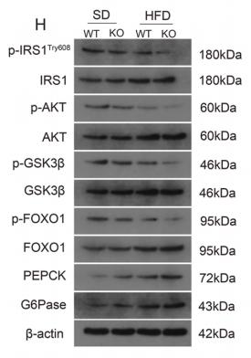
Application: WB Species: mouse Sample: liver
Restrictive clause
Affinity Biosciences tests all products strictly. Citations are provided as a resource for additional applications that have not been validated by Affinity Biosciences. Please choose the appropriate format for each application and consult Materials and Methods sections for additional details about the use of any product in these publications.
For Research Use Only.
Not for use in diagnostic or therapeutic procedures. Not for resale. Not for distribution without written consent. Affinity Biosciences will not be held responsible for patent infringement or other violations that may occur with the use of our products. Affinity Biosciences, Affinity Biosciences Logo and all other trademarks are the property of Affinity Biosciences LTD.