
, using SKP2/p45 Antibody at 1/1000 dilution.
5ug/NC membrane strip.
Exposure for 30s with Affinity™ ECL Kit(#KF8001).
Bands result from membrane strip incubation.")
.
Bands result from membrane strip incubation.")


 and mouse anti-beta tubulin Ab(T0023) for 1 hour at 37°C. An AlexaFluor594 conjugated goat anti-rabbit IgG(H+L) Ab(Red) and an AlexaFluor488 conjugated goat anti-mouse IgG(H+L) Ab(Green) were used as the secondary antibody.
The nuclear counter stain is DAPI (blue).")
| Product: | SKP2/p45 Antibody |
| Catalog: | AF0505 |
| Description: | Rabbit polyclonal antibody to SKP2/p45 |
| Application: | WB IHC IF/ICC |
| Cited expt.: | WB |
| Reactivity: | Human, Mouse |
| Prediction: | Pig, Bovine, Horse, Sheep, Rabbit, Dog, Chicken |
| Mol.Wt.: | 47kDa(Observed); 48kD(Calculated). |
| Uniprot: | Q13309 |
| RRID: | AB_2834158 |
Control Products

Related Downloads
Protocols
Product Info
*The optimal dilutions should be determined by the end user. For optimal experimental results, antibody reuse is not recommended.
*Tips:
WB: For western blot detection of denatured protein samples. IHC: For immunohistochemical detection of paraffin sections (IHC-p) or frozen sections (IHC-f) of tissue samples. IF/ICC: For immunofluorescence detection of cell samples. ELISA(peptide): For ELISA detection of antigenic peptide.
Cite Format: Affinity Biosciences Cat# AF0505, RRID:AB_2834158.
Fold/Unfold
CDK2/Cyclin A associated protein p45; Cyclin A/CDK2 associated protein p45; Cyclin-A/CDK2-associated protein p45; F box protein Skp2; F box/LRR repeat protein 1; F-box protein Skp2; F-box/LRR-repeat protein 1; FBL 1; FBL1; FBXL 1; FBXL1; FLB 1; FLB1; MGC1366; p45; p45skp2; S phase kinase associated protein 2 (p45); S phase kinase associated protein 2; S-phase kinase-associated protein 2; S-phase kinase-associated protein 2 E3 ubiquitin protein ligase; SKP 2; Skp2; SKP2_HUMAN;
Immunogens
A synthesized peptide derived from human SKP2/p45, corresponding to a region within C-terminal amino acids.
- Q13309 SKP2_HUMAN:
- Protein BLAST With
- NCBI/
- ExPASy/
- Uniprot
MHRKHLQEIPDLSSNVATSFTWGWDSSKTSELLSGMGVSALEKEEPDSENIPQELLSNLGHPESPPRKRLKSKGSDKDFVIVRRPKLNRENFPGVSWDSLPDELLLGIFSCLCLPELLKVSGVCKRWYRLASDESLWQTLDLTGKNLHPDVTGRLLSQGVIAFRCPRSFMDQPLAEHFSPFRVQHMDLSNSVIEVSTLHGILSQCSKLQNLSLEGLRLSDPIVNTLAKNSNLVRLNLSGCSGFSEFALQTLLSSCSRLDELNLSWCFDFTEKHVQVAVAHVSETITQLNLSGYRKNLQKSDLSTLVRRCPNLVHLDLSDSVMLKNDCFQEFFQLNYLQHLSLSRCYDIIPETLLELGEIPTLKTLQVFGIVPDGTLQLLKEALPHLQINCSHFTTIARPTIGNKKNQEIWGIKCRLTLQKPSCL
Predictions
Score>80(red) has high confidence and is suggested to be used for WB detection. *The prediction model is mainly based on the alignment of immunogen sequences, the results are for reference only, not as the basis of quality assurance.
High(score>80) Medium(80>score>50) Low(score<50) No confidence
Research Backgrounds
Substrate recognition component of a SCF (SKP1-CUL1-F-box protein) E3 ubiquitin-protein ligase complex which mediates the ubiquitination and subsequent proteasomal degradation of target proteins involved in cell cycle progression, signal transduction and transcription. Specifically recognizes phosphorylated CDKN1B/p27kip and is involved in regulation of G1/S transition. Degradation of CDKN1B/p27kip also requires CKS1. Recognizes target proteins ORC1, CDT1, RBL2, KMT2A/MLL1, CDK9, RAG2, FOXO1, UBP43, and probably MYC, TOB1 and TAL1. Degradation of TAL1 also requires STUB1. Recognizes CDKN1A in association with CCNE1 or CCNE2 and CDK2. Promotes ubiquitination and destruction of CDH1 in a CK1-Dependent Manner, thereby regulating cell migration.
Through the ubiquitin-mediated proteasomal degradation of hepatitis C virus non-structural protein 5A, has an antiviral activity towards that virus.
Ubiquitinated by the APC/C complex, leading to its degradation by the proteasome. Deubiquitinated by USP13.
Acetylation at Lys-68 and Lys-71 increases stability through impairment of APC/C-mediated proteolysis and promotes cytoplasmic retention. Deacetylated by SIRT3.
Cytoplasm. Nucleus.
Research Fields
· Cellular Processes > Cell growth and death > Cell cycle. (View pathway)
· Environmental Information Processing > Signal transduction > FoxO signaling pathway. (View pathway)
· Environmental Information Processing > Signal transduction > mTOR signaling pathway. (View pathway)
· Genetic Information Processing > Folding, sorting and degradation > Ubiquitin mediated proteolysis. (View pathway)
· Human Diseases > Infectious diseases: Viral > Herpes simplex infection.
· Human Diseases > Infectious diseases: Viral > Epstein-Barr virus infection.
· Human Diseases > Cancers: Overview > Pathways in cancer. (View pathway)
· Human Diseases > Cancers: Overview > Viral carcinogenesis.
· Human Diseases > Cancers: Specific types > Small cell lung cancer. (View pathway)
References
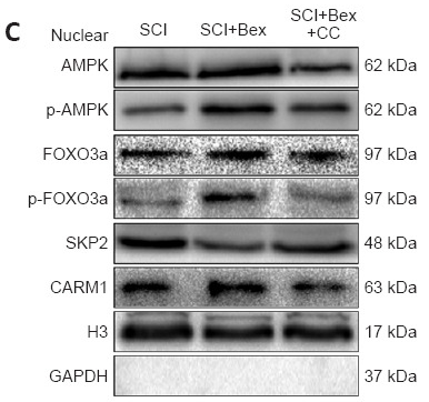
Application: WB Species: Mouse Sample: spinal cord
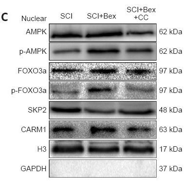
Application: WB Species: Mouse Sample:
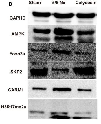
Application: WB Species: rat Sample: muscles

Application: WB Species: human Sample: HEK293T cells

Application: WB Species: human Sample: 5637, T24, EJ and J82 cells and SV-HUC-1
Restrictive clause
Affinity Biosciences tests all products strictly. Citations are provided as a resource for additional applications that have not been validated by Affinity Biosciences. Please choose the appropriate format for each application and consult Materials and Methods sections for additional details about the use of any product in these publications.
For Research Use Only.
Not for use in diagnostic or therapeutic procedures. Not for resale. Not for distribution without written consent. Affinity Biosciences will not be held responsible for patent infringement or other violations that may occur with the use of our products. Affinity Biosciences, Affinity Biosciences Logo and all other trademarks are the property of Affinity Biosciences LTD.