.
Bands result from membrane strip incubation.")
Control Products

Product Info
*The optimal dilutions should be determined by the end user. For optimal experimental results, antibody reuse is not recommended.
*Tips:
WB: For western blot detection of denatured protein samples. IHC: For immunohistochemical detection of paraffin sections (IHC-p) or frozen sections (IHC-f) of tissue samples. IF/ICC: For immunofluorescence detection of cell samples. ELISA(peptide): For ELISA detection of antigenic peptide.
Cite Format: Affinity Biosciences Cat# DF7674, RRID:AB_2841146.
Fold/Unfold
FADD; FADD protein; FADD_HUMAN; Fas (TNFRSF6) associated via death domain; Fas associated via death domain; Fas associating death domain containing protein; Fas associating protein; Fas associating protein with death domain; Fas TNFRSF6 associated via death domain; FAS-associated death domain protein; FAS-associating death domain-containing protein; GIG 3; GIG3; Growth inhibiting gene 3 protein; Growth-inhibiting gene 3 protein; H sapiens mRNA for mediator of receptor induced toxicity; Mediator of receptor induced toxicity; MGC8528; MORT 1; MORT1; Protein FADD;
Immunogens
A synthesized peptide derived from human FADD, corresponding to a region within N-terminal amino acids.
Expressed in a wide variety of tissues, except for peripheral blood mononuclear leukocytes.
- Q13158 FADD_HUMAN:
- Protein BLAST With
- NCBI/
- ExPASy/
- Uniprot
MDPFLVLLHSVSSSLSSSELTELKFLCLGRVGKRKLERVQSGLDLFSMLLEQNDLEPGHTELLRELLASLRRHDLLRRVDDFEAGAAAGAAPGEEDLCAAFNVICDNVGKDWRRLARQLKVSDTKIDSIEDRYPRNLTERVRESLRIWKNTEKENATVAHLVGALRSCQMNLVADLVQEVQQARDLQNRSGAMSPMSWNSDASTSEAS
Predictions
Score>80(red) has high confidence and is suggested to be used for WB detection. *The prediction model is mainly based on the alignment of immunogen sequences, the results are for reference only, not as the basis of quality assurance.
High(score>80) Medium(80>score>50) Low(score<50) No confidence
Research Backgrounds
Apoptotic adaptor molecule that recruits caspase-8 or caspase-10 to the activated Fas (CD95) or TNFR-1 receptors. The resulting aggregate called the death-inducing signaling complex (DISC) performs caspase-8 proteolytic activation. Active caspase-8 initiates the subsequent cascade of caspases mediating apoptosis. Involved in interferon-mediated antiviral immune response, playing a role in the positive regulation of interferon signaling.
Expressed in a wide variety of tissues, except for peripheral blood mononuclear leukocytes.
Contains a death domain involved in the binding of the corresponding domain within Fas receptor.
The interaction between the FAS and FADD death domains is crucial for the formation of the death-inducing signaling complex (DISC).
Research Fields
· Cellular Processes > Cell growth and death > Apoptosis. (View pathway)
· Cellular Processes > Cell growth and death > Apoptosis - multiple species. (View pathway)
· Cellular Processes > Cell growth and death > Necroptosis. (View pathway)
· Environmental Information Processing > Signal transduction > TNF signaling pathway. (View pathway)
· Human Diseases > Drug resistance: Antineoplastic > Platinum drug resistance.
· Human Diseases > Neurodegenerative diseases > Alzheimer's disease.
· Human Diseases > Infectious diseases: Parasitic > Chagas disease (American trypanosomiasis).
· Human Diseases > Infectious diseases: Bacterial > Tuberculosis.
· Human Diseases > Infectious diseases: Viral > Hepatitis B.
· Human Diseases > Infectious diseases: Viral > Human papillomavirus infection.
· Human Diseases > Infectious diseases: Viral > Herpes simplex infection.
· Human Diseases > Cancers: Overview > Pathways in cancer. (View pathway)
· Organismal Systems > Immune system > Toll-like receptor signaling pathway. (View pathway)
· Organismal Systems > Immune system > NOD-like receptor signaling pathway. (View pathway)
· Organismal Systems > Immune system > RIG-I-like receptor signaling pathway. (View pathway)
· Organismal Systems > Immune system > IL-17 signaling pathway. (View pathway)
References
Application: WB Species: human Sample: Caco2 cells
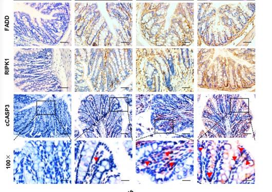
Application: IHC Species: mouse Sample: colon
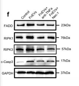
Application: WB Species: rat Sample: intestinal

Application: WB Species: bovine Sample: bEECs

Application: WB Species: human Sample: COV362 cells
Restrictive clause
Affinity Biosciences tests all products strictly. Citations are provided as a resource for additional applications that have not been validated by Affinity Biosciences. Please choose the appropriate format for each application and consult Materials and Methods sections for additional details about the use of any product in these publications.
For Research Use Only.
Not for use in diagnostic or therapeutic procedures. Not for resale. Not for distribution without written consent. Affinity Biosciences will not be held responsible for patent infringement or other violations that may occur with the use of our products. Affinity Biosciences, Affinity Biosciences Logo and all other trademarks are the property of Affinity Biosciences LTD.