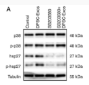
 Antibody at 1/1000 dilution.
5ug/NC membrane strip.
Exposure for 30s with Affinity™ ECL Kit(#KF8003).
Bands result from membrane strip incubation.")


| Product: | Phospho-HSP27 (Ser15) Antibody |
| Catalog: | AF3080 |
| Description: | Rabbit polyclonal antibody to Phospho-HSP27 (Ser15) |
| Application: | WB IHC IF/ICC |
| Cited expt.: | WB |
| Reactivity: | Human, Mouse, Rat |
| Prediction: | Pig, Bovine, Horse, Sheep, Rabbit, Dog |
| Mol.Wt.: | 27kDa(Observed); 23kD(Calculated). |
| Uniprot: | P04792 |
| RRID: | AB_2834517 |
Control Products

Product Info
*The optimal dilutions should be determined by the end user. For optimal experimental results, antibody reuse is not recommended.
*Tips:
WB: For western blot detection of denatured protein samples. IHC: For immunohistochemical detection of paraffin sections (IHC-p) or frozen sections (IHC-f) of tissue samples. IF/ICC: For immunofluorescence detection of cell samples. ELISA(peptide): For ELISA detection of antigenic peptide.
Cite Format: Affinity Biosciences Cat# AF3080, RRID:AB_2834517.
Fold/Unfold
Heat shock 27kDa protein; 28 kDa heat shock protein; CMT2F; DKFZp586P1322; epididymis secretory protein Li 102; Estrogen regulated 24 kDa protein; Estrogen-regulated 24 kDa protein; Heat shock 25kDa protein 1; Heat shock 27 kDa protein; Heat shock 27kD protein 1; Heat shock 27kDa protein 1; Heat shock 28kDa protein 1; Heat Shock Protein 27; Heat shock protein beta 1; Heat shock protein beta-1; heat shock protein family B (small) member 1; HEL-S-102; HMN2B; HS.76067; Hsp 25; HSP 27; Hsp 28; Hsp B1; Hsp25; HSP27; Hsp28; HspB1; HSPB1_HUMAN; SRP27; Stress responsive protein 27; Stress-responsive protein 27;
Immunogens
A synthesized peptide derived from human HSP27 around the phosphorylation site of Ser15.
Detected in all tissues tested: skeletal muscle, heart, aorta, large intestine, small intestine, stomach, esophagus, bladder, adrenal gland, thyroid, pancreas, testis, adipose tissue, kidney, liver, spleen, cerebral cortex, blood serum and cerebrospinal fluid. Highest levels are found in the heart and in tissues composed of striated and smooth muscle.
- P04792 HSPB1_HUMAN:
- Protein BLAST With
- NCBI/
- ExPASy/
- Uniprot
MTERRVPFSLLRGPSWDPFRDWYPHSRLFDQAFGLPRLPEEWSQWLGGSSWPGYVRPLPPAAIESPAVAAPAYSRALSRQLSSGVSEIRHTADRWRVSLDVNHFAPDELTVKTKDGVVEITGKHEERQDEHGYISRCFTRKYTLPPGVDPTQVSSSLSPEGTLTVEAPMPKLATQSNEITIPVTFESRAQLGGPEAAKSDETAAK
Predictions
Score>80(red) has high confidence and is suggested to be used for WB detection. *The prediction model is mainly based on the alignment of immunogen sequences, the results are for reference only, not as the basis of quality assurance.
High(score>80) Medium(80>score>50) Low(score<50) No confidence
Research Backgrounds
Small heat shock protein which functions as a molecular chaperone probably maintaining denatured proteins in a folding-competent state. Plays a role in stress resistance and actin organization. Through its molecular chaperone activity may regulate numerous biological processes including the phosphorylation and the axonal transport of neurofilament proteins.
Phosphorylated upon exposure to protein kinase C activators and heat shock. Phosphorylation by MAPKAPK2 and MAPKAPK3 in response to stress dissociates HSPB1 from large small heat-shock protein (sHsps) oligomers and impairs its chaperone activity and ability to protect against oxidative stress effectively. Phosphorylation by MAPKAPK5 in response to PKA stimulation induces F-actin rearrangement.
Cytoplasm. Nucleus. Cytoplasm>Cytoskeleton>Spindle.
Note: Cytoplasmic in interphase cells. Colocalizes with mitotic spindles in mitotic cells. Translocates to the nucleus during heat shock and resides in sub-nuclear structures known as SC35 speckles or nuclear splicing speckles.
Detected in all tissues tested: skeletal muscle, heart, aorta, large intestine, small intestine, stomach, esophagus, bladder, adrenal gland, thyroid, pancreas, testis, adipose tissue, kidney, liver, spleen, cerebral cortex, blood serum and cerebrospinal fluid. Highest levels are found in the heart and in tissues composed of striated and smooth muscle.
Belongs to the small heat shock protein (HSP20) family.
Research Fields
· Environmental Information Processing > Signal transduction > MAPK signaling pathway. (View pathway)
· Human Diseases > Infectious diseases: Parasitic > Amoebiasis.
· Human Diseases > Infectious diseases: Viral > Epstein-Barr virus infection.
References
Application: WB Species: Human Sample:
Application: WB Species: human Sample:
Restrictive clause
Affinity Biosciences tests all products strictly. Citations are provided as a resource for additional applications that have not been validated by Affinity Biosciences. Please choose the appropriate format for each application and consult Materials and Methods sections for additional details about the use of any product in these publications.
For Research Use Only.
Not for use in diagnostic or therapeutic procedures. Not for resale. Not for distribution without written consent. Affinity Biosciences will not be held responsible for patent infringement or other violations that may occur with the use of our products. Affinity Biosciences, Affinity Biosciences Logo and all other trademarks are the property of Affinity Biosciences LTD.