![Phospho-eIF2 alpha (Ser51)[Ser52] Antibody - Western blot analysis of lysates from RAW264.](http://img.affbiotech.cn/images/202606/af3087_70361_phospho_eif2_alpha_ser51_ser52_antibody_1782125230.jpg "Western blot analysis of lysates from RAW264.7 cells(heat-shock,45min treatment), using Phospho-eIF2 alpha (Ser51)[Ser52] Antibody at 1/1000 dilution.
5ug/NC membrane strip.
Exposure for 5s with Affinity™ ECL Kit(#KF8001).
Bands result from membrane strip incubation.")
![Phospho-eIF2 alpha (Ser51)[Ser52] Antibody - Western blot analysis of lysates from various samples, using Phospho-eIF2 alpha (Ser51)[Ser52] Antibody.](http://img.affbiotech.cn/images/202112/af3087_56149_phospho_eif2_alpha_ser51_ser52_antibody_1640451969.jpg "Western blot analysis of lysates from various samples, using Phospho-eIF2 alpha (Ser51)[Ser52] Antibody.
Lane 1: Rat liver, blocked with antigen-specific peptides.
Lane 2: Rat liver.
Lane 3: Pc12 cells(uv treatment).")
![Phospho-eIF2 alpha (Ser51)[Ser52] Antibody - AF3087 at 1/100 staining Mouse spleen tissue by IHC-P.](http://img.affbiotech.cn/images/201911/thumb_img/af3087_phospho_eif2_alpha_ser51_ser52_antibody_thumb_P_1573714928856.jpg "AF3087 at 1/100 staining Mouse spleen tissue by IHC-P. The sample was formaldehyde fixed and a heat mediated antigen retrieval step in citrate buffer was performed. The sample was then blocked and incubated with the primary antibody at 4°C overnight. An HRP conjugated anti-Rabbit antibody was used as the secondary antibody.")
![Phospho-eIF2 alpha (Ser51)[Ser52] Antibody - AF3087 at 1/100 staining Rat colon tissue by IHC-P.](http://img.affbiotech.cn/images/201911/thumb_img/af3087_phospho_eif2_alpha_ser51_ser52_antibody_thumb_P_1573714929253.jpg "AF3087 at 1/100 staining Rat colon tissue by IHC-P. The sample was formaldehyde fixed and a heat mediated antigen retrieval step in citrate buffer was performed. The sample was then blocked and incubated with the primary antibody at 4°C overnight. An HRP conjugated anti-Rabbit antibody was used as the secondary antibody.")
![Phospho-eIF2 alpha (Ser51)[Ser52] Antibody - AF3087 at 1/100 staining Rat stomach tissue by IHC-P.](http://img.affbiotech.cn/images/201911/thumb_img/af3087_phospho_eif2_alpha_ser51_ser52_antibody_thumb_P_1573714929150.jpg "AF3087 at 1/100 staining Rat stomach tissue by IHC-P. The sample was formaldehyde fixed and a heat mediated antigen retrieval step in citrate buffer was performed. The sample was then blocked and incubated with the primary antibody at 4°C overnight. An HRP conjugated anti-Rabbit antibody was used as the secondary antibody.")
![Phospho-eIF2 alpha (Ser51)[Ser52] Antibody - AF3087 at 1/100 staining Rat ovary tissue by IHC-P.](http://img.affbiotech.cn/images/201911/thumb_img/af3087_phospho_eif2_alpha_ser51_ser52_antibody_thumb_P_1573714929372.jpg "AF3087 at 1/100 staining Rat ovary tissue by IHC-P. The sample was formaldehyde fixed and a heat mediated antigen retrieval step in citrate buffer was performed. The sample was then blocked and incubated with the primary antibody at 4°C overnight. An HRP conjugated anti-Rabbit antibody was used as the secondary antibody.")
![Phospho-eIF2 alpha (Ser51)[Ser52] Antibody - AF3087 at 1/100 staining Human lung cancer by IHC-P.](http://img.affbiotech.cn/images/201911/thumb_img/af3087_phospho_eif2_alpha_ser51_ser52_antibody_thumb_P_1573714929270.jpg "AF3087 at 1/100 staining Human lung cancer by IHC-P. The sample was formaldehyde fixed and a heat mediated antigen retrieval step in citrate buffer was performed. The sample was then blocked and incubated with the primary antibody at 4°C overnight. An HRP conjugated anti-Rabbit antibody was used as the secondary antibody.")
![Phospho-eIF2 alpha (Ser51)[Ser52] Antibody - AF3087 at 1/100 staining Human breast cancer by IHC-P.](http://img.affbiotech.cn/images/201911/thumb_img/af3087_phospho_eif2_alpha_ser51_ser52_antibody_thumb_P_1573714929125.jpg "AF3087 at 1/100 staining Human breast cancer by IHC-P. The sample was formaldehyde fixed and a heat mediated antigen retrieval step in citrate buffer was performed. The sample was then blocked and incubated with the primary antibody at 4°C overnight. An HRP conjugated anti-Rabbit antibody was used as the secondary antibody.")
![Phospho-eIF2 alpha (Ser51)[Ser52] Antibody - AF3087 at 1/100 staining Rat lung tissue by IHC-P.](http://img.affbiotech.cn/images/201911/thumb_img/af3087_phospho_eif2_alpha_ser51_ser52_antibody_thumb_P_1573714929516.jpg "AF3087 at 1/100 staining Rat lung tissue by IHC-P. The sample was formaldehyde fixed and a heat mediated antigen retrieval step in citrate buffer was performed. The sample was then blocked and incubated with the primary antibody at 4°C overnight. An HRP conjugated anti-Rabbit antibody was used as the secondary antibody.")
![Phospho-eIF2 alpha (Ser51)[Ser52] Antibody - AF3087 at 1/100 staining Rat spleen tissue by IHC-P.](http://img.affbiotech.cn/images/201911/thumb_img/af3087_phospho_eif2_alpha_ser51_ser52_antibody_thumb_P_1573714929647.jpg "AF3087 at 1/100 staining Rat spleen tissue by IHC-P. The sample was formaldehyde fixed and a heat mediated antigen retrieval step in citrate buffer was performed. The sample was then blocked and incubated with the primary antibody at 4°C overnight. An HRP conjugated anti-Rabbit antibody was used as the secondary antibody.")
![Phospho-eIF2 alpha (Ser51)[Ser52] Antibody - AF3087 at 1/100 staining Rat liver tissue by IHC-P.](http://img.affbiotech.cn/images/201911/thumb_img/af3087_phospho_eif2_alpha_ser51_ser52_antibody_thumb_P_1573714929599.jpg "AF3087 at 1/100 staining Rat liver tissue by IHC-P. The sample was formaldehyde fixed and a heat mediated antigen retrieval step in citrate buffer was performed. The sample was then blocked and incubated with the primary antibody at 4°C overnight. An HRP conjugated anti-Rabbit antibody was used as the secondary antibody.")
![Phospho-eIF2 alpha (Ser51)[Ser52] Antibody - AF3087 at 1/100 staining Mouse kidney tissue by IHC-P.](http://img.affbiotech.cn/images/201911/thumb_img/af3087_phospho_eif2_alpha_ser51_ser52_antibody_thumb_P_1573714929170.jpg "AF3087 at 1/100 staining Mouse kidney tissue by IHC-P. The sample was formaldehyde fixed and a heat mediated antigen retrieval step in citrate buffer was performed. The sample was then blocked and incubated with the primary antibody at 4°C overnight. An HRP conjugated anti-Rabbit antibody was used as the secondary antibody.")
![Phospho-eIF2 alpha (Ser51)[Ser52] Antibody - AF3087 at 1/100 staining human placenta tissue sections by IHC-P.](http://img.affbiotech.cn/images/201707/thumb_img/1270_thumb_P_1499847414923.jpg "AF3087 at 1/100 staining human placenta tissue sections by IHC-P. The tissue was formaldehyde fixed and a heat mediated antigen retrieval step in citrate buffer was performed. The tissue was then blocked and incubated with the antibody for 1.5 hours at 22°C. An HRP conjugated goat anti-rabbit antibody was used as the secondary antibody.")
![Phospho-eIF2 alpha (Ser51)[Ser52] Antibody - AF3087 at 1/100 staining Mouse heart tissue by IHC-P.](http://img.affbiotech.cn/images/201911/thumb_img/af3087_phospho_eif2_alpha_ser51_ser52_antibody_thumb_P_1573714928655.jpg "AF3087 at 1/100 staining Mouse heart tissue by IHC-P. The sample was formaldehyde fixed and a heat mediated antigen retrieval step in citrate buffer was performed. The sample was then blocked and incubated with the primary antibody at 4°C overnight. An HRP conjugated anti-Rabbit antibody was used as the secondary antibody.")
![Phospho-eIF2 alpha (Ser51)[Ser52] Antibody - AF3087 at 1/100 staining rat ovarian tissue sections by IHC-P.](http://img.affbiotech.cn/images/201707/thumb_img/1270_thumb_P_1499847414116.jpg "AF3087 at 1/100 staining rat ovarian tissue sections by IHC-P. The tissue was formaldehyde fixed and a heat mediated antigen retrieval step in citrate buffer was performed. The tissue was then blocked and incubated with the antibody for 1.5 hours at 22°C. An HRP conjugated goat anti-rabbit antibody was used as the secondary antibody.")
![Phospho-eIF2 alpha (Ser51)[Ser52] Antibody - AF3087 at 1/100 staining rat kidney tissue sections by IHC-P.](http://img.affbiotech.cn/images/201707/thumb_img/1270_thumb_P_1499847414646.jpg "AF3087 at 1/100 staining rat kidney tissue sections by IHC-P. The tissue was formaldehyde fixed and a heat mediated antigen retrieval step in citrate buffer was performed. The tissue was then blocked and incubated with the antibody for 1.5 hours at 22°C. An HRP conjugated goat anti-rabbit antibody was used as the secondary antibody.")
![Phospho-eIF2 alpha (Ser51)[Ser52] Antibody - AF3087 at 1/100 staining rat uterine tissue sections by IHC-P.](http://img.affbiotech.cn/images/201707/thumb_img/1270_thumb_P_1499847414944.jpg "AF3087 at 1/100 staining rat uterine tissue sections by IHC-P. The tissue was formaldehyde fixed and a heat mediated antigen retrieval step in citrate buffer was performed. The tissue was then blocked and incubated with the antibody for 1.5 hours at 22°C. An HRP conjugated goat anti-rabbit antibody was used as the secondary antibody.")
![Phospho-eIF2 alpha (Ser51)[Ser52] Antibody - AF3087 at 1/100 staining mouse lung tissue sections by IHC-P.](http://img.affbiotech.cn/images/201707/thumb_img/1270_thumb_P_1499847414068.jpg "AF3087 at 1/100 staining mouse lung tissue sections by IHC-P. The tissue was formaldehyde fixed and a heat mediated antigen retrieval step in citrate buffer was performed. The tissue was then blocked and incubated with the antibody for 1.5 hours at 22°C. An HRP conjugated goat anti-rabbit antibody was used as the secondary antibody.")
![Phospho-eIF2 alpha (Ser51)[Ser52] Antibody - AF3087 at 1/100 staining mouse kidney tissue sections by IHC-P.](http://img.affbiotech.cn/images/201707/thumb_img/1270_thumb_P_1499847414984.jpg "AF3087 at 1/100 staining mouse kidney tissue sections by IHC-P. The tissue was formaldehyde fixed and a heat mediated antigen retrieval step in citrate buffer was performed. The tissue was then blocked and incubated with the antibody for 1.5 hours at 22°C. An HRP conjugated goat anti-rabbit antibody was used as the secondary antibody.")
![Phospho-eIF2 alpha (Ser51)[Ser52] Antibody - AF3087 at 1/100 staining mouse gastric tissue sections by IHC-P.](http://img.affbiotech.cn/images/201707/thumb_img/1270_thumb_P_1499847414263.jpg "AF3087 at 1/100 staining mouse gastric tissue sections by IHC-P. The tissue was formaldehyde fixed and a heat mediated antigen retrieval step in citrate buffer was performed. The tissue was then blocked and incubated with the antibody for 1.5 hours at 22°C. An HRP conjugated goat anti-rabbit antibody was used as the secondary antibody.")
![Phospho-eIF2 alpha (Ser51)[Ser52] Antibody - AF3087 at 1/100 staining human lung tissue sections by IHC-P.](http://img.affbiotech.cn/images/201707/thumb_img/1270_thumb_P_1499847414469.jpg "AF3087 at 1/100 staining human lung tissue sections by IHC-P. The tissue was formaldehyde fixed and a heat mediated antigen retrieval step in citrate buffer was performed. The tissue was then blocked and incubated with the antibody for 1.5 hours at 22°C. An HRP conjugated goat anti-rabbit antibody was used as the secondary antibody.")
![Phospho-eIF2 alpha (Ser51)[Ser52] Antibody - AF3087 at 1/100 staining human appendiceal tissue sections by IHC-P.](http://img.affbiotech.cn/images/201707/thumb_img/1270_thumb_P_1499847414598.jpg "AF3087 at 1/100 staining human appendiceal tissue sections by IHC-P. The tissue was formaldehyde fixed and a heat mediated antigen retrieval step in citrate buffer was performed. The tissue was then blocked and incubated with the antibody for 1.5 hours at 22°C. An HRP conjugated goat anti-rabbit antibody was used as the secondary antibody.")
![Phospho-eIF2 alpha (Ser51)[Ser52] Antibody - AF3087 staining H2O2 treated Hela cells by IF/ICC.](http://img.affbiotech.cn/images/201911/thumb_img/af3087_phospho_eif2_alpha_ser51_ser52_antibody_thumb_P_1572593579826.jpg "AF3087 staining H2O2 treated Hela cells by IF/ICC. The samples were fixed with PFA and permeabilized in 0.1% Triton X-100,then blocked in 10% serum for 45 minutes at 25°C. Samples were then incubated with primary Ab(AF3087) and mouse anti-beta tubulin Ab(T0023) for 1 hour at 37°C. An AlexaFluor594 conjugated goat anti-rabbit IgG(H+L) Ab(Red) and an AlexaFluor488 conjugated goat anti-mouse IgG(H+L) Ab(Green) were used as the secondary antibody.
The nuclear counter stain is DAPI(blue).")
![Phospho-eIF2 alpha (Ser51)[Ser52] Antibody - AF3087 staining HepG2 cells(4h of LPS treatment) by IF/ICC.](http://img.affbiotech.cn/images/201907/thumb_img/af3087_phospho_eif2_alpha_ser51_antibody_thumb_P_1562948307026.jpg "AF3087 staining HepG2 cells(4h of LPS treatment) by IF/ICC. The samples were fixed with PFA and permeabilized in 0.1% Triton X-100,then blocked in 10% serum for 45 minutes at 25°C. Samples were then incubated with primary Ab(AF3087 1:200) and mouse anti-beta tubulin Ab(T0023 1:200) for 1 hour at 37°C. An AlexaFluor594 conjugated goat anti-rabbit IgG(H+L) Ab(Red) and an AlexaFluor488 conjugated goat anti-mouse IgG(H+L) Ab(Green) were used as the secondary antibody.
The nuclear counter stain is DAPI(blue).")
![Phospho-eIF2 alpha (Ser51)[Ser52] Antibody - peptide-ELISA analysis of AF3087.](http://img.affbiotech.cn/images/pelisa/809/af3087-peptide-elisa.png "peptide-ELISA analysis of AF3087. showing specificity to antigen peptide. Peptides concentration: 1ug/ml.<br>
P-peptide: phospho-peptide. N-peptide: non-phospho-peptide.")
![Phospho-eIF2 alpha (Ser51)[Ser52] Antibody - Figure 3: |The H2-O2 mixture-induced inhibition of ER stress caused by CIH for 35 d: (a) the ER stress markers GRP 78, caspase 12, and CHOP protein expressions; (b–d) the ratios of p-PERK, p-eIF2α/eIF2α, and p-IRE 1/IRE 1 in the left ventricle.](http://img.affbiotech.cn/images/cited_image/202110/cited_img_226.jpg "Figure 3: |The H2-O2 mixture-induced inhibition of ER stress caused by CIH for 35 d: (a) the ER stress markers GRP 78, caspase 12, and CHOP protein expressions; (b–d) the ratios of p-PERK, p-eIF2α/eIF2α, and p-IRE 1/IRE 1 in the left ventricle")
![Phospho-eIF2 alpha (Ser51)[Ser52] Antibody - Figure 1.](http://img.affbiotech.cn/images/cited_image/202112/cite-wx-342-1640949481.jpg "Figure 1. |Rg3 activates ER stress in GBC‑SD cells. (A) GBC‑SD cells were treated with 1, 25, 50 and 100 µM Rg3 for 72 h, and proliferation was determined using a CCK‑8 kit. (B) Rg3 induced p‑eIF2α, ATF4 and Lcn2 expression in GBC‑SD cells. Relative protein expression level of (C) eIF2α, (D) p‑eIF2α, (E) ATF4 and (F) Lcn2 compared with the DMSO group. Relative protein expression was quantified by normalizing to the internal control β‑actin (n=3). *P<0.05, **P<0.01 vs. DMSO group. OD, optical density; ER, endoplasmic reticulum; eIF2α, eukaryotic translation‑initiation factor 2α; ATF4, activating transcription factor 4; p‑, phospho‑; DMSO, dimethylsulfoxide; Lcn2, lipocalin 2.")
![Phospho-eIF2 alpha (Ser51)[Ser52] Antibody - Fig.](http://img.affbiotech.cn/images/cited_image/202202/cite-wx-692-1644226769.jpg "Fig. 2. |Upregulation of MKP-5 alleviates GP-induced apoptosis through the mitochondrial and ER stress pathways. (A, B) MIN6-PC and MIN6-MKP5 cellswere exposed to GP for 24 h, after which the activation of caspase-3, -9, -8, and the expression of PARP-1 were assessed by western blotting (A). Relative Bcl-2/Bax expression was determined by real-time PCR (B).(C) MIN6 cells infected with Ad-MKP5 or Ad-GFP were exposed to GP for 9 h or 12 h, and the expression of MKP-5 and the activation of IRE-1α, eIF-2α and caspase-12 were assessed via western blotting.")
![Phospho-eIF2 alpha (Ser51)[Ser52] Antibody - Fig.](http://img.affbiotech.cn/images/cited_image/202202/cite-wx-785-1644569031.jpg "Fig.6 |Effects of ICA on the PERK signaling in the hippocampus of APP/PS1 mice. The phosphorylation of PERK and eIF2α, as well ATF4 and CHOP proteins expression were increased in APP/PS1 mice, ICA treatment significantly decreased their levels. (A, B) Immunoblots of PERK, p-PERK, eIF2α, p-eIF2α, ATF4 and CHOP.")
![Phospho-eIF2 alpha (Ser51)[Ser52] Antibody - FIGURE 5 | PA treatment attenuated HFD-induced VLDLR expression in rats.](http://img.affbiotech.cn/images/cited_image/202202/cite-wx-960-1644569031.jpg "FIGURE 5 | PA treatment attenuated HFD-induced VLDLR expression in rats. (A) Representative immunoreactive bands of eIF2α, p-eIF2α, ATF4, and VLDLR")
![Phospho-eIF2 alpha (Ser51)[Ser52] Antibody - Figure 3.](http://img.affbiotech.cn/images/cited_image/202202/cite-wx-1135-1645778927.jpg "Figure 3. |DM enhances endoplasmic reticulum stress. Western blot analysis was performed after 24 h of reperfusion. (A) Representative blots and histograms showing (B) p‑PERK/PERK ratio; (C) p‑eIF2α/eIF2α ratio; (D) ATF4 expression; and (E) CHOP expression.")
![Phospho-eIF2 alpha (Ser51)[Ser52] Antibody - Supplementary figure 2.](http://img.affbiotech.cn/images/cited_image/202203/cite-wx-1542-1647827334.jpg "Supplementary figure 2. |miR-185-5p suppresses the activation of the IRE1/XBP1 and PERK/eIF2α/ATF4 branches in TGF-β1-induced HK2 cells.(C, D) Western blot analysis of p-PERK, PERK, p-eIF2α, eIF2α, and ATF4 proteins.Data are presented as the mean ± SD (n = 3) calculated from one-way ANOVA with Tukey’s test.")
![Phospho-eIF2 alpha (Ser51)[Ser52] Antibody - FIGURE 5
ACLY inhibitor triggers ER stress and activates p‐eIF2α/ATF4/CHOP axis in vitro.](http://img.affbiotech.cn/images/cited_image/202208/cite-wx-666-1660890823.jpg "FIGURE 5
ACLY inhibitor triggers ER stress and activates p‐eIF2α/ATF4/CHOP axis in vitro. Western blot analysis of (A) ER stress‐related proteins (p‐eIF2α, eIF2α, ATF4 and CHOP) and (B) UPR signal transduction molecules (p‐PERK, PERK, p‐IRE1α, IRE1α and sXBP1) in HepG2 cells after administration of BMS‐303141. ATF4p‐eIF2α, eIF2α were activated 3 h post‐treatment; CHOP was activated 8 h post‐treatment. (* P < .05, ** P < .01 and *** P < .001, compared with control group) (C) Western blot analysis of protein expression after ATF4 knockdown. (D) Annexin V‐FITC/PI double staining was performed to determine the apoptosis rate of HepG2 cells after ATF4 knockdown via flow cytometry. (* P < .05, ** P < .01 and *** P < .001, compared with con siRNA group). All experiments were repeated 3 times")
![Phospho-eIF2 alpha (Ser51)[Ser52] Antibody - FIGURE 5
(A) Representative images showing p-eIF2α immunohistochemistry in the cortex.](http://img.affbiotech.cn/images/cited_image/202208/cite-wx-705-1660890823.jpg "FIGURE 5
(A) Representative images showing p-eIF2α immunohistochemistry in the cortex. Panels (a’–e’) are magnified areas of panels (a–e). Bars = 200 μm in panels (a–e). Bars = 50 μm in panels (a’–e’). (B) Quantitative analysis of the number of p-eIF2α positive cells. The data are shown as mean ± SD, ∗P < 0.05 vs. control group (n = 6). MD, morphine dependence; AME, acute morphine exposure.")
![Phospho-eIF2 alpha (Ser51)[Ser52] Antibody - Figure 6.](http://img.affbiotech.cn/images/cited_image/202208/cite-wx-189-1661496481.jpg "Figure 6.| Expression of SIRT1, GRP78, p‑PERK, p‑eIF2α, CHOP, caspase‑12 and the mRNA levels of caspase‑12 in the myocardium. (A) Representative blots of SIRT1, GRP78, p‑PERK, p‑eIF2α, CHOP and caspase‑12. Semiquantitative analysis of (B) SIRT1, (C) GRP78, (D) p‑PERK, (E) p‑eIF2α, (F) CHOP and (G) caspase‑12.")
![Phospho-eIF2 alpha (Ser51)[Ser52] Antibody - Figure 6.](http://img.affbiotech.cn/images/cited_image/202209/cite-wx-912-1662106277.jpg "Figure 6.
Expression of SIRT1, GRP78, p-PERK, p-eIF2α, CHOP, caspase-12 and the mRNA levels of caspase-12 in the myocardium. (A) Representative blots of SIRT1, GRP78, p-PERK, p-eIF2α, CHOP and caspase-12. Semiquantitative analysis of (B) SIRT1, (C) GRP78, (D) p-PERK, (E) p-eIF2α, (F) CHOP and (G) caspase-12. (H) The mRNA levels of caspase-12 in the myocardium. Data are presented as the mean ± standard deviation. n=3. ##P<0.01 vs. Sham. *P<0.05 and **P<0.01 vs. MI/R. IOE, Inonotus obliquus extract; SIRT1, NAD-dependent protein deacetylase sirtuin-1; GRP78, glucose-regulated protein 78; PERK, protein kinase R-like endoplasmic reticulum kinase; eIF2α, eukaryotic translation initiation factor 2 subunit α; CHOP, C/EBP homologous protein; p, phosphorylated; MI/R, myocardial ischemia/reperfusion.")
![Phospho-eIF2 alpha (Ser51)[Ser52] Antibody - Figure 1.](http://img.affbiotech.cn/images/cited_image/202209/cite-wx-1092-1663320166.jpg "Figure 1.
Serum from DN mice induced podocyte injury and endoplasmic reticulum stress. (A) MCP5 podocytes were treated either with 10% serum from control C57BL/KsJ dm/m mice or with 10% serum from C57BL/KsJ dm/dm DN mice for 24 h. Western blot image showing activation of PERK-eIF2α-ATF4 in ER stress signaling pathway and increased apoptosis-related molecule cleaved caspase-3 in podocytes treated with serum from DN mice as compared to control mice. (B) Densitometric quantification of protein expression from Figure 1(A). (**p < 0.01, *p < 0.05).")
![Phospho-eIF2 alpha (Ser51)[Ser52] Antibody - Fig.](http://img.affbiotech.cn/images/cited_image/202210/cite-wx-1378-1666341971.jpg "Fig. 4. Activation of the endoplasmic reticulum
stress (ERs) in GnRH neurons and GT1–7 cells
after MC-LR treatment. (A) MC-LR triggered
ultrastructural changes of ERs in hypothalamus
neurons. Drinking water including 15 μg/L MCLR was fed to mice for 180 days without
interruption, and the ultrastructure of ERs
(black arrows) in control and MC-LR treatment
group were detected by electron microscope.
(B) Increase of intracellular Ca2+ concentration
in GT1–7 cells following exposure to MC-LR.
Fluo-3/AM fluorescent probes were used to
measure levels of intracellular Ca2+ through
immunofluorescence assay (scale
bar = 100 µm). (C) The intensity of Fluo-3/AM
was detected by image-pro plus (n = 3). (D)
Mice were given drinking water comprising
MC-LR for 180 sustained days. Expressions of
GRP78, ATF6, ATF4, PERK, CHOP, eIF2α, peIF2α, and LC3B in hypothalamus tissues were
assessed by Western blotting (n = 5 mice/
group). (E) GT1–7 cells were plated in 6-well
plates at 2.0 × 10⁵ cells per well. Cells were
exposed to MC-LR for 24 h at different doses as
indicated. Expressions of GRP78, ATF6, ATF4,
PERK, CHOP, eIF2α, p- eIF2α, and LC3B in
GT1–7 cells were assessed by Western blotting.
(F) GT1–7 cells were plated in 6-well plates at
2.0 × 10⁵ cells per well. Cells were pretreated
with ERs inhibitor 4- Phenyl butyric acid (4-
PBA) at 5 mM for 1 h, followed by the treatment with MC-LR (1000 nM) for 24 h. The
expression levels of ERs related proteins and
LC3B were measured.")
![Phospho-eIF2 alpha (Ser51)[Ser52] Antibody - Figure 4.](http://img.affbiotech.cn/images/cited_image/202211/cite-wx-1672-1668761351.jpg "Figure 4.
PTH activates the PERK-CHOP ER stress pathway as detected by Western blot analysis. (A) HASMCs were treated with 1 × 10⁻⁸–1 × 10⁻⁶ mol/L PTH for three days. (B) Data are the mean ± SD of three independent experiments. (C) HASMCs were treated with 1 × 10⁻⁶ mol/L PTH for 0–14 days. (D) Data are the mean ± SD of three independent experiments. HASMCs cultured in normal medium for the same days as control. *p < .05 versus control.")
![Phospho-eIF2 alpha (Ser51)[Ser52] Antibody - FIGURE 5
CTRP1 inhibited ERS via PERK signal pathway in the cortex of MCAO/R-treated rats.](http://img.affbiotech.cn/images/cited_image/202212/cite-wx-237-1671782324.jpg "FIGURE 5
CTRP1 inhibited ERS via PERK signal pathway in the cortex of MCAO/R-treated rats. (A) Western blot analyzed the expression of PERK, p-PERK, GRP78, ATF6, ATF4, IRE1α, p-IRE1α. n = 4 per group. (B) CTRP1 affected the interaction between PERK and GRP78 after CIRI. n = 3 per group. ****p < 0.0001, ***p < 0.001, **p < 0.01, *p < 0.05 vs. sham group, ####p < 0.0001, ###p < 0.001, ## p < 0.01, #p < 0.05 vs. MCAO/R + LV-NC group.")
![Phospho-eIF2 alpha (Ser51)[Ser52] Antibody - Figure 5 of 9
Figure 5.](http://img.affbiotech.cn/images/cited_image/202301/cite-wx-483-1673940311.jpg "Figure 5 of 9
Figure 5. CBG stimulated ER stress. (A, B) NB4 and NB4-R1 cells were treated with different concentrations of CBG for 24 h, western blotting detects the PERK-eIF2α-ATF4 expression. (C, D) RT-PCR was used to measure the CHOP expression. Results are expressed as mean ± SD (n = 3). Compared with solvent group, *P < 0.05, **P < 0.01, ***P < 0.001. (E, F) NB4 and NB4-R1 cells were incubated with 60 nM CBG, and immunofluorescence was performed to observe CHOP protein. Results are expressed as mean ± SD (n = 3).")
![Phospho-eIF2 alpha (Ser51)[Ser52] Antibody - Figure 4: Effects of 18β-glycyrrhetinic acid on the expression of GRP 78, p-PERK, p-eIF 2α, IκB, and p-NF-κB p65 in the lung tissue.](http://img.affbiotech.cn/images/cited_image/202305/cite-wx-504-1684474098.jpg "Figure 4: Effects of 18β-glycyrrhetinic acid on the expression of GRP 78, p-PERK, p-eIF 2α, IκB, and p-NF-κB p65 in the lung tissue. (a) Representative Western blot band of GRP 78 activation in the lung tissues. (b) Representative Western blot band of p-PERK activation in the lung tissues. (c) Representative Western blot band of p-eIF 2α activation in the lung tissues. (d) Representative Western blot band of IκB activation in the lung tissues. (e) Representative Western blot band of p-NF-κB p65 activation in the lung tissues. Data are expressed as mean ± SEM (n = 6). ##P < 0.01 vs. control group, *P < 0.05, **P < 0.01 versus MCT group. MCT: monocrotaline, 18β-GA: 18β-glycyrrhetinic acid, SEM: Standard error of the mean.")
![Phospho-eIF2 alpha (Ser51)[Ser52] Antibody - Figure 4 Exogenous GRP78 blocks the pro-apoptotic GRP78/eIF2α/CHOP/caspase-3,9 signaling pathway of the UPR in nigral tissue in a rat model of Parkinson’s disease.](http://img.affbiotech.cn/uploads/202406/4d616e23c753167d0211f11cdfd0622f.png "Figure 4 Exogenous GRP78 blocks the pro-apoptotic GRP78/eIF2α/CHOP/caspase-3,9 signaling pathway of the UPR in nigral tissue in a rat model of Parkinson’s disease. Nigral content of (a) GRP78, (b) phosphorylated to total eIF2α, (c) CHOP. (d) Representative immunoblots. Nigral content of (e) cleaved caspase-9, (f) cleaved caspase-3. (g) Representative immunoblots. Western blot analysis of the nigral tissue was conducted with the antibodies against GRP78 (1:1000, rabbit, Abcam, Cambridge, UK), eIF2a (1:750, rabbit, Affinity Biosciences, Zhenjiang, China), pSer51-eIF2a (1:1000, rabbit, Abcam, Cambridge, UK), CHOP (1:1000, rabbit, Affinity Biosciences, Zhenjiang, China), cleaved caspase-9 (1:1000, rabbit, Affinity Biosciences, Zhenjiang, China), and cleaved caspase-3 (1:1000, rabbit, Affinity Biosciences, Zhenjiang, China). Staining with anti-β-Actin antibodies (1:1000, mouse, Santa Cruz Biotechnology, Dallas, TX, USA) was used as the loading control. The results are presented as percentages of the control (panels (a–c,e,f)). Bar charts indicate mean values with standard errors. The dots, squares, triangles and rhombus indicate individual values per rat. Two-way ANOVA test followed by Tukey’s post hoc analysis were performed to determine the effects of GRP78 therapy. Asterisks indicate significant differences between groups according to Tukey’s post hoc tests")
![Phospho-eIF2 alpha (Ser51)[Ser52] Antibody - Figure 8 Combination therapy ameliorated the ER stress-induced PERK-eIF2a pathway and improved insulin signaling.](http://img.affbiotech.cn/uploads/202410/38f4a6e886414fb607b579e1c66e1856.png "Figure 8 Combination therapy ameliorated the ER stress-induced PERK-eIF2a pathway and improved insulin signaling. We detected protein expression levels of hepatic ER stress pathway (a) GRP78, (b) p-PERK, and (c) p-eIF2a (d) ATF4. Concomitantly combination of Met and PSTi8 improved insulin sensitivity in liver as identified by improved insulin signaling. We detected protein phosphorylation of (e) p-(Ser-473) AKT (f) p-(Ser-307) IRS-1. Results are presented as means ± SEM (n = 3). Significance among groups presented as β, Control vs HVCD, δ, HVCD vs HVCD + Met; ε, HVCD vs HVCD + PSTi8; φ, HVCD vs HVCD + Comb. Significance represented as φp")
![Phospho-eIF2 alpha (Ser51)[Ser52] Antibody - Figure 7 HtrA2/Omi protease deficiency activates UPRmt signaling during myogenic differentiation.](http://img.affbiotech.cn/uploads/202411/e656b75aeb81095ecc887dcdb6f90ca3.png "Figure 7 HtrA2/Omi protease deficiency activates UPRmt signaling during myogenic differentiation. (a) Western blot of IMS proteases OMA1, YME1L1, and HtrA2 on D1, D3, and D5 treated with UCF101 (20 μM) or untreated and quantification of western blots relative to GM (n = 3). (b,c) Western blot of protein expression and mRNA transcript levels of the UPRmt upstream signals CHOP, eIF2α (and its phosphorylation), related molecular chaperones HSP10, 60, and 75, and matrix protease LONP1 at D1, D3, and D5 in C2C12 myoblasts treated with UCF101 (20 μM) or untreated, and quantification of WB assays of CHOP and p-eIF2α relative to GM (n = 3). LONP1, lon peptidase 1; CHOP (Ddit3), DNA-damage-inducible transcript 3; HSP, heat shock protein; eIF2α, eukaryotic translation initiation factor 2 subunit alpha; UPRmt, mitochondrial unfold protein response; Clpx, caseinolytic mitochondrial matrix peptidase chaperone subunit X; Clpp, aseinolytic mitochondrial matrix peptidase proteolytic subunit. Student’s t-tests were used. Statistical significance: * p ≤ 0.05, ** p ≤ 0.01, *** p ≤ 0.001.")
![Phospho-eIF2 alpha (Ser51)[Ser52] Antibody - Figure 2.](http://img.affbiotech.cn/uploads/202511/87d5e1742444b7b3ea36276ef42af3b1.png "Figure 2. 5-MTP mitigated ERS-mediated apoptosis in mice exposed to renal I/R injury. (A) The DEGs between renal I/R injury and sham mice (GSE212678). The dotted line indicates the threshold for DEGs, with blue and red dots representing genes with low and high expression in renal I/R injury mice, respectively. (B) GO and KEGG enrichment analyses of DEGs. (C) Representative images of WB assays are shown. (D) The relative protein expression level of ATF6 was determined via normalization to that of GAPDH, and the Sham group was set as the baseline (value of 1). (E–G) The relative phosphorylation levels of PERK, IRE1α, and eIF2α were detected by measuring the ratio of phosphorylated to total proteins, and the Sham group was set as the baseline (value of 1). (H) The relative levels of expression of ERS-mediated apoptosis proteins (ATF4, CHOP, and DR5) in kidney tissues were evaluated and normalized to those of GAPDH, and the Sham group was set as the baseline (value of 1). (I) Relative fluorescence intensity of CHOP (red). (J) Quantification of the TUNEL staining results. (K) CHOP expression in renal tissues was visualized by conducting IF staining (×400, scale bar: 40 μm). (L) Representative TUNEL staining (green) of renal tissues (×400, scale bar: 40 μm). The data are presented as the mean ± SD and evaluated by conducting one-way ANOVA and the Bonferroni correction for multiple comparisons, n = 5 for each group; *P < 0.05, **P < 0.01, and ***P < 0.001.")
![Phospho-eIF2 alpha (Ser51)[Ser52] Antibody - Figure 4 ERS-related protein expression in different SHB plasmid-transfected cells.](http://img.affbiotech.cn/uploads/202511/d8cbb335f9f286f96f2ae0d5c3023402.png "Figure 4 ERS-related protein expression in different SHB plasmid-transfected cells. Western blotting was used to detect ERS-related protein expression levels in HepG2 (a) and Huh7 (b) cells following different SHB plasmid transfections.")
![Phospho-eIF2 alpha (Ser51)[Ser52] Antibody - Fig.](http://img.affbiotech.cn/uploads/202603/57c5632ac569069628846feb41371a29.png "Fig. 1. Serine (or cysteine) peptidase inhibitor, clade A, member 3C (Serpina3c) knockdown (3cKD) in adipocytes enhanced endoplasmic reticulum overoxidation and endoplasmic reticulum stress (ERS), and promoted the expression of pro-inflammatory cytokines and adipocyte apoptosis under lipotoxicity injury. (A) Western blotting of Serpina3c protein level in 3cKD 3T3-L1 adipocytes and its control LV3 group, as well as Serpina3c overexpression (3cOV) 3T3-L1 adipocytes and its control LV5 group. (B) Cell counting kit-8 (CCK8) assay measured the cell viability (%) of LV3 group and 3cKD group after 500 μM palmitic acid (PA)treated for 24 or 48 hours. (C-J) In these experiments, LV3, 3cKD, LV5, and 3cOV groups were treated by 500 μM PA for 48 hours. (C, D) The relative mRNA levels of indicated genes in 3T3-L1 adipocytes. (E) The protein levels of indicated genes in 3T3L1 adipocytes and quantification of the relative protein band density (n=3 for each group). (F) Hydrogen peroxide (H2O2) level in 3T3-L1 adipocytes was determined. (G) The protein levels of ERS makers in 3T3-L1 adipocytes and quantification of the relative protein band density (n=3 for each group). (H) Reactive oxygen species (ROS) level in 3T3-L1 adipocytes was detected. (I) The protein levels of mitogen-activated protein kinase signaling pathway in 3T3-L1 adipocytes and quantification of the relative protein band density (n=3 for each group). (J) Caspase-3 activity in 3T3-L1 adipocytes was determined. Data were presented as mean±standard error of the mean (n=5 for each group unless otherwise mentioned). NS, no significance; IL-6, interleukin-6; CCL2 or CCL5, C-C motif chemokine ligand 2 or 5; CXLC1 or CXCL10, C-X-C motif chemokine ligand 1 or 10; HIF1α, hypoxia-inducible factor 1α; Ero1α, endoplasmic reticulum oxidoreductase 1α; PDIA3 or PDIA4, protein disulfide isomerase family A member 3 or 4; GRP78, glucose regulated protein 78; CHOP, C/EBP homologous protein; p-eIF2α, phosphorylated eukaryotic initiation factor 2α; c-ATF6, cleaved activating transcription factor 6; XBP1S, spliced X-box binding protein 1; p-JNK, phosphorylated c-Jun N-terminal kinase; p-ERK, phosphorylated extracellular signal-regulated kinase.")
![Phospho-eIF2 alpha (Ser51)[Ser52] Antibody - Figure 6 HLJDD Reduces H/R-Induced ER Stress Proteins and mRNA via AKT.](http://img.affbiotech.cn/uploads/202604/d81a63b9cdd51a2f8aff135748119862.png "Figure 6 HLJDD Reduces H/R-Induced ER Stress Proteins and mRNA via AKT. (A−F). Western blotting and quantification of p-AKT, AKT, GRP78, p-PERK, PERK, p-eIF2α, eIF2α, and CHOP (n=3). (G−J). mRNA levels of GRP78, PERK, eIF2α, and CHOP (n=3).")
![Phospho-eIF2 alpha (Ser51)[Ser52] Antibody - Fig.](http://img.affbiotech.cn/uploads/202604/4c5c5d4b7bd99a941c31310723aab0c3.png "Fig. 8 FNDC5 modulated ferroptosis by the eIF2α-ATF4-CHOP pathway. (A, B) The cells were treated with HG with or without FNDC5 overexpression. Protein levels of eif2a, ATF4, and CHOP. *P")
| Product: | Phospho-eIF2 alpha (Ser51)[Ser52] Antibody |
| Catalog: | AF3087 |
| Description: | Rabbit polyclonal antibody to Phospho-eIF2 alpha (Ser51)[Ser52] |
| Application: | WB IHC IF/ICC |
| Cited expt.: | WB, IHC |
| Reactivity: | Human, Mouse, Rat |
| Prediction: | Pig, Bovine, Horse, Sheep, Rabbit, Dog, Chicken, Xenopus |
| Mol.Wt.: | 38kDa(Observed); 36kD(Calculated). |
| Uniprot: | P05198 |
| RRID: | AB_2834524 |
Control Products

Related Downloads
Protocols
Product Info
*The optimal dilutions should be determined by the end user. For optimal experimental results, antibody reuse is not recommended.
*Tips:
WB: For western blot detection of denatured protein samples. IHC: For immunohistochemical detection of paraffin sections (IHC-p) or frozen sections (IHC-f) of tissue samples. IF/ICC: For immunofluorescence detection of cell samples. ELISA(peptide): For ELISA detection of antigenic peptide.
Cite Format: Affinity Biosciences Cat# AF3087, RRID:AB_2834524.
Fold/Unfold
EIF 2 alpha; EIF 2; EIF 2A; EIF 2alpha; eIF-2-alpha; eIF-2A; EIF-2alpha; EIF2 alpha; EIF2; EIF2A; EIF2S1; Eukaryotic translation initiation factor 2 subunit 1 alpha 35kDa; Eukaryotic translation initiation factor 2 subunit 1 alpha; Eukaryotic translation initiation factor 2 subunit 1; Eukaryotic translation initiation factor 2 subunit alpha; IF2A_HUMAN;
Immunogens
A synthesized peptide derived from human eIF2 alpha around the phosphorylation site of Ser51.
- P05198 IF2A_HUMAN:
- Protein BLAST With
- NCBI/
- ExPASy/
- Uniprot
MPGLSCRFYQHKFPEVEDVVMVNVRSIAEMGAYVSLLEYNNIEGMILLSELSRRRIRSINKLIRIGRNECVVVIRVDKEKGYIDLSKRRVSPEEAIKCEDKFTKSKTVYSILRHVAEVLEYTKDEQLESLFQRTAWVFDDKYKRPGYGAYDAFKHAVSDPSILDSLDLNEDEREVLINNINRRLTPQAVKIRADIEVACYGYEGIDAVKEALRAGLNCSTENMPIKINLIAPPRYVMTTTTLERTEGLSVLSQAMAVIKEKIEEKRGVFNVQMEPKVVTDTDETELARQMERLERENAEVDGDDDAEEMEAKAED
Predictions
Score>80(red) has high confidence and is suggested to be used for WB detection. *The prediction model is mainly based on the alignment of immunogen sequences, the results are for reference only, not as the basis of quality assurance.
High(score>80) Medium(80>score>50) Low(score<50) No confidence
Research Backgrounds
Functions in the early steps of protein synthesis by forming a ternary complex with GTP and initiator tRNA. This complex binds to a 40S ribosomal subunit, followed by mRNA binding to form a 43S pre-initiation complex. Junction of the 60S ribosomal subunit to form the 80S initiation complex is preceded by hydrolysis of the GTP bound to eIF-2 and release of an eIF-2-GDP binary complex. In order for eIF-2 to recycle and catalyze another round of initiation, the GDP bound to eIF-2 must exchange with GTP by way of a reaction catalyzed by eIF-2B.
Substrate for at least 4 kinases: EIF2AK1/HRI, EIF2AK2/PKR, EIF2AK3/PERK and EIF2AK4/GCN2. Phosphorylation stabilizes the eIF-2/GDP/eIF-2B complex and prevents GDP/GTP exchange reaction, thus impairing the recycling of eIF-2 between successive rounds of initiation and leading to global inhibition of translation. Phosphorylated; phosphorylation on Ser-52 by the EIF2AK4/GCN2 protein kinase occurs in response to amino acid starvation and UV irradiation (By similarity).
Cytoplasm>Stress granule.
Note: Colocalizes with NANOS3 in the stress granules.
Belongs to the eIF-2-alpha family.
Research Fields
· Cellular Processes > Transport and catabolism > Autophagy - animal. (View pathway)
· Cellular Processes > Cell growth and death > Apoptosis. (View pathway)
· Genetic Information Processing > Translation > RNA transport.
· Genetic Information Processing > Folding, sorting and degradation > Protein processing in endoplasmic reticulum. (View pathway)
· Human Diseases > Endocrine and metabolic diseases > Non-alcoholic fatty liver disease (NAFLD).
· Human Diseases > Infectious diseases: Viral > Hepatitis C.
· Human Diseases > Infectious diseases: Viral > Measles.
· Human Diseases > Infectious diseases: Viral > Influenza A.
· Human Diseases > Infectious diseases: Viral > Herpes simplex infection.
References
Application: WB Species: mouse Sample: liver

Application: WB Species: Mouse Sample:

Application: WB Species: Mouse Sample: H22 cell
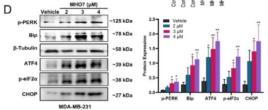
Application: WB Species: human Sample: MDA-MB-231 cells

Application: WB Species: human Sample: MCF-7,MDA-MB-231
Restrictive clause
Affinity Biosciences tests all products strictly. Citations are provided as a resource for additional applications that have not been validated by Affinity Biosciences. Please choose the appropriate format for each application and consult Materials and Methods sections for additional details about the use of any product in these publications.
For Research Use Only.
Not for use in diagnostic or therapeutic procedures. Not for resale. Not for distribution without written consent. Affinity Biosciences will not be held responsible for patent infringement or other violations that may occur with the use of our products. Affinity Biosciences, Affinity Biosciences Logo and all other trademarks are the property of Affinity Biosciences LTD.