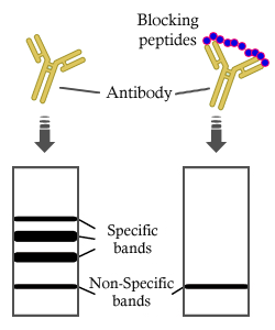
Product: Phospho-NCF2 (Thr233) Antibody
Catalog: AF4343
Description: Rabbit polyclonal antibody to Phospho-NCF2 (Thr233)
Application: WB IF/ICC
Cited expt.: WB
Reactivity: Human, Mouse, Rat
Prediction: Pig, Bovine, Horse, Sheep, Rabbit, Dog, Chicken, Xenopus
Mol.Wt.: 60kDa; 60kD(Calculated).
Uniprot: P19878
RRID: AB_2844422

View similar products>>

   Size Price Inventory
 100ul $350 In stock
 200ul $450 In stock

Lead Time: Same day delivery

For pricing and ordering contact:
Local distributors

Product Info

Source:
Rabbit
Application:
WB 1:500-1:2000, IF/ICC 1:100-1:500
*The optimal dilutions should be determined by the end user. For optimal experimental results, antibody reuse is not recommended.
*Tips:

WB: For western blot detection of denatured protein samples. IHC: For immunohistochemical detection of paraffin sections (IHC-p) or frozen sections (IHC-f) of tissue samples. IF/ICC: For immunofluorescence detection of cell samples. ELISA(peptide): For ELISA detection of antigenic peptide.

Reactivity:
Human,Mouse,Rat
Prediction:
Pig(100%), Bovine(100%), Horse(92%), Sheep(100%), Rabbit(92%), Dog(100%), Chicken(92%), Xenopus(82%)
Clonality:
Polyclonal
Specificity:
Phospho-NCF2 (Thr233) Antibody detects endogenous levels of NCF2 only when phosphorylated at Thr233.
RRID:
AB_2844422
Cite Format: Affinity Biosciences Cat# AF4343, RRID:AB_2844422.
Conjugate:
Unconjugated.
Purification:
The antibody is from purified rabbit serum by affinity purification via sequential chromatography on phospho-peptide and non-phospho-peptide affinity columns.
Storage:
Rabbit IgG in phosphate buffered saline , pH 7.4, 150mM NaCl, 0.02% sodium azide and 50% glycerol. Store at -20 °C. Stable for 12 months from date of receipt.
Alias:

Fold/Unfold

67 kDa neutrophil oxidase factor; Chronic granulomatous disease autosomal 2; FLJ93058; NADPH oxidase activator 2; NCF-2; Ncf2; NCF2_HUMAN; Neutrophil cytosol factor 2; Neutrophil cytosolic factor 2 (65kD, chronic granulomatous disease, autosomal 2); Neutrophil NADPH oxidase factor 2; NOXA2; P67 PHOX; p67-phox; p67phox;

Immunogens

Immunogen:

A synthesized peptide derived from human NCF2 around the phosphorylation site of Thr233.

Uniprot:
Gene(ID):
Sequence:
MSLVEAISLWNEGVLAADKKDWKGALDAFSAVQDPHSRICFNIGCMYTILKNMTEAEKAFTRSINRDKHLAVAYFQRGMLYYQTEKYDLAIKDLKEALIQLRGNQLIDYKILGLQFKLFACEVLYNIAFMYAKKEEWKKAEEQLALATSMKSEPRHSKIDKAMECVWKQKLYEPVVIPVGKLFRPNERQVAQLAKKDYLGKATVVASVVDQDSFSGFAPLQPQAAEPPPRPKTPEIFRALEGEAHRVLFGFVPETKEELQVMPGNIVFVLKKGNDNWATVMFNGQKGLVPCNYLEPVELRIHPQQQPQEESSPQSDIPAPPSSKAPGRPQLSPGQKQKEEPKEVKLSVPMPYTLKVHYKYTVVMKTQPGLPYSQVRDMVSKKLELRLEHTKLSYRPRDSNELVPLSEDSMKDAWGQVKNYCLTLWCENTVGDQGFPDEPKESEKADANNQTTEPQLKKGSQVEALFSYEATQPEDLEFQEGDIILVLSKVNEEWLEGECKGKVGIFPKVFVEDCATTDLESTRREV

Predictions

Predictions:

Score>80(red) has high confidence and is suggested to be used for WB detection. *The prediction model is mainly based on the alignment of immunogen sequences, the results are for reference only, not as the basis of quality assurance.

Species
Results
Score
Pig
100
Bovine
100
Sheep
100
Dog
100
Horse
92
Chicken
92
Rabbit
92
Xenopus
82
Zebrafish
55
Model Confidence:
High(score>80) Medium(80>score>50) Low(score<50) No confidence

Research Backgrounds

Function:

NCF2, NCF1, and a membrane bound cytochrome b558 are required for activation of the latent NADPH oxidase (necessary for superoxide production).

Subcellular Location:

Cytoplasm.

Extracellular region or secreted Cytosol Plasma membrane Cytoskeleton Lysosome Endosome Peroxisome ER Golgi apparatus Nucleus Mitochondrion Manual annotation Automatic computational assertionSubcellular location
Family&Domains:

The OPR/PB1 domain mediates the association with NCF4/p40-PHOX.

Belongs to the NCF2/NOXA1 family.

Research Fields

· Cellular Processes > Transport and catabolism > Phagosome.   (View pathway)

· Human Diseases > Infectious diseases: Parasitic > Leishmaniasis.

· Organismal Systems > Development > Osteoclast differentiation.   (View pathway)

· Organismal Systems > Immune system > Leukocyte transendothelial migration.   (View pathway)

References

1). NCF4 attenuates colorectal cancer progression by modulating inflammasome activation and immune surveillance. Nature communications, 2024 (PubMed: 38886341) [IF=16.6]

Application: WB    Species: human    Sample:
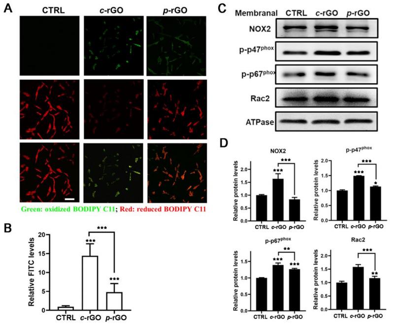
Fig. 1: ASC-interacting protein NCF4 is associated with colorectal cancer development. a Immunoblot analysis of ASC from the immunoprecipitated products generated with immunoprecipitation with an ASC antibody from the lysates of WT and Asc–/– BMDMs infected with F. novicida (100 MOI) for 12 h. b Mass spectrometry analysis of the IP product in (a). The peptides of ASC, NCF4, NCF1, and NCF2 were detected in the IP product with ASC antibody from WT and Asc–/– BMDMs in two repeated experiments. c Immunoblot analysis of Myc-ASC co-IP with FLAG-NCF4, FLAG-NCF4-PX, FLAG-NCF4-SH3 and FLAG-NCF4-PB1 from lysates of HEK293T cells transfected with the indicated plasmids. d Immunoblot analysis of FLAG-NCF4 co-IP with V5-ASC, V5-ASC-PYD, and V5-ASC-CARD from lysates of HEK293T cells transfected with the indicated plasmids. e Co-IP analysis of endogenous ASC interacting with total and phosphorylated NCF4, NCF1, and NCF2 in WT and Asc–/– BMDMs treated with the NLRP3 activator LPS plus ATP (LPS, 500 ng/mL for 4 h and ATP, 5 mM for 15 min). f Gene expression analysis of NCF1, NCF2, and NCF4 in total (left) and paired (right, n = 41) colorectal tumors (n = 480) and control tissues (n = 41) from 480 colorectal cancer (CRC) patients of pooled colon and rectal adenocarcinoma datasets in the TCGA database. g Correlation analysis between the gene expression of NCF4 and survival rate of CRC patients. (High, n = 466; Low, n = 131) Data are from 2 (a, b) or representative of 3 independent experiments with similar results (c–e). Wilcoxon signed rank test for (f), Log-rank (Mantel–Cox) test for (g), p-value is indicated in the graph. Source data are provided as a Source Data file.

2). Oxidation of Reduced Graphene Oxide via Cellular Redox Signaling Modulates Actin-Mediated Neurotransmission. ACS Nano, 2020 (PubMed: 32057235) [IF=15.8]

Application: WB    Species: rat    Sample: PC12

Figure S6 Oxidized rGO increased lipid peroxidation and NOX2 activation in PC12 cells. Oxidized rGO (c-rGO) was collected after pristine rGO (p-rGO) was cultured with PC12 cells for 3 h. (A-B) PC12 cells were pretreated with NAC for 30 min before being treated with two kinds of rGO (20 µg/mL) for 1 h and stained with BODIPY 581/591 C11 reagent. The lipid peroxidation degrees were quantified via FITC fluorescence intensity, with n = 10 fields per experimental condition from 3 independent tests. Scale bar: 100 μm. (C-D) After PC12 cells were treated as described in (A), plasma membrane proteins were isolated to detect the expression levels of NOX2, p-p47phox, p-p67phox and Rac2. The protein levels were quantified after normalization to ATPase. The results represent the mean ± SD of three independent experiments. *p<0.05, **p<0.01, ***p<0.001 compared with the control group and with the p-rGO group.

Restrictive clause


Affinity Biosciences tests all products strictly. Citations are provided as a resource for additional applications that have not been validated by Affinity Biosciences. Please choose the appropriate format for each application and consult Materials and Methods sections for additional details about the use of any product in these publications.

For Research Use Only.
Not for use in diagnostic or therapeutic procedures. Not for resale. Not for distribution without written consent. Affinity Biosciences will not be held responsible for patent infringement or other violations that may occur with the use of our products. Affinity Biosciences, Affinity Biosciences Logo and all other trademarks are the property of Affinity Biosciences LTD.