, using PCNA Antibody at 1/1000 dilution.
5ug/NC membrane strip.
Exposure for 20s with Affinity™ ECL Kit(#KF8003).
Bands result from membrane strip incubation.")
, blocked with antigen-specific peptides.
Lane 2: Hela cells(lps 4h treatment).
Lane 3: Pc12 cells(uv treatment).")
, blocked with antigen-specific peptides,
Lane 2: Raji cells(UV treatment),
Lane 3: 293 cells(heat-shock treatment).")


 Effects of BCL2L10 on protein expression of PCNA and CDC25C by Western blot.")
 The mRNA expression level of HSPA12B in MDS cells after si-HSPA12B and HSPA12B-OE transfection. (C)
The protein expression level of HSPA12B was accessed by western blot using GAPDH protein and PCNA (proliferation marker) as an internal control after si-HSPA12B
transfection. Data collected from three independently replicated wells. (D) The HSPA12B protein expression level accessed by western blot using GAPDH protein as
an internal control after HSPA12B-OE transfection. Data collected from three independently replicated wells. (E-F) The CCK-8 assay displayed absorbance at 450 nm in
MDS cells after si-HSPA12B and HSPA12B-OE transfection. Data were collected and calculated as mean ± SD from three independently replicated wells. (G-H) The EdU
positive cells in red after si-HSPA12B and HSPA12B-OE transfection. Nuclei were stained with DAPI (blue). Data were collected from three independently replicated
wells. Scale bar, 100 µm. Significance in (A-B) and (E-F): **P < 0.01.")

 The silencing of mortalin reversed the TGR5-induced increase in CC cell viability,
as assessed by CCK-8 assays. (B) The silencing of mortalin reversed the TGR5-induced increase in PCNA expression, as shown by western blotting. Glyceraldehyde
phosphate dehydrogenase (GAPDH) was used as a normalizing control. (C) The silencing of mortalin reversed the TGR5-induced increase in colony formation.
*P < 0.05, **P < 0.01 and ***P < 0.001, compared with the control group.")
 The mRNA levels of TRPC5 in different groups. (b) The protein levels of TRPC5 and PCNA were determined by Western blot analysis. GAPDH was loading control. The representative images were shown from 5 independently performed tests. (c) Densitometry analysis of TRPC5 protein expression. (d) Densitometry analysis of PCNA protein expression. Values are expressed as means ± SD (n = 5). ∗∗P < 0.01, compared to control group; ##P < 0.01, compared to Ang II group. Control: control group; Ang II: Ang II group; 10ISL: Ang II 10−6 mol/l + 10 μmol/l ISL group; 20ISL: Ang II 10−6 mol/l + 20 μmol/l ISL group; 40ISL: Ang II 10−6 mol/l + 40 μmol/l ISL group; 60ISL: Ang II 10−6 mol/l + 60 μmol/l ISL group.")
 HE staining and PCNA immunohistochemistry of tumor xenograft sections, 100×; (E) relative ratio of PCNA-positive cells in the engrafted tumors with SW1990- LNC-NC cell or SW1990-LNC-KD cell. **P<0.01, ***P<0.001.")
, cytoplasmic Nrf2 (B) and nuclear Nrf2 (C) protein expression in rat liver of control and NP treatment (low, middle and high dose) groups for 7,
14 and 28 days. Protein levels were normalized against those of GAPDH and PCNA. Representative immunoblots are also shown below the graphs. Data are presented
as mean ± S.D.(n = 3). * p<0.05 and * * p < 0.01 versus C group. & p < 0.05 versus NPL group.")


. Representative photographs are shown (a). Positive staining was quantified and is presented as the mean ± SD (b, c). d Serum levels of HGF were detected by ELISA (4h, n = 3/group; 12h and 24h, n = 4/group). Data are presented as the mean ± SD. *P < 0.05, **P < 0.01, ***P < 0.001")
 Positive expression of Ki67, PCNA, P53, and PTEN in tumor tissues was detected by an immunohistochemistry assay (magnification: ×200). (b) Protein expression levels of P53, P21, PTEN, BRCA1, BIRC5, and CTGF in tumor tissues were detected by Western blot assay, and GAPDH was used as the internal control. OC: ovarian cancer.")
 protein expression level in HCAECs and coronary artery endothelial cells of streptozotocin-induced diabetic mice. PCNA protein expression levels in HCAECs cultured in HG (for 1, 3, 7, and 14 days). Representative western blot images (A) and summary data (B–E) of PCNA protein levels in HCAECs cultured in NG medium (5.6 mM+19.5 mM α-mannitol) or HG medium (25 mM). β-tubulin was used as the loading control. (F) Representative images of coronary artery endothelium immunostaining (brown; eg, red arrowheads) for protein expression levels of PCNA. (G) Quantification of PCNA protein expression levels in the coronary artery endothelium of a mouse model of DM and control mice. (H) Results of Cell Count Kit-8 assay detection of living HCAECs exposed to HG for 1, 3, 7, or 14 days. Values are mean±SEM (n=6–8 samples). *P<0.05, **P<0.01, ***P<0.001 compared with NG-cultured cells or control group. DM, diabetes mellitus; HCAECs, human coronary artery endothelial cells; HG, high glucose; IOD, integrated optical density; NG, normal glucose; OD, optical density.")
 protein expression level in HCAECs and coronary artery endothelial cells of streptozotocin-induced diabetic mice. PCNA protein expression levels in HCAECs cultured in HG (for 1, 3, 7, and 14 days). Representative western blot images (A) and summary data (B–E) of PCNA protein levels in HCAECs cultured in NG medium (5.6 mM+19.5 mM α-mannitol) or HG medium (25 mM). β-tubulin was used as the loading control. (F) Representative images of coronary artery endothelium immunostaining (brown; eg, red arrowheads) for protein expression levels of PCNA. (G) Quantification of PCNA protein expression levels in the coronary artery endothelium of a mouse model of DM and control mice. (H) Results of Cell Count Kit-8 assay detection of living HCAECs exposed to HG for 1, 3, 7, or 14 days. Values are mean±SEM (n=6–8 samples). *P<0.05, **P<0.01, ***P<0.001 compared with NG-cultured cells or control group. DM, diabetes mellitus; HCAECs, human coronary artery endothelial cells; HG, high glucose; IOD, integrated optical density; NG, normal glucose; OD, optical density.")
 GA treatment increased the expression of the Keap1 gene but decreased that of the Nrf2 gene in H9C2 cells. (b, c) GA treatment increased the protein expression of HO-1, Nrf2, and Keap1 in the cytoplasm but decreased the protein expression of Nrf2 in the nucleus of H9C2 cells. H2O2 was used as a positive control. Values reflect the mean ± SD of three independent experiments.∗P < 0.05 vs. the control group.")
 Expression of HOXC6 after transfection of siRNA or overexpression plasmid measured by PCR. (b) CCK-8 assay to evaluate the effect of HOXC6 knockdown or overexpression on cell proliferation in LN229 cells. ∗p < 0.05, ∗∗p < 0.01. (c) CCK-8 assay to evaluate the effect of HOXC6 knockdown or overexpression on cell proliferation in T98G cells. From left to right: NC, OE, and SI. (d) Flow cytometry analysis to evaluate the effects of HOXC6 on cell cycle progression in LN229 cells. (e) Flow cytometry analysis to evaluate the effects of HOXC6 on cell cycle progression in T98G cells. From left to right: NC, OE, and SI. (f) Western blot analysis to evaluate the effects of HOXC6 on expression level of cell cycle related proteins.")
 cell growth and colony formation and promotes apoptosis in vitro. A and B. The CCK-8 assay showed that circ-ITCH inhibited the proliferation of C4-2 and DU145 cells. The data are the means ± S.D. of triplicate experiments (*P < 0.05, **P < 0.01, Student's t test). C and D. Additionally, colony formation assays showed that circ-ITCH inhibited colony formation in C4-2 and DU145 cells (*P < 0.05, **P < 0.01, ***P < 0.001, Student's t test). E and F. Cell apoptosis assays showed that circ-ITCH overexpression increased the rate of apoptosis in C4-2 and DU145 cells. The data are the mean ± SEM of three experiments (*P < 0.05, **P < 0.01, ***P < 0.001, Student's t test). G. Western blot showing the protein expression of the proliferation-associated genes PCNA and KI67 and the apoptosis-associated genes Caspase-3 and Caspase-9. The results are consistent with the qRT–PCR data.")

 Cell Counting Kit-8 assay was used to determine the effects of miR-638 mimics or miR-638 inhibitor on ovarian cancer cell line viability. *P")
 laser (810 nm) and LED (808 ± 3 nm) irradiated groups compared to non-irradiated burn control. PCNA and TGF-β2 are represented by red and green color respectively. Blue color represented nuclear counterstain with DAPI. Scale bar, 50 µm. Respective surface plots with peaks representing the expression of PCNA and TGF-β2 in burn control, pulsed laser and LED groups. Integrated fluorescence intensity was quantified by Fiji (ImageJ) software. Change in expression depicted as net fluorescence intensity (% control). Values are presented as mean ± SE, n = 6 animals per group. *p < 0.05 compared to non-irradiated burn control.")
 Representative images of CD31 and VEGFR2 staining. Quantification of (B) CD31 and (C) VEGFR2-positive area (n=5). (D) The expression of VEGFR2 was detected by western blotting. (E) The statistics of VEGFR2 were analyzed via ImageJ. (F) Immunohistochemical staining for PCNA days 2 and 3 after 70% hepatectomy. Quantification of PCNA positive nuclei from (G) SEC and (H) hepatocytes (n=5). The asterisk (*) represented the central vein, the arrows (→) represented positive SEC, the triangle (△) represented positive hepatocytes. A total of three independent experiments were pooled in the statistical analysis. All data are expressed as the means ± standard deviation. *P")
 The expression levels of EGR1, PTEN and other proteins in the liver tissues of the normal group and the model group, as analyzed by WB. (B) Expression of EGR1, PTEN and other proteins in liver tissues of the model and LNT-treated groups, as analyzed by WB. (C) Immunohistochemical analysis of EGR1, PTEN and Ki-67 in the tissue sections. Magnification, ×400. Scale bars, 200 µm (black). *P")
 patients. (A) Hematoxylin and eosin (H&E) staining of prostate sections from BPH patients (n = 6). (B) The relative protein levels of proliferating cell nuclear antigen (PCNA) in clinical prostate tissue by immunohistochemistry (IHC) staining and western blot analysis (n = 6). (C) The relative protein levels of Ki67 in clinical prostate tissue by IHC staining (n = 6). (D) The relative protein levels of cell cycle protein 1 (CyclinD1) in clinical prostate tissue by western blot analysis (n = 6). (E) Hierarchical clustered heat map of steroid hormones in the serum of normal men and BPH patients (n = 100). (F) 3D scatter plot of steroid hormones in the serum of normal men and BPH patients. (G) Variable importance for the projection (VIP) value of steroid hormones in the serum of normal men and BPH patients. (H) Hierarchical clustered heat maps of steroid hormones in the prostate tissue of normal men (n = 8) and BPH patients (n = 19). (I) 3D scatter plot of steroid hormones in the prostate tissue of normal men and BPH patients. (J) VIP value of steroid hormones in the prostate of normal men and BPH patients. Arrows indicate anatomical regions: prostatic epithelium or the acini (yellow arrow) and prostatic stroma (blue arrow). Data are mean ± standard error of mean (SEM). ∗∗P < 0.01, compared to N as indicated. A: androsterone; A4: androstenedione; DHEA: dehydroepiandrosterone; DHT: dihydrotestosterone; E1: estrone; E2: estradiol; T: testosterone.")
 patients. (A) Hematoxylin and eosin (H&E) staining of prostate sections from BPH patients (n = 6). (B) The relative protein levels of proliferating cell nuclear antigen (PCNA) in clinical prostate tissue by immunohistochemistry (IHC) staining and western blot analysis (n = 6). (C) The relative protein levels of Ki67 in clinical prostate tissue by IHC staining (n = 6). (D) The relative protein levels of cell cycle protein 1 (CyclinD1) in clinical prostate tissue by western blot analysis (n = 6). (E) Hierarchical clustered heat map of steroid hormones in the serum of normal men and BPH patients (n = 100). (F) 3D scatter plot of steroid hormones in the serum of normal men and BPH patients. (G) Variable importance for the projection (VIP) value of steroid hormones in the serum of normal men and BPH patients. (H) Hierarchical clustered heat maps of steroid hormones in the prostate tissue of normal men (n = 8) and BPH patients (n = 19). (I) 3D scatter plot of steroid hormones in the prostate tissue of normal men and BPH patients. (J) VIP value of steroid hormones in the prostate of normal men and BPH patients. Arrows indicate anatomical regions: prostatic epithelium or the acini (yellow arrow) and prostatic stroma (blue arrow). Data are mean ± standard error of mean (SEM). ∗∗P < 0.01, compared to N as indicated. A: androsterone; A4: androstenedione; DHEA: dehydroepiandrosterone; DHT: dihydrotestosterone; E1: estrone; E2: estradiol; T: testosterone.")
 on dihydrotestosterone (DHT)-induced proliferation in vitro. (A) The relative protein levels of androgen receptor (AR) in benign prostate hyperplasia (BPH)-1 cells cultured with DHT. (B) The relative protein levels of proliferating cell nuclear antigen (PCNA) in BPH-1 cells cultured with DHT by immunofluorescence (IF) and western blot analysis. (C) The relative protein levels of Ki67 in BPH-1 cells cultured with DHT by IF. (D) BPH-1 cell proliferation activity detected by 5-Ethynyl-20-deoxyuridine (EdU) assay. (E) Cell viability in DHT treated BPH-1 cells. (F) The relative protein levels of cell cycle protein 1 (CyclinD1) in BPH-1 cells cultured with DHT. (G) The relative protein levels of phosphorylation-AMP-activated protein kinase (p-AMPK) in BPH-1 cells cultured with DHT and Met. (H) The relative protein levels of AR in BPH-1 cells cultured with DHT and Met. (I) The relative protein levels of PCNA in BPH-1 cells co-cultured with DHT and Met by IF and western blot analysis. (J) The relative protein levels of Ki67 in BPH-1 cells cultured with DHT and Met by IF. (K) BPH-1 cell proliferation activity detected by EdU assay. (L) Cell viability in DHT and Met cultured BPH-1 cells. (M) The relative protein levels of CyclinD1 in BPH-1 cells cultured with DHT and Met. Data are mean ± standard error of mean (SEM) for cell groups of three. ∗P < 0.05, ∗∗P < 0.01, compared to normal control (NC) as indicated; #P < 0.05, ##P < 0.01, compared to DHT as indicated. t-AMPK: total-AMP-activated protein kinase.")
 Detection of mRNA level of SOX18 following overexpression of SOX18 in DPCs. (b,c) Detection of mRNA level of SOX18 following knockdown of SOX18 in DPCs. (d,e) Detection of mRNA level of PCNA, CDK2, and protein level of PCNA following overexpression or knockdown of SOX18 in DPCs. (f,g) CCK-8 assay following overexpression or knockdown of SOX18 in DPCs, respectively. (h,i) Cell cycle assay following overexpression or knockdown of SOX18 in DPCs, respectively. (j,k) EdU assay following overexpression or knockdown of SOX18 in DPCs, respectively. The scale is 100 µm. The data are presented as means ± SEM (standard error of the mean) (n = 3). The statistical significance was assessed using the unpaired Student’s t-test.")
. (A) Representative images of hepatic PCNA and Ki67 immunohistochemical staining of liver sections and the statistical quantification of the positive cell number/field were shown. mRNA expression of mitogenic genes (B) Cyclin A2, (C) Cyclin D1 and (D) Cyclin E1 in liver tissues were determined using RT-qPCR. (E) Western blotting of CCND1 and PCNA in liver tissues and quantification of the CCND1 and PCNA protein expression in both Nlrp3fl/fl and Nlrp3Δhep mice. Data are presented as the mean ± SD. *P")
. (A) Representative images of hepatic PCNA and Ki67 immunohistochemical staining of liver sections and the statistical quantification of the positive cell number/field were shown. mRNA expression of mitogenic genes (B) Cyclin A2, (C) Cyclin D1 and (D) Cyclin E1 in liver tissues were determined using RT-qPCR. (E) Western blotting of CCND1 and PCNA in liver tissues and quantification of the CCND1 and PCNA protein expression in both Nlrp3fl/fl and Nlrp3Δhep mice. Data are presented as the mean ± SD. *P")
 Relative mRNA expression of Wnt pathway related genes was measured in dermal fibroblast cells 24 h after transfection with NC or miR-140-y mimics by qPCR. (B–C) Relative protein expression of Wnt pathway related genes was measured in dermal fibroblast cells 72 h after transfection with NC or miR-140-y mimics by Western blot. (D–E) Representative observations of the EdU assay of dermal fibroblast cells after transfection with NC and miR-140-y mimics (Bars: 100 µm). Red illustrate the EdU staining and blue display the cell nuclei stained with Hoechst 33342. (F) The mRNA expression of proliferation related gene (PCNA). (G-H) Protein expression level of proliferation related gene (PCNA). The data were shown as mean ± SEM, n = 3, * p < 0.05, ** p < 0.01.")
 Mechanism of the PKCα function on Sertoli cell proliferation and differentiation through the MAPK/ERK signaling pathway. (B) Immunofluorescence staining of SOX9 in goose Sertoli cells. (C) Chemical structure of PKCα (C2-4) inhibitor peptide. (D) Quantification of mRNA expression levels of the MAPK/ERK signaling pathway hub genes. (E) Quantification of protein expression levels of the MAPK/ERK signaling pathway hub protein and proliferation and apoptosis related proteins. The data is displayed as the mean ± SD of three replicates, with statistical significance at p < 0.05. 18S rRNA gene expression was employed as a reference gene.")
, CREBBP (red), Scale bars = 50 μm. B Experimental design. C, D Representative western blots (C) and quantitative data (D) of Col-III, FSP1, and PCNA in NRK-49 F cells after stimulation with NRK-52E–delivered exosomes. *p")
 Immunofluorescence was employed to detect protein expression in rat brain tissue, followed by statistical analysis: (a) brain sections treated with DAPI (blue), PCNA (red), TH (green), Tuj-1 (orange), 7 and 14 days after AS-IV intervention, observed under a 10x microscope (scale bar = 100 μm); (b) staining of the brain sections with DAPI (blue), GFAP (green), caspase-3 (red) (scale bar = 100 μm), arrows represent caspase-3 protein fluorescence; (c) statistical comparison of fluorescence intensities of PCNA by the independent samples t test for PCNA; (d) fluorescence intensity of TH by independent samples t test; (e) fluorescence intensity of PCNA by independent samples t test; (f) fluorescence intensity of caspase-3 by independent samples t test; the data are expressed as mean ± SEM; n ≥ 3, ( ∗P < 0.05 vs. control group, ∗∗P < 0.005 vs. control group, #P < 0.05 vs. model group, ##P < 0.005 vs. model group, and ###P < 0.001 vs. model group).")
 Protein changes in the rat brain were investigated and subjected to statistical analysis. (a) protein expression in the rat’s brain 14 days after modeling; (b) TH, PCNA, and SHH protein expression; (c) TH mRNA expression level in the brain; (d) the TH PCNA mRNA expression level in the brain; (e) Tuj-1 mRNA expression level in the brain; The data are expressed as mean ± SEM; n ≥ 3, ( ∗P < 0.05 vs. control group, ∗∗P < 0.005 vs. control group, #P < 0.05 vs. model group, ##P < 0.005 vs. model group, and ###P < 0.001 vs. model group).")
 The proliferation of HCC cells with upregulated or downregulated HOXB4 expression; (c-d) EdU assay detected the proliferation of HCC cells with upregulated or downregulated HOXB4 expression. Scale bar, 50 μm; (e-f) Colony formation ability of HCC cells with upregulated or downregulated HOXB4 expression; (g) Representative immunoblots of PCNA in Li-7 cells with upregulated or downregulated HOXB4 expression; (h) Cell apoptosis of Li-7 cells with upregulated HOXB4 expression according to flow cytometry analysis; (i) Representative immunoblots of Bax, Bcl-2, cleaved caspase 3, and cleaved caspase 9 protein levels in Li-7 cells with upregulated HOXB4 expression. Data are expressed as mean ± SD, N = 3. **p")
 deficiency aggravated Sugen5416 +hypoxia-induced pulmonary hypertension (PH) in mice. A, Two pairs of primers were used to characterize MrgDSMKO mouse tails (wild-type [WT], heterozygous [HE], and homozygous [HO]). B, MrgD analysis by Western blotting in the pulmonary artery obtained from MrgDSMKO mice (n=4 male mice per group). C, Immunofluorescence costaining of MrgD (green), α-SMA (α-smooth muscle actin; red), and cell nuclei (blue) in distal pulmonary artery sections from control and MrgDSMKO mice. Scale bar, 20 µm. D, Measurement of right ventricular systolic pressure (RVSP), right ventricle (RV)/(left ventricle [LV]+septum [S]), pulmonary acceleration time to ejection time ratio (PAT/ET), tricuspid annular plane systolic excursion (TAPSE), and RV internal diameter (RVID; n=6 male mice per group). E, Hematoxylin and eosin (HE), Masson trichrome, and immunohistochemistry images of α-SMA and PCNA (proliferating cell nuclear antigen). Scale bar, 20 µm. F, Measurement of the medial wall thickness (n=6 male mice per group). G, Corresponding analysis of the muscularization percentage. Nonmuscularized vessels; partial, vessels with partial muscularization; full, vessels with complete muscularization (n=6 male mice per group). H, Measurement of PCNA+ cells surrounding blood vessels (n=6 male mice per group). I, Quantitative data from Masson trichrome staining (n=6 male mice per group). J, Images and quantitative analysis of Western blotting of PCNA, collagen I, and collagen III in the lung tissue from MrgDSMKO mice treated with hypoxia and Sugen5416 (n=6 male mice per group). All quantitative data are represented as mean±SEM. Nonparametric Mann-Whitney U test (B) and 2-way ANOVA with the Tukey multiple comparison test between multiple groups (D, F–J), and a Welch ANOVA followed by a 2-step linear step-up procedure of Benjamini, Krieger, and Yekutieli (D).")
| Product: | PCNA Antibody |
| Catalog: | AF0239 |
| Description: | Rabbit polyclonal antibody to PCNA |
| Application: | WB IHC IF/ICC |
| Cited expt.: | WB, IHC, IF/ICC |
| Reactivity: | Human, Mouse, Rat |
| Prediction: | Pig, Zebrafish, Bovine, Horse, Sheep, Rabbit, Dog, Chicken |
| Mol.Wt.: | 36kDa(Observed); 29kD(Calculated). |
| Uniprot: | P12004 |
| RRID: | AB_2833414 |
Control Products

Related Downloads
Protocols
Product Info
*The optimal dilutions should be determined by the end user. For optimal experimental results, antibody reuse is not recommended.
*Tips:
WB: For western blot detection of denatured protein samples. IHC: For immunohistochemical detection of paraffin sections (IHC-p) or frozen sections (IHC-f) of tissue samples. IF/ICC: For immunofluorescence detection of cell samples. ELISA(peptide): For ELISA detection of antigenic peptide.
Cite Format: Affinity Biosciences Cat# AF0239, RRID:AB_2833414.
Fold/Unfold
ATLD2; cb16; Cyclin; DNA polymerase delta auxiliary protein; etID36690.10; fa28e03; fb36g03; HGCN8729; MGC8367; Mutagen-sensitive 209 protein; OTTHUMP00000030189; OTTHUMP00000030190; PCNA; Pcna/cyclin; PCNA_HUMAN; PCNAR; Polymerase delta accessory protein; Proliferating cell nuclear antigen; wu:fa28e03; wu:fb36g03;
Immunogens
A synthesized peptide derived from human PCNA.
- P12004 PCNA_HUMAN:
- Protein BLAST With
- NCBI/
- ExPASy/
- Uniprot
MFEARLVQGSILKKVLEALKDLINEACWDISSSGVNLQSMDSSHVSLVQLTLRSEGFDTYRCDRNLAMGVNLTSMSKILKCAGNEDIITLRAEDNADTLALVFEAPNQEKVSDYEMKLMDLDVEQLGIPEQEYSCVVKMPSGEFARICRDLSHIGDAVVISCAKDGVKFSASGELGNGNIKLSQTSNVDKEEEAVTIEMNEPVQLTFALRYLNFFTKATPLSSTVTLSMSADVPLVVEYKIADMGHLKYYLAPKIEDEEGS
Predictions
Score>80(red) has high confidence and is suggested to be used for WB detection. *The prediction model is mainly based on the alignment of immunogen sequences, the results are for reference only, not as the basis of quality assurance.
High(score>80) Medium(80>score>50) Low(score<50) No confidence
Research Backgrounds
Auxiliary protein of DNA polymerase delta and is involved in the control of eukaryotic DNA replication by increasing the polymerase's processibility during elongation of the leading strand. Induces a robust stimulatory effect on the 3'-5' exonuclease and 3'-phosphodiesterase, but not apurinic-apyrimidinic (AP) endonuclease, APEX2 activities. Has to be loaded onto DNA in order to be able to stimulate APEX2. Plays a key role in DNA damage response (DDR) by being conveniently positioned at the replication fork to coordinate DNA replication with DNA repair and DNA damage tolerance pathways. Acts as a loading platform to recruit DDR proteins that allow completion of DNA replication after DNA damage and promote postreplication repair: Monoubiquitinated PCNA leads to recruitment of translesion (TLS) polymerases, while 'Lys-63'-linked polyubiquitination of PCNA is involved in error-free pathway and employs recombination mechanisms to synthesize across the lesion.
Phosphorylated. Phosphorylation at Tyr-211 by EGFR stabilizes chromatin-associated PCNA.
Acetylated by CREBBP and p300/EP300; preferentially acetylated by CREBBP on Lys-80, Lys-13 and Lys-14 and on Lys-77 by p300/EP300 upon loading on chromatin in response to UV irradiation. Lysine acetylation disrupts association with chromatin, hence promoting PCNA ubiquitination and proteasomal degradation in response to UV damage in a CREBBP- and EP300-dependent manner. Acetylation disrupts interaction with NUDT15 and promotes degradation.
Ubiquitinated. Following DNA damage, can be either monoubiquitinated to stimulate direct bypass of DNA lesions by specialized DNA polymerases or polyubiquitinated to promote recombination-dependent DNA synthesis across DNA lesions by template switching mechanisms. Following induction of replication stress, monoubiquitinated by the UBE2B-RAD18 complex on Lys-164, leading to recruit translesion (TLS) polymerases, which are able to synthesize across DNA lesions in a potentially error-prone manner. An error-free pathway also exists and requires non-canonical polyubiquitination on Lys-164 through 'Lys-63' linkage of ubiquitin moieties by the E2 complex UBE2N-UBE2V2 and the E3 ligases, HLTF, RNF8 and SHPRH. This error-free pathway, also known as template switching, employs recombination mechanisms to synthesize across the lesion, using as a template the undamaged, newly synthesized strand of the sister chromatid. Monoubiquitination at Lys-164 also takes place in undamaged proliferating cells, and is mediated by the DCX(DTL) complex, leading to enhance PCNA-dependent translesion DNA synthesis. Sumoylated during S phase.
Methylated on glutamate residues by ARMT1/C6orf211.
Nucleus.
Note: Colocalizes with CREBBP, EP300 and POLD1 to sites of DNA damage (PubMed:24939902). Forms nuclear foci representing sites of ongoing DNA replication and vary in morphology and number during S phase. Together with APEX2, is redistributed in discrete nuclear foci in presence of oxidative DNA damaging agents.
Belongs to the PCNA family.
Research Fields
· Cellular Processes > Cell growth and death > Cell cycle. (View pathway)
· Cellular Processes > Cellular community - eukaryotes > Tight junction. (View pathway)
· Genetic Information Processing > Replication and repair > DNA replication.
· Genetic Information Processing > Replication and repair > Base excision repair.
· Genetic Information Processing > Replication and repair > Nucleotide excision repair.
· Genetic Information Processing > Replication and repair > Mismatch repair.
· Human Diseases > Infectious diseases: Viral > Hepatitis B.
· Human Diseases > Infectious diseases: Viral > HTLV-I infection.
References
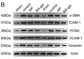
Application: WB Species: Rat Sample:

Application: IF/ICC Species: Rat Sample:
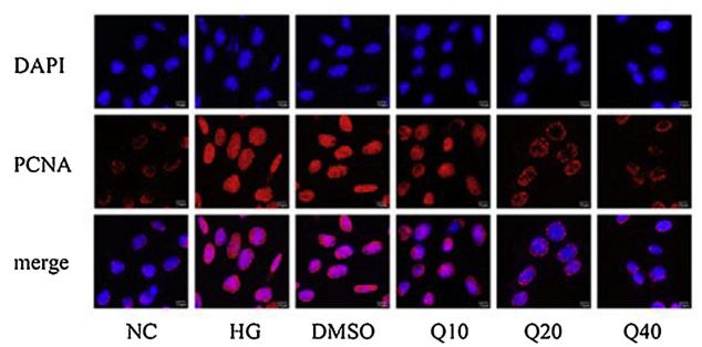
Application: IF/ICC Species: mouse Sample: glomerular mesangial cells
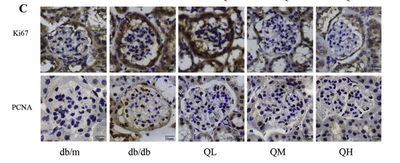
Application: IHC Species: mouse Sample: renal cortex
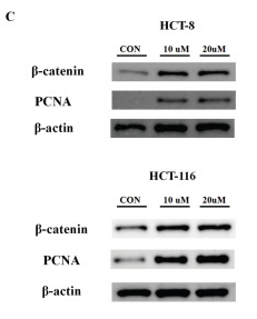
Application: WB Species: Human Sample: HCT 8 cells and HCT 116 cells
Restrictive clause
Affinity Biosciences tests all products strictly. Citations are provided as a resource for additional applications that have not been validated by Affinity Biosciences. Please choose the appropriate format for each application and consult Materials and Methods sections for additional details about the use of any product in these publications.
For Research Use Only.
Not for use in diagnostic or therapeutic procedures. Not for resale. Not for distribution without written consent. Affinity Biosciences will not be held responsible for patent infringement or other violations that may occur with the use of our products. Affinity Biosciences, Affinity Biosciences Logo and all other trademarks are the property of Affinity Biosciences LTD.