Product: KLF2 Antibody
Catalog: DF13602
Description: Rabbit polyclonal antibody to KLF2
Application: WB IHC
Cited expt.: WB
Reactivity: Human, Mouse, Rat
Prediction: Chicken, Xenopus
Mol.Wt.: 37kD, 52kD; 37kD(Calculated).
Uniprot: Q9Y5W3
RRID: AB_2846621

View similar products>>

   Size Price Inventory
 100ul $280 In stock
 200ul $350 In stock

Lead Time: Same day delivery

For pricing and ordering contact:
Local distributors

Product Info

Source:
Rabbit
Application:
WB 1:500-1:2000, IHC 1:50-1:200
*The optimal dilutions should be determined by the end user. For optimal experimental results, antibody reuse is not recommended.
*Tips:

WB: For western blot detection of denatured protein samples. IHC: For immunohistochemical detection of paraffin sections (IHC-p) or frozen sections (IHC-f) of tissue samples. IF/ICC: For immunofluorescence detection of cell samples. ELISA(peptide): For ELISA detection of antigenic peptide.

Reactivity:
Human,Mouse,Rat
Prediction:
Chicken(100%), Xenopus(83%)
Clonality:
Polyclonal
Specificity:
KLF2 Antibody detects endogenous levels of total KLF2.
RRID:
AB_2846621
Cite Format: Affinity Biosciences Cat# DF13602, RRID:AB_2846621.
Conjugate:
Unconjugated.
Purification:
The antiserum was purified by peptide affinity chromatography using SulfoLink™ Coupling Resin (Thermo Fisher Scientific).
Storage:
Rabbit IgG in phosphate buffered saline , pH 7.4, 150mM NaCl, 0.02% sodium azide and 50% glycerol. Store at -20 °C. Stable for 12 months from date of receipt.
Alias:

Fold/Unfold

KLF 2; Klf2; KLF2_HUMAN; Krueppel-like factor 2; Kruppel like factor 2; Kruppel like factor; Kruppel like factor LKLF; Kruppel-like factor 2 (lung); Lklf; Lung krueppel like factor; Lung krueppel-like factor; Lung Kruppel like zinc finger transcription factor;

Immunogens

Immunogen:

A synthesized peptide derived from human KLF2, corresponding to a region within the internal amino acids.

Uniprot:
Gene(ID):
Sequence:
MALSEPILPSFSTFASPCRERGLQERWPRAEPESGGTDDDLNSVLDFILSMGLDGLGAEAAPEPPPPPPPPAFYYPEPGAPPPYSAPAGGLVSELLRPELDAPLGPALHGRFLLAPPGRLVKAEPPEADGGGGYGCAPGLTRGPRGLKREGAPGPAASCMRGPGGRPPPPPDTPPLSPDGPARLPAPGPRASFPPPFGGPGFGAPGPGLHYAPPAPPAFGLFDDAAAAAAALGLAPPAARGLLTPPASPLELLEAKPKRGRRSWPRKRTATHTCSYAGCGKTYTKSSHLKAHLRTHTGEKPYHCNWDGCGWKFARSDELTRHYRKHTGHRPFQCHLCDRAFSRSDHLALHMKRHM

Predictions

Predictions:

Score>80(red) has high confidence and is suggested to be used for WB detection. *The prediction model is mainly based on the alignment of immunogen sequences, the results are for reference only, not as the basis of quality assurance.

Species
Results
Score
Chicken
100
Xenopus
83
Pig
0
Horse
0
Bovine
0
Sheep
0
Dog
0
Zebrafish
0
Rabbit
0
Model Confidence:
High(score>80) Medium(80>score>50) Low(score<50) No confidence

Research Backgrounds

Function:

Transcription factor that binds to the CACCC box in the promoter of target genes such as HBB/beta globin or NOV and activates their transcription. Might be involved in transcriptional regulation by modulating the binding of the RARA nuclear receptor to RARE DNA elements.

PTMs:

Ubiquitinated. Polyubiquitination involves WWP1 and leads to proteasomal degradation of this protein (By similarity).

Subcellular Location:

Nucleus.

Extracellular region or secreted Cytosol Plasma membrane Cytoskeleton Lysosome Endosome Peroxisome ER Golgi apparatus Nucleus Mitochondrion Manual annotation Automatic computational assertionSubcellular location
Family&Domains:

The 9aaTAD motif is a transactivation domain present in a large number of yeast and animal transcription factors.

Belongs to the krueppel C2H2-type zinc-finger protein family.

Research Fields

· Environmental Information Processing > Signal transduction > FoxO signaling pathway.   (View pathway)

· Environmental Information Processing > Signal transduction > Apelin signaling pathway.   (View pathway)

References

1). TRβ activation confers AT2-to-AT1 cell differentiation and anti-fibrosis during lung repair via KLF2 and CEBPA. Nature communications, 2024 (PubMed: 39375377) [IF=16.6]

Application: WB    Species: Mouse    Sample:

Fig. 6: TRβ modulates KLF2 and CEBPA to drive the expression of AT1 cell marker genes. a Principal component analysis (PCA) of TFs in bulk RNA-Seq of mouse lung (same as Fig. 3c, n = 3). The lower and upper bounds of the boxplot correspond to the first and third quartiles (the 25th and 75th percentiles); whiskers represent minima/maxima or 1.5*IQR. b Histogram showing the contributions of TFs in PC.2 and PC.5. c, d Protein and mRNA levels (n = 3) of KLF2 and CEBPA in the mouse lung, as shown in Fig. 3a. e qPCR analysis of KLF2 and CEBPA expression in isolated AT2 cells from mice (n = 3). f, g Immunoblotting and qPCR analysis (n = 3) of KLF2 and CEBPA in A549 and MLE12 with a GC-1 gradient. h The mRNA level of Klf2 and Cebpa in primary AT2 cells after 10 nM GC-1 treatment for 6 h (n = 3). i–l Protein (i, k) and mRNA (j, l) tests of AT1 cell markers, KLF2, and CEBPA expression in A549 after KLF2 (i, j) and CEBPA (k, l) overexpression (n = 3). m Luciferase activity of KLF2, CEBPA, and AT1-marker promoters cloned in pGL3.0 after THRB transfection for 36 h with or without GC-1 10 nM for 12 h (n = 3). Values were normalized to the transfection vector. n, o Luciferase activity of indicated promoters after KLF2 (n) and CEBPA (o) transfection with 600 ng plasmid for 36 h (n = 3). p–r TRβ (p), KLF2 (q), and CEBPA (r) bind to the promoter regions (pro) of KLF2, CEBPA, and AT1 cell genes in ChIP q-PCR assays (n = 3). KRT5 promoter as negative control. Values were normalized to IgG. s Luciferase activity of AT1-maker promoters with KLF2 (300 ng) and CEBPA (300 ng) co-transfection for 36 h, as shown in (n, o) (n = 3). t IF image of KLF2 and CEBPA after co-transfection in A549. Scale bar, 10 μm. u CoIP of KLF2 and CEBPA overexpressed in A549. v Illustration of the regulatory processes of GC-1 on AT1 cell markers. The value of n indicates biologically independent samples. Similar results were repeated in two biologically independent experiments. The statistical tests used were one-way ANOVA (d, e, and g), two-tailed unpaired Student’s t test (h), and two-way ANOVA (j, l, m–s). Data are mean ± SEM.

2). DNMT aberration-incurred GPX4 suppression prompts osteoblast ferroptosis and osteoporosis. Bone research, 2024 (PubMed: 39617773) [IF=14.3]

3). FGF2 Alleviates Microvascular Ischemia-Reperfusion Injury by KLF2-mediated Ferroptosis Inhibition and Antioxidant Responses. International journal of biological sciences, 2023 (PubMed: 37705747) [IF=8.2]

Application: WB    Species: Mouse    Sample:

Figure 6 FGF2 reduces oxidative stress and ferroptosis via KLF2-NFE2L2 pathway in vivo. (A) Skeletal muscle specimens were used for the KLF2 and NFE2L2 WB assay. (B) Protein quantification of KLF2 and NFE2L2 from (A), normalised to GAPDH band density and histone 3 band density, respectively. (C) Skeletal muscle specimens showing HO-1, SOD1, SLC7A11, 4-HNE, and GPX4 on a WB assay. (D) Quantification of the GAPDH band density-normalized HO-1 and SOD1 protein levels from (C). (E) Quantification of protein level of SLC7A11, 4-HNE and GPX4 from (C) with normalized to GAPDH band density. (F) DAPI staining shows the nuclei in these micrographs of skeletal muscle slices that have been immunostained for KLF2 and CD31. Scale bars: 100 μm. (G) Micrographs of skeletal muscle slices with NFE2L2 and CD31 immunostaining; DAPI labelling shows nuclei. Scale bars: 100 μm. (H) Quantification of cells that co-express KLF2 and CD31; proportions of co-expressing cells to all CD31-positive cells are shown. (I) Quantification of NFE2L2 and CD31 double-positive cells, the ratio of co-expressing cells to all CD31-positive cells is shown. (J) Micrographs of skeletal muscle slices with nuclei identified by DAPI labelling and DHE and 4-HNE staining. Scale bars: 100 μm. Based on the immunofluorescence data in (J), the ROS level was quantified in (K). Based on the immunofluorescence results in (J), 4-HNE fluorescence intensity was quantified in (L). (M) Content of Fe2+ was plotted as a histogram. (N) The concentration of MDA is shown as a histogram. (O) The concentration of GSH is shown as a histogram. Data are provided as mean ± SD (n = 3-5 per group). Significance: ns, not significant; *P < 0.05.

4). Netrin-1 protects blood-brain barrier (BBB) integrity after cerebral ischemia-reperfusion by activating the Kruppel-like factor 2 (KLF2)/occludin pathway. Journal of biochemical and molecular toxicology, 2024 (PubMed: 38229322) [IF=3.2]

5). Bioinformatics analysis of KLF2 as a potential prognostic factor in ccRCC and association with epithelial‑mesenchymal transition. Experimental and Therapeutic Medicine, 2022 (PubMed: 35978925) [IF=2.4]

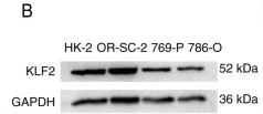
Application: WB    Species: Human    Sample: HK-2 cells

Figure 3 KLF2 inhibits ccRCC migration in vitro and KLF2 protein expression is decreased in high pathological grade tissue (II + III). (A) Expression of KLF2 mRNA in ccRCC and normal HK-2 cells. (B) Western blotting was used for assessment of KLF2 protein levels in ccRCC and normal HK-2 cells. (C) Western blotting assessment of KLF2 OE in 769-P and 786-O cells. (D) Migration of 769-P and 786-O cells after wounding (magnification, x40). (E) compared with NC. (F) Immunohistochemistry and (G) analysis of the KLF2 expression in ccRCC tissue (n=20). **P<0.01 vs. stage I and ***P<0.001 vs. NC. KLF, Krüppel-like factor; ccRCC, clear cell renal cell carcinoma; NC, negative control; OE, overexpression.

Restrictive clause


Affinity Biosciences tests all products strictly. Citations are provided as a resource for additional applications that have not been validated by Affinity Biosciences. Please choose the appropriate format for each application and consult Materials and Methods sections for additional details about the use of any product in these publications.

For Research Use Only.
Not for use in diagnostic or therapeutic procedures. Not for resale. Not for distribution without written consent. Affinity Biosciences will not be held responsible for patent infringement or other violations that may occur with the use of our products. Affinity Biosciences, Affinity Biosciences Logo and all other trademarks are the property of Affinity Biosciences LTD.