
, using PKC delta Antibody at 1/1000 dilution.
5ug/NC membrane strip.
Exposure for 20s with Affinity™ ECL Kit(#KF8001).
Bands result from membrane strip incubation.")

| Product: | PKC delta Antibody |
| Catalog: | AF6408 |
| Description: | Rabbit polyclonal antibody to PKC delta |
| Application: | WB IHC IF/ICC |
| Cited expt.: | WB, IHC |
| Reactivity: | Human, Mouse |
| Prediction: | Pig, Bovine, Horse, Sheep, Dog, Chicken |
| Mol.Wt.: | 78kDa(Observed); 78kD(Calculated). |
| Uniprot: | Q05655 |
| RRID: | AB_2835238 |
Control Products

Product Info
*The optimal dilutions should be determined by the end user. For optimal experimental results, antibody reuse is not recommended.
*Tips:
WB: For western blot detection of denatured protein samples. IHC: For immunohistochemical detection of paraffin sections (IHC-p) or frozen sections (IHC-f) of tissue samples. IF/ICC: For immunofluorescence detection of cell samples. ELISA(peptide): For ELISA detection of antigenic peptide.
Cite Format: Affinity Biosciences Cat# AF6408, RRID:AB_2835238.
Fold/Unfold
CVID9; D14Ertd420e; Kinase PKC delta; KPCD; KPCD_HUMAN; MAY 1; MAY1; MGC49908; nPKC delta; nPKC-delta; PCKd; PKC d; PKC delta; PKCD; PKCdelta; PRKC D; PRKC delta; Prkcd; Protein Kinase C delta; Protein kinase C delta type; Protein kinase C delta VIII; Protein Kinase Cdelta; Tyrosine protein kinase PRKCD;
Immunogens
A synthesized peptide derived from human PKC delta, corresponding to a region within C-terminal amino acids.
- Q05655 KPCD_HUMAN:
- Protein BLAST With
- NCBI/
- ExPASy/
- Uniprot
MAPFLRIAFNSYELGSLQAEDEANQPFCAVKMKEALSTERGKTLVQKKPTMYPEWKSTFDAHIYEGRVIQIVLMRAAEEPVSEVTVGVSVLAERCKKNNGKAEFWLDLQPQAKVLMSVQYFLEDVDCKQSMRSEDEAKFPTMNRRGAIKQAKIHYIKNHEFIATFFGQPTFCSVCKDFVWGLNKQGYKCRQCNAAIHKKCIDKIIGRCTGTAANSRDTIFQKERFNIDMPHRFKVHNYMSPTFCDHCGSLLWGLVKQGLKCEDCGMNVHHKCREKVANLCGINQKLLAEALNQVTQRASRRSDSASSEPVGIYQGFEKKTGVAGEDMQDNSGTYGKIWEGSSKCNINNFIFHKVLGKGSFGKVLLGELKGRGEYFAIKALKKDVVLIDDDVECTMVEKRVLTLAAENPFLTHLICTFQTKDHLFFVMEFLNGGDLMYHIQDKGRFELYRATFYAAEIMCGLQFLHSKGIIYRDLKLDNVLLDRDGHIKIADFGMCKENIFGESRASTFCGTPDYIAPEILQGLKYTFSVDWWSFGVLLYEMLIGQSPFHGDDEDELFESIRVDTPHYPRWITKESKDILEKLFEREPTKRLGVTGNIKIHPFFKTINWTLLEKRRLEPPFRPKVKSPRDYSNFDQEFLNEKARLSYSDKNLIDSMDQSAFAGFSFVNPKFEHLLED
Predictions
Score>80(red) has high confidence and is suggested to be used for WB detection. *The prediction model is mainly based on the alignment of immunogen sequences, the results are for reference only, not as the basis of quality assurance.
High(score>80) Medium(80>score>50) Low(score<50) No confidence
Research Backgrounds
Calcium-independent, phospholipid- and diacylglycerol (DAG)-dependent serine/threonine-protein kinase that plays contrasting roles in cell death and cell survival by functioning as a pro-apoptotic protein during DNA damage-induced apoptosis, but acting as an anti-apoptotic protein during cytokine receptor-initiated cell death, is involved in tumor suppression as well as survival of several cancers, is required for oxygen radical production by NADPH oxidase and acts as positive or negative regulator in platelet functional responses. Negatively regulates B cell proliferation and also has an important function in self-antigen induced B cell tolerance induction. Upon DNA damage, activates the promoter of the death-promoting transcription factor BCLAF1/Btf to trigger BCLAF1-mediated p53/TP53 gene transcription and apoptosis. In response to oxidative stress, interact with and activate CHUK/IKKA in the nucleus, causing the phosphorylation of p53/TP53. In the case of ER stress or DNA damage-induced apoptosis, can form a complex with the tyrosine-protein kinase ABL1 which trigger apoptosis independently of p53/TP53. In cytosol can trigger apoptosis by activating MAPK11 or MAPK14, inhibiting AKT1 and decreasing the level of X-linked inhibitor of apoptosis protein (XIAP), whereas in nucleus induces apoptosis via the activation of MAPK8 or MAPK9. Upon ionizing radiation treatment, is required for the activation of the apoptosis regulators BAX and BAK, which trigger the mitochondrial cell death pathway. Can phosphorylate MCL1 and target it for degradation which is sufficient to trigger for BAX activation and apoptosis. Is required for the control of cell cycle progression both at G1/S and G2/M phases. Mediates phorbol 12-myristate 13-acetate (PMA)-induced inhibition of cell cycle progression at G1/S phase by up-regulating the CDK inhibitor CDKN1A/p21 and inhibiting the cyclin CCNA2 promoter activity. In response to UV irradiation can phosphorylate CDK1, which is important for the G2/M DNA damage checkpoint activation. Can protect glioma cells from the apoptosis induced by TNFSF10/TRAIL, probably by inducing increased phosphorylation and subsequent activation of AKT1. Is highly expressed in a number of cancer cells and promotes cell survival and resistance against chemotherapeutic drugs by inducing cyclin D1 (CCND1) and hyperphosphorylation of RB1, and via several pro-survival pathways, including NF-kappa-B, AKT1 and MAPK1/3 (ERK1/2). Can also act as tumor suppressor upon mitogenic stimulation with PMA or TPA. In N-formyl-methionyl-leucyl-phenylalanine (fMLP)-treated cells, is required for NCF1 (p47-phox) phosphorylation and activation of NADPH oxidase activity, and regulates TNF-elicited superoxide anion production in neutrophils, by direct phosphorylation and activation of NCF1 or indirectly through MAPK1/3 (ERK1/2) signaling pathways. May also play a role in the regulation of NADPH oxidase activity in eosinophil after stimulation with IL5, leukotriene B4 or PMA. In collagen-induced platelet aggregation, acts a negative regulator of filopodia formation and actin polymerization by interacting with and negatively regulating VASP phosphorylation. Downstream of PAR1, PAR4 and CD36/GP4 receptors, regulates differentially platelet dense granule secretion; acts as a positive regulator in PAR-mediated granule secretion, whereas it negatively regulates CD36/GP4-mediated granule release. Phosphorylates MUC1 in the C-terminal and regulates the interaction between MUC1 and beta-catenin. The catalytic subunit phosphorylates 14-3-3 proteins (YWHAB, YWHAZ and YWHAH) in a sphingosine-dependent fashion (By similarity). Phosphorylates ELAVL1 in response to angiotensin-2 treatment.
Autophosphorylated and/or phosphorylated at Thr-507, within the activation loop; phosphorylation at Thr-507 is not a prerequisite for enzymatic activity. Autophosphorylated at Ser-299, Ser-302 and Ser-304. Upon TNFSF10/TRAIL treatment, phosphorylated at Tyr-155; phosphorylation is required for its translocation to the endoplasmic reticulum and cleavage by caspase-3. Phosphorylated at Tyr-313, Tyr-334 and Tyr-567; phosphorylation of Tyr-313 and Tyr-567 following thrombin stimulation potentiates its kinase activity. Phosphorylated by protein kinase PDPK1; phosphorylation is inhibited by the apoptotic C-terminal cleavage product of PKN2.
Proteolytically cleaved into a catalytic subunit and a regulatory subunit by caspase-3 during apoptosis which results in kinase activation.
Cytoplasm. Cytoplasm>Perinuclear region. Nucleus. Cell membrane>Peripheral membrane protein.
The C1 domain, containing the phorbol ester/DAG-type region 1 (C1A) and 2 (C1B), is the diacylglycerol sensor.
The C2 domain is a non-calcium binding domain. It binds proteins containing phosphotyrosine in a sequence-specific manner.
Belongs to the protein kinase superfamily. AGC Ser/Thr protein kinase family. PKC subfamily.
Research Fields
· Cellular Processes > Transport and catabolism > Autophagy - animal. (View pathway)
· Human Diseases > Endocrine and metabolic diseases > Type II diabetes mellitus.
· Human Diseases > Endocrine and metabolic diseases > Insulin resistance.
· Organismal Systems > Immune system > Chemokine signaling pathway. (View pathway)
· Organismal Systems > Circulatory system > Vascular smooth muscle contraction. (View pathway)
· Organismal Systems > Immune system > NOD-like receptor signaling pathway. (View pathway)
· Organismal Systems > Immune system > Fc gamma R-mediated phagocytosis. (View pathway)
· Organismal Systems > Nervous system > Neurotrophin signaling pathway. (View pathway)
· Organismal Systems > Sensory system > Inflammatory mediator regulation of TRP channels. (View pathway)
· Organismal Systems > Endocrine system > Estrogen signaling pathway. (View pathway)
References
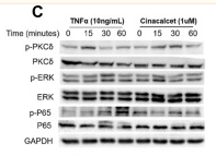
Application: WB Species: Mouse Sample:
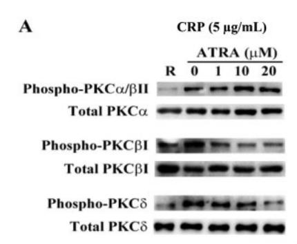
Application: WB Species: human Sample: human platelets
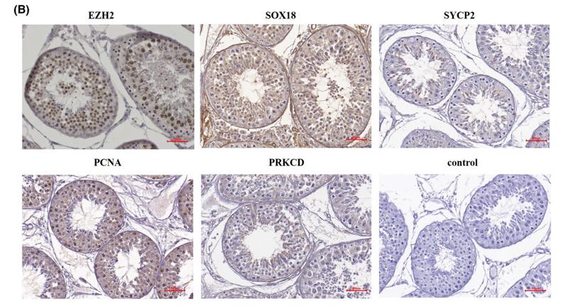
Application: IHC Species: sheep Sample: testicular cells
Restrictive clause
Affinity Biosciences tests all products strictly. Citations are provided as a resource for additional applications that have not been validated by Affinity Biosciences. Please choose the appropriate format for each application and consult Materials and Methods sections for additional details about the use of any product in these publications.
For Research Use Only.
Not for use in diagnostic or therapeutic procedures. Not for resale. Not for distribution without written consent. Affinity Biosciences will not be held responsible for patent infringement or other violations that may occur with the use of our products. Affinity Biosciences, Affinity Biosciences Logo and all other trademarks are the property of Affinity Biosciences LTD.