
.
Bands result from membrane strip incubation.")


 Relative mRNA expression levels of miR‐448‐3p and DPP‐4 in high glucose‐treated aortic
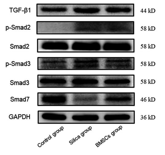
endothelial cells. (b) The protein levels of DPP‐4, CD31, VE‐cadherin, α‐SMA, and vimentin in each group. (c) Relative protein levels of TGF‐β1,
Smad2, p‐Smad2 (S465/S467), Smad3, p‐Smad3 (S423/S425), and Smad7 in all the groups. Data were expressed as means ± standard deviation
(SD), n = 3. *p < .05 compared with the OC group. DPP‐4, dipeptidyl peptidase‐4; EndMT, endothelial–mesenchymal transition; HG, high glucose;
miR, microRNA; mRNA, messenger RNA; TGF‐β, transforming growth factor‐β; α‐SMA, α‐smooth muscle actin")

 SMAD2/3 protein levels in Nuclear was analyzed by Western blotting.(B) SMAD4 protein levels in Nuclear was analyzed by Western blotting.(C) Expression of SMAD7 was analyzed by Western blotting. The data represent the mean ± SD of three independent experiments. Each bar represents the mean of three independent experiments. *p\0.05 versus control.")
 were infected with adenovirus encoding shRNA targeting TFPI2 (TFPI2 shRNA) or overexpressing TFPI2 (TFPI2 OE), followed by stimulation of 5 ng/ml TGF-β2 for 48 h. A, the expression of SMAD7, TGFBR1, TGFBR2, SMAD2/3, and phospho-SMAD2/3 (p-SMAD2/3) was determined by Western blot. Semiquantitative analysis of (B) SMAD7, (C) TGFBR1, and (D) TGFBR2, as well as (E and F) the ratio of p-SMAD2/3 to SMAD2/3. G and H, immunofluorescent staining of SMAD2/3 in hRGECs. Yellow arrows indicated nuclear translocation of SMAD2/3. Data are shown as the mean ± SD (n = 3). ∗p < 0.05, ∗∗p < 0.01, ∗∗∗p < 0.001. TGF-β, transforming growth factor beta; TFP12, tissue factor pathway inhibitor 2.")
, TβRI (b), TβRII (c), p-Smad2 (d), p-Smad3 (e), and Smad7 (f) protein and their protein band ((g), (h)) in the lung tissue of mice in each group. NC, normal control group; BLM, bleomycin-induced systemic sclerosis model group; PESV-L, low-dose PESV intervention group; PESV-M, medium-dose PESV intervention group; PESV-H, high-dose PESV intervention group; DXM, dexamethasone intervention group.")
 Relative mRNA expression of DDIT4. (B) Protein bands of DDIT4. (C) DDIT4/β-actin protein expression levels. Data are presented as the mean ± SD (n=11). *P")

| Product: | Smad7 Antibody |
| Catalog: | AF5147 |
| Description: | Rabbit polyclonal antibody to Smad7 |
| Application: | WB IHC |
| Cited expt.: | WB, IHC |
| Reactivity: | Human, Mouse, Rat |
| Prediction: | Pig, Bovine, Rabbit, Dog |
| Mol.Wt.: | 46 kDa(Observed); 46kD(Calculated). |
| Uniprot: | O15105 |
| RRID: | AB_2837633 |
Control Products

Related Downloads
Protocols
Product Info
*The optimal dilutions should be determined by the end user. For optimal experimental results, antibody reuse is not recommended.
*Tips:
WB: For western blot detection of denatured protein samples. IHC: For immunohistochemical detection of paraffin sections (IHC-p) or frozen sections (IHC-f) of tissue samples. IF/ICC: For immunofluorescence detection of cell samples. ELISA(peptide): For ELISA detection of antigenic peptide.
Cite Format: Affinity Biosciences Cat# AF5147, RRID:AB_2837633.
Fold/Unfold
CRCS3; FLJ16482; hSMAD 7; hSMAD7; MAD (mothers against decapentaplegic Drosophila) homolog 7; MAD; Mad homolog 7; MAD homolog 8; MAD mothers against decapentaplegic homolog 7; MADH 7; MADH 8; MADH6; MADH8; Mothers Against Decapentaplegic Drosophila Homolog of 6; Mothers Against Decapentaplegic Drosophila Homolog of 7; Mothers against decapentaplegic homolog 7; Mothers against decapentaplegic homolog 8; Mothers against DPP homolog 7; Mothers against DPP homolog 8; SMA- AND MAD-RELATED PROTEIN 7; SMAD 7; SMAD; SMAD family member 7; SMAD, mothers against DPP homolog 7 (Drosophila); SMAD, mothers against DPP homolog 7; SMAD6; Smad7; SMAD7_HUMAN;
Immunogens
A synthesized peptide derived from human Smad7, corresponding to a region within N-terminal amino acids.
- O15105 SMAD7_HUMAN:
- Protein BLAST With
- NCBI/
- ExPASy/
- Uniprot
MFRTKRSALVRRLWRSRAPGGEDEEEGAGGGGGGGELRGEGATDSRAHGAGGGGPGRAGCCLGKAVRGAKGHHHPHPPAAGAGAAGGAEADLKALTHSVLKKLKERQLELLLQAVESRGGTRTACLLLPGRLDCRLGPGAPAGAQPAQPPSSYSLPLLLCKVFRWPDLRHSSEVKRLCCCESYGKINPELVCCNPHHLSRLCELESPPPPYSRYPMDFLKPTADCPDAVPSSAETGGTNYLAPGGLSDSQLLLEPGDRSHWCVVAYWEEKTRVGRLYCVQEPSLDIFYDLPQGNGFCLGQLNSDNKSQLVQKVRSKIGCGIQLTREVDGVWVYNRSSYPIFIKSATLDNPDSRTLLVHKVFPGFSIKAFDYEKAYSLQRPNDHEFMQQPWTGFTVQISFVKGWGQCYTRQFISSCPCWLEVIFNSR
Predictions
Score>80(red) has high confidence and is suggested to be used for WB detection. *The prediction model is mainly based on the alignment of immunogen sequences, the results are for reference only, not as the basis of quality assurance.
High(score>80) Medium(80>score>50) Low(score<50) No confidence
Research Backgrounds
Antagonist of signaling by TGF-beta (transforming growth factor) type 1 receptor superfamily members; has been shown to inhibit TGF-beta (Transforming growth factor) and activin signaling by associating with their receptors thus preventing SMAD2 access. Functions as an adapter to recruit SMURF2 to the TGF-beta receptor complex. Also acts by recruiting the PPP1R15A-PP1 complex to TGFBR1, which promotes its dephosphorylation. Positively regulates PDPK1 kinase activity by stimulating its dissociation from the 14-3-3 protein YWHAQ which acts as a negative regulator.
Phosphorylation on Ser-249 does not affect its stability, nuclear localization or inhibitory function in TGFB signaling; however it affects its ability to regulate transcription (By similarity). Phosphorylated by PDPK1.
Ubiquitinated by WWP1 (By similarity). Polyubiquitinated by RNF111, which is enhanced by AXIN1 and promotes proteasomal degradation. In response to TGF-beta, ubiquitinated by SMURF1; which promotes its degradation.
Acetylation prevents ubiquitination and degradation mediated by SMURF1.
Nucleus. Cytoplasm.
Note: Interaction with NEDD4L or RNF111 induces translocation from the nucleus to the cytoplasm (PubMed:16601693). TGF-beta stimulates its translocation from the nucleus to the cytoplasm. PDPK1 inhibits its translocation from the nucleus to the cytoplasm in response to TGF-beta (PubMed:17327236).
Ubiquitous with higher expression in the lung and vascular endothelium.
Belongs to the dwarfin/SMAD family.
Research Fields
· Environmental Information Processing > Signal transduction > TGF-beta signaling pathway. (View pathway)
· Environmental Information Processing > Signal transduction > Hippo signaling pathway. (View pathway)
References

Application: IHC Species: Rat Sample:

Application: WB Species: Rat Sample:
Application: WB Species: Mice Sample: H22 cells
Application: WB Species: rat Sample: lung
Application: WB Species: Human Sample: Hypertrophic scar tissue
Application: WB Species: human Sample: HSFs

Application: WB Species: Mouse Sample: TCMK-1 cells
Restrictive clause
Affinity Biosciences tests all products strictly. Citations are provided as a resource for additional applications that have not been validated by Affinity Biosciences. Please choose the appropriate format for each application and consult Materials and Methods sections for additional details about the use of any product in these publications.
For Research Use Only.
Not for use in diagnostic or therapeutic procedures. Not for resale. Not for distribution without written consent. Affinity Biosciences will not be held responsible for patent infringement or other violations that may occur with the use of our products. Affinity Biosciences, Affinity Biosciences Logo and all other trademarks are the property of Affinity Biosciences LTD.