
 The expression of p-Tau and Tau in control rats and CUMS rats in the sixth week and tenth week of the CUMS period.(B) The expression of PSD-95 and SYN in control rats and CUMS rats in the sixth week and tenth week of the CUMS period. Bars represent mean ± S.E.M of the different groups (n = 3/group). (compared with control rats, *P < 0.05; **P < 0.01).")
 and spleen (d–f). Morris water maze test for day 1 escape latency (g) and day 2 escape latency (h). Western blot analysis for FTL and Tf in brain (i–k) and hippocampus (l–n), and for SYN1, NMDAR, PSD-95 in hippocampus (o–r). The quantification of western blotting was provided in supplementary material. FTL ferritin light chain, Tf transferrin, SYN1 synapsin 1, NMDAR N-methyl-D-aspartate receptor, PSD-95 postsynaptic density protein 95. Data of Western blot analysis (mean ± SD) are expressed as the ratio of the relative contents between the value from IDA group and NG group and six iron treatment groups (n = 3). The relative contents of target proteins were quantified using the ratio between the optical density (OD) of target protein and the amount of the housekeeping protein GAPDH. **p < 0.01, compared with NG, #p < 0.05, ##p < 0.01, compared with IDAG. One-way ANOVA followed by Tukey multiple comparison test was used for comparison among 8 different groups.")
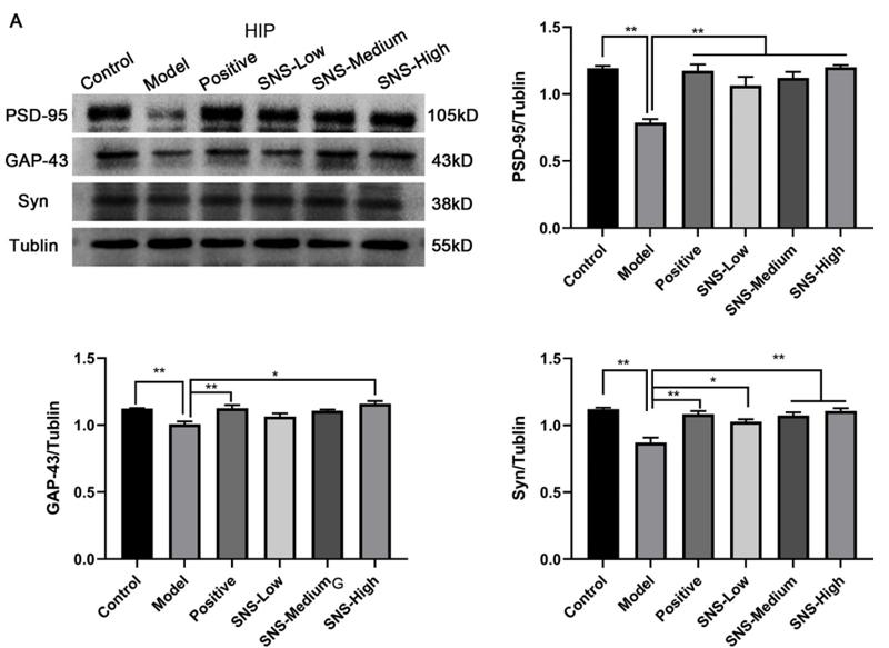
, including PSD-95 (B), GAP-43 (C), and SYN (D). The values represent the mean ± SEM, n = 5. *p < 0.05, **p < 0.01 vs. CON, #p < 0.05, ##p < 0.01 vs. MS. CON, control; MS, maternal separation; CUMS, chronic unpredictable mild stress.")
 CHIP knock-down protein was designed by siRNA in N2a cells. Approximately 5 µl Stub1-siRNA was able to achieve effective knock-down CHIP expression, and consequently the expression level of synapsin I S9 was decreased with no change in synaptic protein SNAP25 and PSD95 expression.")
 Relative level of the Syn protein. (B) Western blotting was used to detect Syn expression. (C) Relative level of the PSD-95 protein. (D) Western blotting was used to detect PSD-95 expression. *p < 0.05, **p < 0.01, and ***p < 0.001 compared with the corresponding SHAM group. #p < 0.05 and ###p < 0.001 compared with the corresponding SED-MCAO group. +p < 0.05 compared with the corresponding EX-MCAO group. Means ± standard deviations, n = 6/group.")
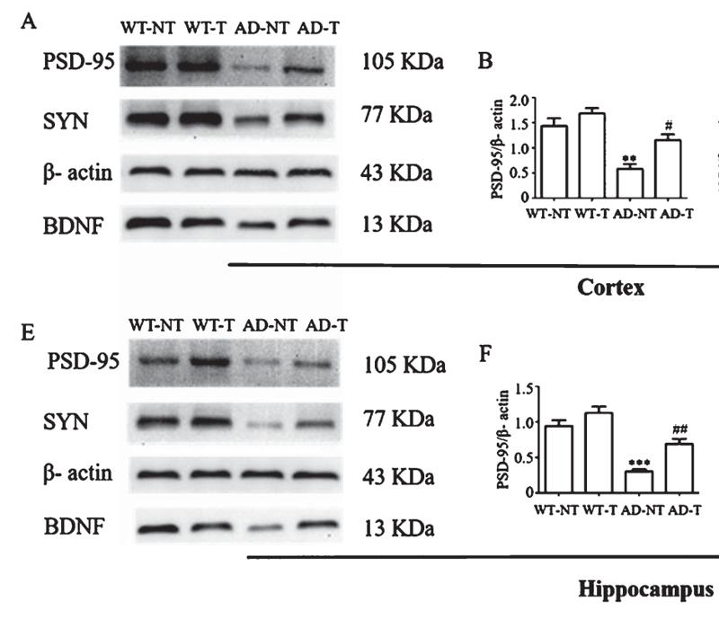
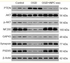
 The bands of synaptic-plasticity proteins of SYN, PSD-95, and GAP-43 in the hippocampus by WB. Statistical results indicate the relative protein levels expressed by SYN, GAP-43, and PSD-95. (B) The bands of synaptic-plasticity proteins of SYN, PSD-95, and GAP-43 in cortex by WB. Statistical results indicate the relative protein levels expressed by SYN, GAP-43, and PSD-95. Statistical analyses are performed by two-way ANOVA followed by t-test. Data are presented as mean ± SEM, *p < 0.05, **p < 0.01, n = 3 per group.")
 Quantified protein levels of cell cycle arrest-related proteins of p53, p21, p16/p14, and Rb in mouse hippocampus tissues. Data are shown as the mean ± SD, n = 3 per group. Significance was determined by one-way ANOVA, followed by the LSD post hoc test or Dunnett’s T3 test for the comparison of multiple groups; *p < 0.05, **p < 0.01, and ***p < 0.001. (C,D) Quantified protein levels of the DNA damage-related protein ATM and the cognitive-related proteins synapsin I, synaptophysin, and PSD95 in mouse hippocampus tissues. Data are shown as the mean ± SD, n = 3 per group. Significance was determined by one-way ANOVA, followed by the LSD post hoc test or Dunnett’s T3 test for the comparison of multiple groups; *p < 0.05, **p < 0.01, and ***p < 0.001. (E,F) Quantified protein levels of the cell senescence-related proteins CREB, p-CREB, ERK, p-ERK, AKT, and p-AKT in mouse hippocampus tissues. Data are shown as the mean ± SD, n = 3 per group. Significance was determined by one-way ANOVA, followed by the LSD post hoc test or Dunnett’s T3 test for the comparison of multiple groups; *p < 0.05, **p < 0.01, and ***p < 0.001. (G,H) Quantified levels of the c-H2AX and TP53BP1 proteins in bone marrow mesenchymal stem cells. Data are shown as the mean ± SD, n = 3 per group. Significance was determined by one-way ANOVA, followed by the LSD post hoc test or Dunnett’s T3 test for the comparison of multiple groups; *p < 0.05, **p < 0.01, and ***p < 0.001.")
 Western blot analysis of PSD95 (postsynaptic density protein 95), an essential postsynaptic scaffold protein that decreased in the DM group. (B) Western blot analysis of SYT1 (synaptotagmin 1), the primary regulator of synaptic transmission that functions as an integrin in the synaptic vesicular membrane; its expression decreased in DM rats (*P < 0.05, ***P < 0.001, and ****P < 0.0001).")
. B Quantification of NeuN-positive area in A. C Western blot analysis of hippocampal Syn and PSD-95 expression levels. D Quantification of protein expression levels from C. E Representative HE and Nissl staining in the CA1 region (scale bar, 50 μm). F Representative Golgi staining of hippocampal neurons. G Quantification of dendritic spine density per 10 μm from F. Data are presented as mean ± SEM (n = 5 per group for IHC and WB; n = 10 per group for Golgi staining). Shapiro–Wilk and Bartlett’s tests were used to assess normality and variance homogeneity. All data were analyzed using one-way ANOVA with Tukey’s post hoc test. *P")
| Product: | PSD95 Antibody |
| Catalog: | AF5283 |
| Description: | Rabbit polyclonal antibody to PSD95 |
| Application: | WB IHC |
| Cited expt.: | WB, IHC |
| Reactivity: | Human, Mouse, Rat |
| Prediction: | Zebrafish, Bovine, Horse, Sheep, Rabbit |
| Mol.Wt.: | 105 kDa(Observed); 80kD(Calculated). |
| Uniprot: | P78352 |
| RRID: | AB_2827690 |
Control Products

Related Downloads
Protocols
Product Info
*The optimal dilutions should be determined by the end user. For optimal experimental results, antibody reuse is not recommended.
*Tips:
WB: For western blot detection of denatured protein samples. IHC: For immunohistochemical detection of paraffin sections (IHC-p) or frozen sections (IHC-f) of tissue samples. IF/ICC: For immunofluorescence detection of cell samples. ELISA(peptide): For ELISA detection of antigenic peptide.
Cite Format: Affinity Biosciences Cat# AF5283, RRID:AB_2827690.
Fold/Unfold
Discs large homolog 4; Disks large homolog 4; DLG 4; Dlg4; DLG4_HUMAN; FLJ97752; FLJ98574; Human post synaptic density protein 95; Post synaptic density protein 95; Postsynaptic density protein 95; PSD 95; PSD-95; PSD95; SAP 90; SAP-90; SAP90; Synapse associated protein 90; Synapse-associated protein 90; Tax interaction protein 15;
Immunogens
A synthesized peptide derived from human PSD95, corresponding to a region within C-terminal amino acids.
- P78352 DLG4_HUMAN:
- Protein BLAST With
- NCBI/
- ExPASy/
- Uniprot
MDCLCIVTTKKYRYQDEDTPPLEHSPAHLPNQANSPPVIVNTDTLEAPGYELQVNGTEGEMEYEEITLERGNSGLGFSIAGGTDNPHIGDDPSIFITKIIPGGAAAQDGRLRVNDSILFVNEVDVREVTHSAAVEALKEAGSIVRLYVMRRKPPAEKVMEIKLIKGPKGLGFSIAGGVGNQHIPGDNSIYVTKIIEGGAAHKDGRLQIGDKILAVNSVGLEDVMHEDAVAALKNTYDVVYLKVAKPSNAYLSDSYAPPDITTSYSQHLDNEISHSSYLGTDYPTAMTPTSPRRYSPVAKDLLGEEDIPREPRRIVIHRGSTGLGFNIVGGEDGEGIFISFILAGGPADLSGELRKGDQILSVNGVDLRNASHEQAAIALKNAGQTVTIIAQYKPEEYSRFEAKIHDLREQLMNSSLGSGTASLRSNPKRGFYIRALFDYDKTKDCGFLSQALSFRFGDVLHVIDASDEEWWQARRVHSDSETDDIGFIPSKRRVERREWSRLKAKDWGSSSGSQGREDSVLSYETVTQMEVHYARPIIILGPTKDRANDDLLSEFPDKFGSCVPHTTRPKREYEIDGRDYHFVSSREKMEKDIQAHKFIEAGQYNSHLYGTSVQSVREVAEQGKHCILDVSANAVRRLQAAHLHPIAIFIRPRSLENVLEINKRITEEQARKAFDRATKLEQEFTECFSAIVEGDSFEEIYHKVKRVIEDLSGPYIWVPARERL
Predictions
Score>80(red) has high confidence and is suggested to be used for WB detection. *The prediction model is mainly based on the alignment of immunogen sequences, the results are for reference only, not as the basis of quality assurance.
High(score>80) Medium(80>score>50) Low(score<50) No confidence
Research Backgrounds
Interacts with the cytoplasmic tail of NMDA receptor subunits and shaker-type potassium channels. Required for synaptic plasticity associated with NMDA receptor signaling. Overexpression or depletion of DLG4 changes the ratio of excitatory to inhibitory synapses in hippocampal neurons. May reduce the amplitude of ASIC3 acid-evoked currents by retaining the channel intracellularly. May regulate the intracellular trafficking of ADR1B. Also regulates AMPA-type glutamate receptor (AMPAR) immobilization at postsynaptic density keeping the channels in an activated state in the presence of glutamate and preventing synaptic depression.
Palmitoylated. Palmitoylation is required for targeting to postsynaptic density, plasma membrane and synapses (By similarity). Palmitoylation may play a role in glutamate receptor GRIA1 synapse clustering (By similarity). Depalmitoylated by ABHD17A and ABHD17B and to a lesser extent by ABHD17C, ABHD12, ABHD13, LYPLA1 and LYPLA2. Undergoes rapid synaptic palmitoylation/depalmitoylation cycles during neuronal development which slow down in mature neurons (By similarity).
Ubiquitinated by MDM2 in response to NMDA receptor activation, leading to proteasome-mediated degradation of DLG4 which is required for AMPA receptor endocytosis.
Cell membrane>Lipid-anchor>Cytoplasmic side. Cell junction>Synapse>Postsynaptic density. Cell junction>Synapse. Cytoplasm. Cell projection>Axon. Cell projection>Dendritic spine. Cell projection>Dendrite. Cell junction>Synapse>Presynapse.
Note: High levels in postsynaptic density of neurons in the forebrain. Also in presynaptic region of inhibitory synapses formed by cerebellar basket cells on axon hillocks of Purkinje cells.
Brain.
The PDZ domain 3 mediates interaction with ADR1B.
The L27 domain near the N-terminus of isoform 2 is required for HGS/HRS-dependent targeting to postsynaptic density.
Belongs to the MAGUK family.
Research Fields
· Environmental Information Processing > Signal transduction > Hippo signaling pathway. (View pathway)
· Human Diseases > Neurodegenerative diseases > Huntington's disease.
· Human Diseases > Substance dependence > Cocaine addiction.
· Organismal Systems > Nervous system > Glutamatergic synapse.
References

Application: WB Species: Mouse Sample:
Application: WB Species: rat Sample: HIP

Application: WB Species: Mouse Sample:

Application: WB Species: Mouse Sample:
Application: WB Species: Rat Sample: neural progenitor cells
Restrictive clause
Affinity Biosciences tests all products strictly. Citations are provided as a resource for additional applications that have not been validated by Affinity Biosciences. Please choose the appropriate format for each application and consult Materials and Methods sections for additional details about the use of any product in these publications.
For Research Use Only.
Not for use in diagnostic or therapeutic procedures. Not for resale. Not for distribution without written consent. Affinity Biosciences will not be held responsible for patent infringement or other violations that may occur with the use of our products. Affinity Biosciences, Affinity Biosciences Logo and all other trademarks are the property of Affinity Biosciences LTD.