, using Glut 4 Antibody at 1/1000 dilution.")
, using Glut 4 Antibody at 1/1000 dilution.
5ug/NC membrane strip.
Exposure for 10s with Affinity™ ECL Kit(#KF8003).
Bands result from membrane strip incubation.")
 3T3-L1 preadipocytes were successfully differentiated into adipocytes. (B) Expression levels of CYP2E1 protein and mRNA were reduced after treatment with PAG-EA and PAF-EA for 48 h in E47 cells. (C) PAG-EA and PAF-EA significantly increased the GLUT4 protein and mRNA expression after 48 h. The CYP2E1 and GLUT4 mRNA values are represented as a fold change. *p < 0.05 and **p < 0.01 represent statistical significance against the control.")
 The wild-type C57BL/6 mice were assigned into exercise and sedentary groups,
(C–I) and then intraperitoneally injected with 0.5 mg/kg MOTS-c or saline daily for eight weeks. (C, D) The skeletal muscle and plasma concentrations of MOTS-c
were detected by ELISA kits after normalizing to total protein content. (E-I) Protein expression of PGC-1α and GLUT4 as well as phosphorylation levels of AMPK and
ACC in mouse muscle (n = 6–8 in each group) were determined by immunoblotting. Each bar represents mean ± SD of triplicates. *P < 0.05, **P < 0.01, remarkably
different from control mice.")
. (a) GLUT4 expression in scWAT. (b) Representative western blots of GLUT4 expression in scWAT. (c) GLUT4 expression in iBAT. (d) Representative western blots of GLUT4 expression in iBAT. (e) The expression of GLUT4 in PM of the scWAT. (f) Representative western blots of PM GLUT4 expression in scWAT. (g) The expression of GLUT4 in PM of the iBAT. (h) Representative western blots of PM GLUT4 expression in iBAT. The corresponding control levels are arbitrarily assigned at a value of 1. The values are expressed as mea±SD (n = 6). For statistical significance, *p<0.05 shows the comparison to the BL group; #p<0.05 shows the comparison to the OLZ group.")
 The expression levels of GLUT3 and GLUT4 proteins were assessed by WB (n = 6). (d, e) Glucose uptake assay was performed by using 2-NBDG (n = 6, scale bar = 10 μm). (f, g) Immunofluorescence was used to detect the level of GLUT3 (cytoskeleton was labeled with β-tubulin) (n = 6, scale bar = 50 μm). (h, i) The expression of GLUT4 was detected by immunofluorescence (cytoskeleton was labeled with phalloidin) (n = 6, scale bar = 50 μm). Data are represented as the means ± SD. ∗p < 0.05, ∗∗p < 0.01, and ∗∗∗p < 0.001.")
 Insulin receptor substrate 1 (IRS-1), (B) the phosphorylated Akt (p-Akt) /total Akt, (C) the phosphorylated mTOR/total mTOR, (D) Glucose transporter type 4 (GLUT4). The total protein expressions are normalized to β-actin and presented as the fold differences compared to the YC group which was set as one (n = 3). (E) Muscular glucose uptake activity and (F) glycogen content. Data are expressed as means ± SD for each group. The letters were significantly different compared with the YC group (a p < 0.05, aa p < 0.01, and aaa p < 0.001), MC groups (b p < 0.05, bb p < 0.01, and bbb p < 0.001), CURSD groups (c p < 0.05, and ccc p < 0.001). # is significantly different between without insulin vs Insulin.")
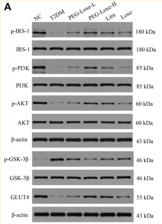
 Representative bands of IRS-1, p-PI3K, PI3K, p-AKT, AKT and GLUT4 were shown.")
 Representative micrographs of heart sections from the ischemic border zones of the three groups. (B,D) Protein expressional levels of GLUT4 and PFKFB3 in the Sham, MIC and MIE groups. n = 4 rats per group. ***P")
| Product: | Glut 4 Antibody |
| Catalog: | AF5386 |
| Description: | Rabbit polyclonal antibody to Glut 4 |
| Application: | WB IHC |
| Cited expt.: | WB, IHC |
| Reactivity: | Human, Mouse, Rat |
| Prediction: | Bovine, Horse, Sheep, Rabbit |
| Mol.Wt.: | 54 kDa(Observed); 55kD(Calculated). |
| Uniprot: | P14672 |
| RRID: | AB_2837871 |
Control Products

Related Downloads
Protocols
Product Info
*The optimal dilutions should be determined by the end user. For optimal experimental results, antibody reuse is not recommended.
*Tips:
WB: For western blot detection of denatured protein samples. IHC: For immunohistochemical detection of paraffin sections (IHC-p) or frozen sections (IHC-f) of tissue samples. IF/ICC: For immunofluorescence detection of cell samples. ELISA(peptide): For ELISA detection of antigenic peptide.
Cite Format: Affinity Biosciences Cat# AF5386, RRID:AB_2837871.
Fold/Unfold
insulin-responsive; Glucose transporter GLUT 4; Glucose transporter type 4; Glucose transporter type 4 insulin responsive; GLUT 4; GLUT-4; GLUT4; GTR4_HUMAN; Insulin responsive glucose transporter type 4; kug; SLC 2A4; SLC2A4; solute carrier family 2 (facilitated glucose transporter) member 4; Solute carrier family 2 member 4; Solute carrier family 2, facilitated glucose transporter member 4;
Immunogens
A synthesized peptide derived from human Glut 4, corresponding to a region within the internal amino acids.
- P14672 GLUT4_HUMAN:
- Protein BLAST With
- NCBI/
- ExPASy/
- Uniprot
MPSGFQQIGSEDGEPPQQRVTGTLVLAVFSAVLGSLQFGYNIGVINAPQKVIEQSYNETWLGRQGPEGPSSIPPGTLTTLWALSVAIFSVGGMISSFLIGIISQWLGRKRAMLVNNVLAVLGGSLMGLANAAASYEMLILGRFLIGAYSGLTSGLVPMYVGEIAPTHLRGALGTLNQLAIVIGILIAQVLGLESLLGTASLWPLLLGLTVLPALLQLVLLPFCPESPRYLYIIQNLEGPARKSLKRLTGWADVSGVLAELKDEKRKLERERPLSLLQLLGSRTHRQPLIIAVVLQLSQQLSGINAVFYYSTSIFETAGVGQPAYATIGAGVVNTVFTLVSVLLVERAGRRTLHLLGLAGMCGCAILMTVALLLLERVPAMSYVSIVAIFGFVAFFEIGPGPIPWFIVAELFSQGPRPAAMAVAGFSNWTSNFIIGMGFQYVAEAMGPYVFLLFAVLLLGFFIFTFLRVPETRGRTFDQISAAFHRTPSLLEQEVKPSTELEYLGPDEND
Predictions
Score>80(red) has high confidence and is suggested to be used for WB detection. *The prediction model is mainly based on the alignment of immunogen sequences, the results are for reference only, not as the basis of quality assurance.
High(score>80) Medium(80>score>50) Low(score<50) No confidence
Research Backgrounds
Insulin-regulated facilitative glucose transporter, which plays a key role in removal of glucose from circulation. Response to insulin is regulated by its intracellular localization: in the absence of insulin, it is efficiently retained intracellularly within storage compartments in muscle and fat cells. Upon insulin stimulation, translocates from these compartments to the cell surface where it transports glucose from the extracellular milieu into the cell.
Sumoylated.
Cell membrane>Multi-pass membrane protein. Endomembrane system>Multi-pass membrane protein. Cytoplasm>Perinuclear region.
Note: Localizes primarily to the perinuclear region, undergoing continued recycling to the plasma membrane where it is rapidly reinternalized (PubMed:8300557). The dileucine internalization motif is critical for intracellular sequestration (PubMed:8300557). Insulin stimulation induces translocation to the cell membrane (By similarity).
Skeletal and cardiac muscles; brown and white fat.
The dileucine internalization motif is critical for intracellular sequestration.
Belongs to the major facilitator superfamily. Sugar transporter (TC 2.A.1.1) family. Glucose transporter subfamily.
Research Fields
· Environmental Information Processing > Signal transduction > FoxO signaling pathway. (View pathway)
· Environmental Information Processing > Signal transduction > AMPK signaling pathway. (View pathway)
· Human Diseases > Endocrine and metabolic diseases > Type II diabetes mellitus.
· Human Diseases > Endocrine and metabolic diseases > Insulin resistance.
· Organismal Systems > Endocrine system > Insulin signaling pathway. (View pathway)
· Organismal Systems > Endocrine system > Adipocytokine signaling pathway.
References

Application: WB Species: Mouse Sample:

Application: WB Species: Mouse Sample:

Application: WB Species: Mice Sample: hippocampal homogenates

Application: WB Species: Rat Sample: PC12 cells
Application: WB Species: Mice Sample: epididymal white adipose tissue

Application: WB Species: Rat Sample:
Application: WB Species: Mouse Sample:

Application: WB Species: Mouse Sample: TCMK-1 cells
Restrictive clause
Affinity Biosciences tests all products strictly. Citations are provided as a resource for additional applications that have not been validated by Affinity Biosciences. Please choose the appropriate format for each application and consult Materials and Methods sections for additional details about the use of any product in these publications.
For Research Use Only.
Not for use in diagnostic or therapeutic procedures. Not for resale. Not for distribution without written consent. Affinity Biosciences will not be held responsible for patent infringement or other violations that may occur with the use of our products. Affinity Biosciences, Affinity Biosciences Logo and all other trademarks are the property of Affinity Biosciences LTD.