


 by IF/ICC. The samples were fixed with PFA and permeabilized in 0.1% Triton X-100,then blocked in 10% serum for 45 minutes at 25°C. Samples were then incubated with primary Ab(AF5388) and mouse anti-beta tubulin Ab(T0023) for 1 hour at 37°C. An AlexaFluor594 conjugated goat anti-rabbit IgG(H+L) Ab(Red) and an AlexaFluor488 conjugated goat anti-mouse IgG(H+L) Ab(Green) were used as the secondary antibody.
The nuclear counter stain is DAPI(blue).")
. Expression of self-renewal and apoptosis-associated proteins were evaluated by western blotting (B). Representative of three independent experiments. Relative protein levels were quantified by densitometry and expressed as optical density ratio with GAPDH serving as internal standards (C). *, P<0.05, **, P<0.01 vs. control.")
| Product: | Nanog Antibody |
| Catalog: | AF5388 |
| Description: | Rabbit polyclonal antibody to Nanog |
| Application: | WB IHC IF/ICC |
| Cited expt.: | WB, IHC |
| Reactivity: | Human, Mouse, Rat |
| Prediction: | Bovine, Horse, Sheep, Rabbit |
| Mol.Wt.: | 35 kDa(Observed); 35kD(Calculated). |
| Uniprot: | Q9H9S0 |
| RRID: | AB_2814953 |
Control Products

Product Info
*The optimal dilutions should be determined by the end user. For optimal experimental results, antibody reuse is not recommended.
*Tips:
WB: For western blot detection of denatured protein samples. IHC: For immunohistochemical detection of paraffin sections (IHC-p) or frozen sections (IHC-f) of tissue samples. IF/ICC: For immunofluorescence detection of cell samples. ELISA(peptide): For ELISA detection of antigenic peptide.
Cite Format: Affinity Biosciences Cat# AF5388, RRID:AB_2814953.
Fold/Unfold
Embryonic stem cell specific homeobox protein (Nanog); ENK; FLJ12581; hNanog; Homeobox protein NANOG; Homeobox transcription factor Nanog; homeobox transcription factor Nanog-delta 48; NANOG; Nanog homeobox; NANOG_HUMAN;
Immunogens
A synthesized peptide derived from human Nanog, corresponding to a region within the internal amino acids.
Expressed in testicular carcinoma and derived germ cell tumors (at protein level). Expressed in fetal gonads, ovary and testis. Also expressed in ovary teratocarcinoma cell line and testicular embryonic carcinoma. Not expressed in many somatic organs and oocytes.
- Q9H9S0 NANOG_HUMAN:
- Protein BLAST With
- NCBI/
- ExPASy/
- Uniprot
MSVDPACPQSLPCFEASDCKESSPMPVICGPEENYPSLQMSSAEMPHTETVSPLPSSMDLLIQDSPDSSTSPKGKQPTSAEKSVAKKEDKVPVKKQKTRTVFSSTQLCVLNDRFQRQKYLSLQQMQELSNILNLSYKQVKTWFQNQRMKSKRWQKNNWPKNSNGVTQKASAPTYPSLYSSYHQGCLVNPTGNLPMWSNQTWNNSTWSNQTQNIQSWSNHSWNTQTWCTQSWNNQAWNSPFYNCGEESLQSCMQFQPNSPASDLEAALEAAGEGLNVIQQTTRYFSTPQTMDLFLNYSMNMQPEDV
Predictions
Score>80(red) has high confidence and is suggested to be used for WB detection. *The prediction model is mainly based on the alignment of immunogen sequences, the results are for reference only, not as the basis of quality assurance.
High(score>80) Medium(80>score>50) Low(score<50) No confidence
Research Backgrounds
Transcription regulator involved in inner cell mass and embryonic stem (ES) cells proliferation and self-renewal. Imposes pluripotency on ES cells and prevents their differentiation towards extraembryonic endoderm and trophectoderm lineages. Blocks bone morphogenetic protein-induced mesoderm differentiation of ES cells by physically interacting with SMAD1 and interfering with the recruitment of coactivators to the active SMAD transcriptional complexes. Acts as a transcriptional activator or repressor. Binds optimally to the DNA consensus sequence 5'-TAAT[GT][GT]-3' or 5'-[CG][GA][CG]C[GC]ATTAN[GC]-3'. Binds to the POU5F1/OCT4 promoter. Able to autorepress its expression in differentiating (ES) cells: binds to its own promoter following interaction with ZNF281/ZFP281, leading to recruitment of the NuRD complex and subsequent repression of expression. When overexpressed, promotes cells to enter into S phase and proliferation.
Nucleus.
Expressed in testicular carcinoma and derived germ cell tumors (at protein level). Expressed in fetal gonads, ovary and testis. Also expressed in ovary teratocarcinoma cell line and testicular embryonic carcinoma. Not expressed in many somatic organs and oocytes.
Belongs to the Nanog homeobox family.
Research Fields
· Cellular Processes > Cellular community - eukaryotes > Signaling pathways regulating pluripotency of stem cells. (View pathway)
· Human Diseases > Cancers: Overview > Proteoglycans in cancer.
References
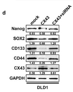
Application: WB Species: Human Sample: CRC cells

Application: WB Species: human Sample: OVCAR8 cells
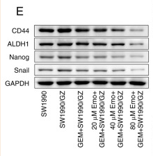
Application: WB Species: Human Sample: SW1990/GZ cells
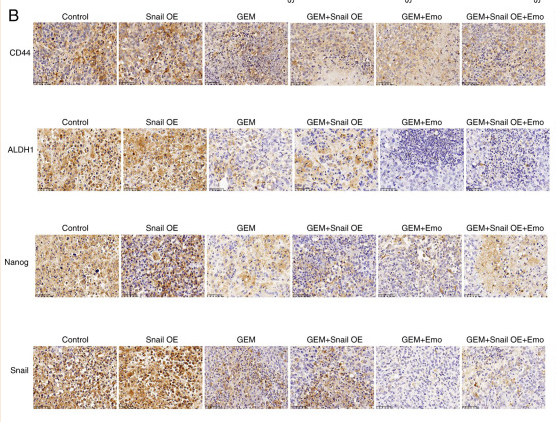
Application: IHC Species: Human Sample: SW1990/GZ cells
Restrictive clause
Affinity Biosciences tests all products strictly. Citations are provided as a resource for additional applications that have not been validated by Affinity Biosciences. Please choose the appropriate format for each application and consult Materials and Methods sections for additional details about the use of any product in these publications.
For Research Use Only.
Not for use in diagnostic or therapeutic procedures. Not for resale. Not for distribution without written consent. Affinity Biosciences will not be held responsible for patent infringement or other violations that may occur with the use of our products. Affinity Biosciences, Affinity Biosciences Logo and all other trademarks are the property of Affinity Biosciences LTD.