

 and week 4 (n = 6).")
 mRNA levels of SOX-2, OCT4, and ALDH1 were determined by qRT–PCR using specific primers, with GAPDH serving as the internal control. (D, E) mRNA levels of HIF-1α and COX-2 were determined by qRT–PCR using specific primers, with GAPDH serving as the internal control. (F) The concentration of PGE2 secreted by A549 cells was determined by ELISA. Each bar indicates the mean ± SD of n = 3 experiments. * indicates P < 0.05, ** indicates P < 0.01. (G) SOX-2, OCT4, ALDH1, HIF-1α, and COX-2 protein levels were determined by Western blot, with GAPDH serving as the loading control.")
.")
 The effect of melatonin on BMSC proliferation measured by CCK-8 assays. (B, C) Images and quantification of ALP activity after 7 days of osteogenic induction (scale bars, 200 μm). (D, E) Calcium mineralization was assessed via ARS staining and quantification (scale bars, 200 μm). (F) mRNA expression levels of osteogenesis-related markers in BMSCs following treatment with/without melatonin. (G, H) Protein expression levels of osteogenesis-related markers (OCN, RUNX2, and ALP). (I, J) Immunofluorescent images of BMSCs stained for ALP and OCN (scale bars, 100 μm). All the experiments were repeated at least 3 times independently. CCK-8, cell counting kit-8; BMSCs, bone marrow mesenchymal stem cells; ARS, alizarin red S; ALP, alkaline phosphatase; OCN, osteocalcin; RUNX2, runt-related transcription factor 2. The data are presented as means ± SEM. * p < 0.05, ** p < 0.01 vs. control group, # p < 0.05, ## p < 0.01 vs. 100 nM melatonin group.")
 The effect of melatonin on BMSC proliferation measured by CCK-8 assays. (B, C) Images and quantification of ALP activity after 7 days of osteogenic induction (scale bars, 200 μm). (D, E) Calcium mineralization was assessed via ARS staining and quantification (scale bars, 200 μm). (F) mRNA expression levels of osteogenesis-related markers in BMSCs following treatment with/without melatonin. (G, H) Protein expression levels of osteogenesis-related markers (OCN, RUNX2, and ALP). (I, J) Immunofluorescent images of BMSCs stained for ALP and OCN (scale bars, 100 μm). All the experiments were repeated at least 3 times independently. CCK-8, cell counting kit-8; BMSCs, bone marrow mesenchymal stem cells; ARS, alizarin red S; ALP, alkaline phosphatase; OCN, osteocalcin; RUNX2, runt-related transcription factor 2. The data are presented as means ± SEM. * p < 0.05, ** p < 0.01 vs. control group, # p < 0.05, ## p < 0.01 vs. 100 nM melatonin group.")
 ALP staining of rBMSCs treated with different concentrations of DA after 7 days of differentiation. Optical photos (upper photo) and microscopic images (lower photo). (b) ALP activity of rBMSCs treated with different concentrations of DA after 7 days of differentiation. (c) Calcium nodules assessed by Alizarin Red staining after 14-day osteogenic induction. Optical photos (upper photo) and microscopic images (lower photo). (d) Semiquantification of Alizarin Red staining. (e) The effect of DA on osteogenic-related mRNA expression in rBMSCs evaluated by qRT-PCR assay after 7 days of differentiation. (f, g) The expression levels of osteogenic-related proteins (COL1A1, ALP, RUNX2, OPN, and OCN) detected by western blot. (f) and quantified (g) in the indicated groups with 7-day osteogenic induction. (h) Oil Red “O” staining of rBMSCs treated with different concentrations of DA after 14-day adipogenic induction. (i) The effect of DA on adipogenic-related mRNA (Adipoq, Cebp, Fabp4, Pparr, and Plin1) expression in rBMSCs evaluated by qRT-PCR assay after 14-day adipogenic induction. Scale bar, 100 μm. ∗p < 0.05 and ∗∗p < 0.01.")
 enhanced osteogenesis via Erk1/2 MAPK signaling pathway: (A) Schematic representation of the relative linear location in which the FGFR2 mutation is identified as illustrated by the large red arrow shown in the context of the protein structure. (B) CCK-8 assay was carried out to assess cell proliferation. Proliferation of MC3T3-E1 cells was more active in the MT group. (C) Relative expressions of osteogenic marker measured by qPCR. The expressions of Alp, ColIα2, Runx2, Opn and Ocn mRNA in differentiated MC3T3-e1 were remarkably increased in the MT group. Western blot analysis showed that the level of ALP, COLI and RUNX2 were also increased in the MT group. (D) ALP staining, alizarin red staining and quantitative tests. ALP staining showed increased crystal violet-staining cells in the MT group compared with the WT group. Quantitative experiment demonstrated that ALP activity is more active in the MT group. Alizarin red staining and quantitative test showed there were more mineralized nodules and mineral content in the MT group than in the WT group. (E) Western blot analysis demonstrated that the levels of p-FGFR2 and p-Erk1/2 were increased in the MT group. There was no significant change in the expression of key proteins in other downstream pathways. The western blot results of FGF/FGFR2-Erk1/2 was circled by the red frame. p values were significant at * p < 0.05, ** p < 0.01, *** p < 0.001 and **** p < 0.0001.")
 Relative expression of miR-27a-3p in BMSCs from OVX rats compared with Sham rats. (B) Relative expressions of miR-27a-3p in BMSCs on day 5, 7 and 9 after osteogenesis induction compared with preinduction. (C) Relative expressions of miR-27a-3p in BMSCs transfected with miR-27a-3p mimic/inhibitor. (D–F) The protein and mRNA levels of ALP, OCN, OSX and RUNX2 in BMSCs transfected with miR-27a-3p mimic/inhibitor. (G) Relative expression of miR-196b-5p in BMSCs from OVX rats compared with Sham rats. (H) Relative expressions of miR-196b-5p in BMSCs on day 5, 7 and 9 after osteogenesis induction compared with preinduction. (I) Relative expressions of miR-196b-5p in BMSCs transfected with miR-196b-5p mimic/inhibitor. (J–L) The protein and mRNA levels of ALP, OCN, OSX and RUNX2 in BMSCs transfected with miR-196b-5p mimic/inhibitor. The expression levels of miRNA and mRNA were determined by qRT-PCR, and protein expressions were detected by western blot. U6 is used for the normalization of miRNA and β-actin is used for normalization of mRNA. β-actin served as the loading control in the western blot analysis. Data are presented as the mean ± SD, and all experiments were repeated three times. * P < 0.05, ** p < 0.01, *** P < 0.001, ns: not significant.")
 A heatmap identified the differently expressed miRNAs between M2 macrophages and M1 macrophages using GSE 110339 from the Gene Expression Omnibus (GEO) dataset (fold change > 1 or < − 1, Benjamini-Hochberg-corrected p). (B) Expression of the differentially expressed miR-486-5p between M2 macrophages-derived exosomes (M2D-Exos) and monocyte-derived exosomes using GSE97467 from the GEO dataset. (C) The miR-486-5p levels in bone marrow-derived macrophages (BMDMs), M1 macrophages, and M2 macrophages were measured by qRT-PCR analysis. (D) The morphology of M2D-Exos was shown by transmission electron microscopy (TEM). Scale bars, 200 nm. (E) The particle size distribution in purified M2D-Exos determined by nanoparticle tracking analysis (NTA). (F) Laser scanning confocal microscopy analysis of the internalization of PKH26-labelled M2D-Exos by BMMSCs, Scale bars, 50 μm. (G) Overexpression of miR-486-5p was detected in the BMMSCs treated with M2D-Exos by qRT-PCR analysis. (H) qRT-PCR analysis was used following the addition of PBS, M2D-Exosinhibitor-NC (exosomes from M2 macrophages transfected with the NC inhibitor) or M2D-ExosmiR-486-5p inhibitor (exosomes from M2 macrophages transfected with the miR-486-5p inhibitor) to assess miR-486-5p expression in the mimic NC- or miR-486-5p-transfected BMMSCs. (I, J) The expression of osteogenic differentiation proteins and mRNAs were assessed by Western blot and qRT-PCR. (K) An ALP activity assay was performed to analyse ALP activity on days 0, 3, and 7. (L) Alizarin red staining of BMMSCs after different transfections for 21 days. Alkaline phosphatase staining of BMMSCs following different treatments for 14 days. Scale bars, 200 μm. (M) Western blot analysis was used to assess the expression of adipogenic differentiation proteins, including LPL, CEBPα, PPARγ, and CEBPβ. (N) qRT-PCR analysis of AP, LPL, CEBPα, CEBPβ, and PPARγ gene levels; (O, P) Oil red O staining and extraction were performed to detect lipid droplet formation on day 10 of adipogenic differentiation. Scale bars, 200 μm. Data are expressed as the mean ± SEM, *p < 0.05, **p < 0.01, ***p < 0.005.")
 Representative images of AR staining of mice from each group, scale bar = 400 μm and 100 μm respectively. (B) Representative images of von kossa staining of mice from each group, scale bar = 400 μm and 100 μm respectively. (C) Representative images of ALP staining of mice from each group, scale bar = 400 μm and 100 μm respectively. (D) Representative images of p16 staining of mice from each group, scale bar = 400 μm and 100 μm respectively. (E) Quantification of positive area of Alizarin red staining, n = 6. (F) Quantification of positive area of von kossa staining, n = 6. (G) Quantification of the relative expression of ALP, n = 6. (H) Quantification of the relative expression of p16, n = 6.")
 CCK-8 assay detected the viability of osteoblastic precursor cells after treated with Loc14 in gradient concentration (0–20 μM) for 24h. (B) Representative images of ALP staining in control and Loc14 (5 μM)-treated osteoblasts. (C–D) WB analysis of ALP in control and Loc14 (5 μM)-treated osteoblasts at day 7 of osteogenic induction. Full-length gels are presented in Supplementary material 5: Figure S7 (E-G) Representative images and quantitative analysis of double calcein labeled femurs. MAR: Mineral apposition rate, BFR: Bone formation rate. Scale bar = 20 μm. (H) Representative images of femurs stained for TRAP and counterstained with Weigert hematoxylin solution. Red arrow heads indicate osteoblasts. Scale bar = 20 μm. Data were presented as mean ± SD (n = 3 per group, ***p < 0.001). (For interpretation of the references to color in this figure legend, the reader is referred to the Web version of this article.)")
 Relative expression of ALP, RUNX2 mRNA in Saos-2 on days 6 of differentiation after candidate gene knockdown (N = 3). (C–E) Protein expression and quantification of ALP and RUNX2 in Saos-2 on day 6 of differentiation (N = 3). (F) Alizarin red staining of Saos-2 on day 14 of osteogenic differentiation (Scale bar: 200 μm). Values represent the mean ± SD. *: P < 0.05, **: P < 0.01, ***: P < 0.001, ****: P < 0.0001.")
 with or without activin receptor-like kinase 5 (ALK5) kinase inhibitor (10 μM SB-505124). (A) Western blot and protein expression levels of secreted phosphoprotein 1 (SPP1), alkaline phosphatase (ALPL), vascular endothelial growth factor (VEGFA), and type Ⅹ Collagen α1 (COL10A1). (B) mRNA expression levels of SPP1, ALPL, VEGFA, and COL10A1. All experiments were repeated at least thrice. Data are means ± SEM (n = 3 in each group) *p < 0.05; **p < 0.01.")
 Osteogenic differentiation-related markers (ALP, OCN, OPN and RUNX2) were assessed by immunohistochemical staining in infection human clinical tissues and healthy human clinical tissues. (b) Osteogenic differentiation-related markers (ALP, OCN, OPN and RUNX2) were assessed by immunohistochemical staining in infected bone marrow cavity tissues and healthy tissues of rats. (c) Alkaline phosphatase (ALP) staining and alizarin red staining. (d) Semi-quantitative analysis of osteogenic differentiation-related markers (ALP, OCN, OPN and RUNX2) in the human clinical tissues. (e) Semi-quantitative analysis of osteogenic differentiation-related markers (ALP, OCN, OPN and RUNX2) in the bone marrow cavity tissue of rats. (f) Western blot of osteogenesis-related proteins (RUNX2 and ALP). (g, h) Semi-quantitative analysis of ALP staining and Alizarin red staining. n = 3 independent experiments per group,")
 Representative Western blot images of TNAP, IL-10, IL-6, and NF-κB in the injured cerebral cortex. (D–G) Compared with those in the Sham group, the cortex expression levels of TNAP and IL-10 were significantly reduced in the Model group (p < 0.01, n = 6). Compared with those in the Sham group, the levels of IL-6 (p < 0.05, p < 0.01, n = 6) and NF-κB (p < 0.001, p < 0.05, n = 6) were significantly increased in the Model group. (B,C) On day 7 (p < 0.001, r = 0.96, n = 12) and day 35 post-HIE-modeling (p < 0.001, r = 0.91, n = 12), there was a positive correlation in the levels of TNAP between the peripheral plasma and the brain. *Denotes statistical significance.")
 Fluorescence images showing cellular uptake of PKH26-labeled apoEVs and BT-apoEVs by BMSCs (Scale bar = 20 μm). (B) Live/dead staining of BMSCs treated with PBS, apoEVs, and BT-apoEVs (Scale bar = 200 μm). (C) ALP staining of BMSCs after treatment for 7 days (Scale bar = 200 μm). (D) Alizarin Red S staining of the mineralized matrix after treatment for 21 days (Scale bar = 200 μm). (E) In vitro osteogenic-related gene expression of BMSCs measured by qPCR assay (n = 3). (F) Western blotting analyzed the expression of osteogenic markers.")
 The relative expression levels of miR-25-3p in ONFH-BMSCs and control BMSCs were examined by RT-qPCR analysis. (B) The BMSCs were transfected with miR-25-3p mimics or its control mimics NC, and the relative expression levels of miR-25-3p were determined by RT-qPCR analysis. (C) The BMSCs were treated with miR-25-3p inhibitor or its control inhibitor NC, and the relative expression levels of miR-25-3p were determined by RT-qPCR analysis. (D and E) The cell proliferation ability of BMSCs with corresponding treatment was revealed by the cell-cycle analysis, with the cell population of G1, G2, and S cell-cycle phase indicated. #p < 0.05 versus the corresponding NC group for comparing cell percentage in S phase; &p < 0.05 versus the corresponding NC group for comparing cell percentage in G1 phase; n = 3. (F and G) The protein expression levels of osteogenic marker genes in BMSCs of different groups were detected by western blot analysis. The quantitative analysis showed the protein expression levels of ALP, BMP2, RUNX2, and OCN were increased in the miRNA mimics group compared with the mimics NC group and were decreased in the miRNA inhibitor group compared with the inhibitor NC group. (H and I) The osteogenic differentiation ability of BMSCs at the 7th and 14th day after corresponding treatment was evaluated by alizarin red staining. Data were shown as mean ± SD. ∗∗p < 0.01, ∗∗∗p < 0.001, n = 3. Data between two groups were analyzed by Student's t test. Data among multiple groups were analyzed by one-way ANOVA test.")
 and (b) Representative images of immunofluorescence costaining RUNX2 (red) and OSX (green) of the bone tissue in each group of implants after 8 weeks and the quantitative analysis of fluorescence intensity. (c and d) Representative images of immunofluorescence costaining β-catenin (red), ALP (green) and AXIN2 (pink) of the bone tissue in each group of implants after 8 weeks and the quantitative analysis of fluorescence intensity. (e and f) Representative images of immunofluorescence costaining Collagen-I (red), OPN (green) and OCN (pink) of the bone tissue in each group of implants after 8 weeks and the quantitative analysis of fluorescence intensity.")
 Representative images of protein bands. (b-e) TGF-β1, (c) p-ERK1/2 / ERK1/2, (d) p-Smad2/Smad2, (e) pSmad3/Smad3, (f) type II collagen, (g) ALP, (h) TRACP, and (i) BMP7 were measured by western blot. (n=3, means ± SD) **P")
 of rPTX3 treatment. B Cell morphology of MC3T3-E1 after treatment with different doses of rPTX3 (0, 50, 100, 200, 500, and 1000 ng/ml). Scale bar, 100 μm. C WB analysis of osteogenesis markers (ALP, Runx2 and COL1 at day 7, OCN and OPN at day 14) and apoptosis markers (Bcl-2 and Bax at 24 h) in Dex-stimulated MC3T3-E1 treated with or without rPTX3 and related quantification. Actin was used as an internal control. D, E Immunofluorescence staining and quantitative analysis of osteogenesis markers OCN (day 14), Runx2 (day 7). F Cell death/live analysis in Dex stimulated MC3T3-E1 treated with or without rPTX3 and related quantification. G Flow cytometry analysis in Dex stimulated MC3T3-E1 treated with or without rPTX3 and quantification. (Apoptotic cells: Q2 + Q3). H, I ALP staining and ARS staining in Dex stimulated MC3T3-E1 treated with or without rPTX3 and quantification. Control: Standard OIM, n = 3; Dex: Standard OIM co-cultured with dexamethasone (10 μM), n = 3; Dex+rPTX3: Standard OIM co-cultured with dexamethasone (10 μM) and rPTX3 (200 ng/mL), n = 3. Statistical analysis: Dunnett’s post-hoc tests (n = 3 independent experiments). Error bars: standard deviation, SD. The images provided in all figures represent typical examples from each experimental group.")
 Construction of IR BMSCs and validations for diminished osteogenic and migrative potential of IR BMSCs. (a) Among 2, 4, and 6 Gy, notable changes were observed starting from 2 Gy, as determined by ALP activity assay. (b) CCK-8 assay revealed no significant differences in cell proliferation between BMSCs and IR BMSCs (2 Gy radiation). (c) Flow cytometry analysis of apoptosis levels showed no significant difference between IR BMSCs and its control. (d) Expression of ALP, OSX, and RUNX2. (e) The results of ALP, OSX, and RUNX2 expression. (f) Histograms showed the quantification of band intensities. (g) ALP staining results. (h) ARS staining results. (i) Would healing assay showed IR BMSCs’ decreased migrative potential. (j) Transwell assay results. Scalar bar = 100 µm. Data are represented as mean ± SEM, n = 3")
 in the Control group, marked increase in expression (black arrows) in L-MTZ, increased expression (black arrows) in S-MTZ. BMP-2: expression findings between the groups, moderate expression (black arrow) in the Control group, increase in expression (black arrows) in L-MTZ, marked expression (black arrow) in S-MTZ. Runx2: moderate expression (black arrows) in the Control group, increase in expression (black arrows) in L-MTZ, marked expression (black arrows) in S-MTZ. ALP: moderate expression (black arrow) in the Control group, marked increase in expression (black arrows) in L-MTZ, moderate expression (black arrow) in S-MTZ. OCN: moderate expression (black arrow) in the control group, markedly increased expression (black arrow) in L-MTZ, and increased expression (black arrow) in S-MTZ. RANKL: Similar RANKL expressions (black arrows) in (A) Control group, (B) L-MTZ, and (C) S-MTZ, Streptavidin biotin peroxidase method, Scale bars = 50 μm Representative histopathological images among the groups. (A) Marked fibrous tissue, moderate new bone formation (white arrow with a black border), and residual graft materials (RG) in the Control group. (B) Decreased fibrous tissue, moderate residual graft material (RG), and marked new bone formation (white arrow with a black border) in L-MTZ. (C) Decreased fibrous tissue, moderately increased new bone formation (white arrow with a black border), and residual graft materials (RG) in S-MTZ. HE, Scale bars = 200 μm")
 The mRNA levels of Plxnb1 and Plxnb2 in osteoblasts increased after SN incubation, and the anti-Sema4D antibody inhibited the expression. SN, supernatant of γδ T cells. n = 6, *p")
 ALP staining (100 × ); (B) Alizarin red staining (100 × ); (C–I) Relative expression levels of mRNA and protein of ALP, Runx2 and OCN (n = 3), ∗∗∗p < 0.001, ∗∗p < 0.01, ∗p < 0.05. (For interpretation of the references to colour in this figure legend, the reader is referred to the Web version of this article.)")
 qRT-PCR analysis of Odaph mRNA levels in: LV18-NC ameloblasts (LV-NC, transfected with empty vector lentivirus LV18-NC); ODAPH-overexpressing cells (LV-Odaph, transfected with pCMV-ODAPH) after 72-hour transfection. (B) Western blot of ODAPH protein expression in LV-NC and LV-Odaph groups (β-actin as loading control). (C) qRT-PCR analysis of Alpl mRNA levels at day 7 of mineralization induction (DMEM-F12 + 50 μg/mL ascorbic acid, 10 mM/L β-glycerophosphate and 10 nM/L dexamethasone). (D) Representative western blot of ALP protein expression at day 7 of mineralization induction. (E) Densitometric quantification of ALP protein levels normalized to β-actin. (F) ALP staining (BCIP/NBT, purple) in: LV-NC and LV-Odaph groups at days 7 and 14 of mineralization induction. Insets show 10 × magnified views of stained ameloblasts. Scale bars = 300 μm.")

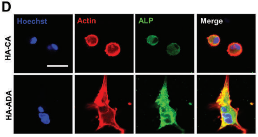
, red (actin), blue (Nucleus/DAPI). d OCN immunofluorescent staining of rBMSC in conditioned medium for 14 days: green (ALP), red (actin), blue (Nucleus/DAPI). e Alizarin Red staining of rBMSC cultured in conditioned medium for 14 days. f Quantitative analysis of Alizarin Red staining. g ALP, BMP-2 and OCN western blotting bands of rBMSC in conditioned medium for 14 days. h–j The expressions of ALP, BMP-2 and OCN proteins were quantitatively analyzed by Image J. (n = 3; *, # and + represent P")
| Product: | Alkaline Phosphatase Antibody |
| Catalog: | DF6225 |
| Description: | Rabbit polyclonal antibody to Alkaline Phosphatase |
| Application: | WB IHC IF/ICC |
| Cited expt.: | WB, IHC, IF/ICC |
| Reactivity: | Human, Mouse, Rat |
| Prediction: | Pig, Bovine, Horse, Sheep, Rabbit, Dog |
| Mol.Wt.: | 57kDa(Observed); 57kD(Calculated). |
| Uniprot: | P05186 |
| RRID: | AB_2838191 |
Control Products

Product Info
*The optimal dilutions should be determined by the end user. For optimal experimental results, antibody reuse is not recommended.
*Tips:
WB: For western blot detection of denatured protein samples. IHC: For immunohistochemical detection of paraffin sections (IHC-p) or frozen sections (IHC-f) of tissue samples. IF/ICC: For immunofluorescence detection of cell samples. ELISA(peptide): For ELISA detection of antigenic peptide.
Cite Format: Affinity Biosciences Cat# DF6225, RRID:AB_2838191.
Fold/Unfold
AKP2; Alkaline phosphatase liver/bone/kidney; Alkaline phosphatase liver/bone/kidney isozyme; Alkaline phosphatase tissue nonspecific isozyme; Alkaline phosphatase, tissue-nonspecific isozyme; Alkaline phosphomonoesterase; Alpl; AP TNAP; AP-TNAP; APTNAP; BAP; FLJ40094; FLJ93059; Glycerophosphatase; HOPS; Liver/bone/kidney type alkaline phosphatase; MGC161443; MGC167935; PHOA; PPBT_HUMAN; Tissue non specific alkaline phosphatase; Tissue nonspecific ALP; TNAP; TNSALP;
Immunogens
A synthesized peptide derived from human ALPL, corresponding to a region within the internal amino acids.
- P05186 PPBT_HUMAN:
- Protein BLAST With
- NCBI/
- ExPASy/
- Uniprot
MISPFLVLAIGTCLTNSLVPEKEKDPKYWRDQAQETLKYALELQKLNTNVAKNVIMFLGDGMGVSTVTAARILKGQLHHNPGEETRLEMDKFPFVALSKTYNTNAQVPDSAGTATAYLCGVKANEGTVGVSAATERSRCNTTQGNEVTSILRWAKDAGKSVGIVTTTRVNHATPSAAYAHSADRDWYSDNEMPPEALSQGCKDIAYQLMHNIRDIDVIMGGGRKYMYPKNKTDVEYESDEKARGTRLDGLDLVDTWKSFKPRYKHSHFIWNRTELLTLDPHNVDYLLGLFEPGDMQYELNRNNVTDPSLSEMVVVAIQILRKNPKGFFLLVEGGRIDHGHHEGKAKQALHEAVEMDRAIGQAGSLTSSEDTLTVVTADHSHVFTFGGYTPRGNSIFGLAPMLSDTDKKPFTAILYGNGPGYKVVGGERENVSMVDYAHNNYQAQSAVPLRHETHGGEDVAVFSKGPMAHLLHGVHEQNYVPHVMAYAACIGANLGHCAPASSAGSLAAGPLLLALALYPLSVLF
Predictions
Score>80(red) has high confidence and is suggested to be used for WB detection. *The prediction model is mainly based on the alignment of immunogen sequences, the results are for reference only, not as the basis of quality assurance.
High(score>80) Medium(80>score>50) Low(score<50) No confidence
Research Backgrounds
This isozyme plays a key role in skeletal mineralization by regulating levels of diphosphate (PPi).
N-glycosylated.
Cell membrane>Lipid-anchor.
Belongs to the alkaline phosphatase family.
Research Fields
· Metabolism > Metabolism of cofactors and vitamins > Thiamine metabolism.
· Metabolism > Metabolism of cofactors and vitamins > Folate biosynthesis.
· Metabolism > Global and overview maps > Metabolic pathways.
References
Application: IF/ICC Species: Human Sample: hMSCs

Application: IHC Species: human Sample:

Application: IF/ICC Species: human Sample:

Application: WB Species: Rat Sample:
Application: WB Species: Rat Sample: BMSCs
Restrictive clause
Affinity Biosciences tests all products strictly. Citations are provided as a resource for additional applications that have not been validated by Affinity Biosciences. Please choose the appropriate format for each application and consult Materials and Methods sections for additional details about the use of any product in these publications.
For Research Use Only.
Not for use in diagnostic or therapeutic procedures. Not for resale. Not for distribution without written consent. Affinity Biosciences will not be held responsible for patent infringement or other violations that may occur with the use of our products. Affinity Biosciences, Affinity Biosciences Logo and all other trademarks are the property of Affinity Biosciences LTD.