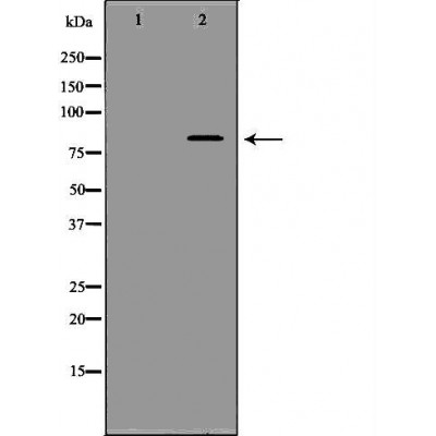
APBB1 Antibody - #DF6708
| Product: | APBB1 Antibody |
| Catalog: | DF6708 |
| Description: | Rabbit polyclonal antibody to APBB1 |
| Application: | WB IHC |
| Reactivity: | Human, Mouse, Rat |
| Prediction: | Bovine, Horse, Rabbit, Dog |
| Mol.Wt.: | 77kDa; 77kD(Calculated). |
| Uniprot: | O00213 |
| RRID: | AB_2838670 |
Related Downloads
Protocols
Product Info
*The optimal dilutions should be determined by the end user. For optimal experimental results, antibody reuse is not recommended.
*Tips:
WB: For western blot detection of denatured protein samples. IHC: For immunohistochemical detection of paraffin sections (IHC-p) or frozen sections (IHC-f) of tissue samples. IF/ICC: For immunofluorescence detection of cell samples. ELISA(peptide): For ELISA detection of antigenic peptide.
Cite Format: Affinity Biosciences Cat# DF6708, RRID:AB_2838670.
Fold/Unfold
Adaptor protein FE65a2; Amyloid beta (A4) precursor protein binding family B member 1; Amyloid beta A4 precursor protein binding family B; Amyloid beta A4 precursor protein binding family B member 1; Amyloid beta A4 precursor protein-binding family B member 1; Amyloid beta precursor protein binding family B member 1; APBB 1; APBB1; APBB1_HUMAN; FE 65; Fe65 protein; Protein Fe65; RIR; stat like protein;
Immunogens
A synthesized peptide derived from human APBB1, corresponding to a region within C-terminal amino acids.
Highly expressed in brain; strongly reduced in post-mortem elderly subjects with Alzheimer disease.
- O00213 APBB1_HUMAN:
- Protein BLAST With
- NCBI/
- ExPASy/
- Uniprot
MSVPSSLSQSAINANSHGGPALSLPLPLHAAHNQLLNAKLQATAVGPKDLRSAMGEGGGPEPGPANAKWLKEGQNQLRRAATAHRDQNRNVTLTLAEEASQEPEMAPLGPKGLIHLYSELELSAHNAANRGLRGPGLIISTQEQGPDEGEEKAAGEAEEEEEDDDDEEEEEDLSSPPGLPEPLESVEAPPRPQALTDGPREHSKSASLLFGMRNSAASDEDSSWATLSQGSPSYGSPEDTDSFWNPNAFETDSDLPAGWMRVQDTSGTYYWHIPTGTTQWEPPGRASPSQGSSPQEESQLTWTGFAHGEGFEDGEFWKDEPSDEAPMELGLKEPEEGTLTFPAQSLSPEPLPQEEEKLPPRNTNPGIKCFAVRSLGWVEMTEEELAPGRSSVAVNNCIRQLSYHKNNLHDPMSGGWGEGKDLLLQLEDETLKLVEPQSQALLHAQPIISIRVWGVGRDSGRERDFAYVARDKLTQMLKCHVFRCEAPAKNIATSLHEICSKIMAERRNARCLVNGLSLDHSKLVDVPFQVEFPAPKNELVQKFQVYYLGNVPVAKPVGVDVINGALESVLSSSSREQWTPSHVSVAPATLTILHQQTEAVLGECRVRFLSFLAVGRDVHTFAFIMAAGPASFCCHMFWCEPNAASLSEAVQAACMLRYQKCLDARSQASTSCLPAPPAESVARRVGWTVRRGVQSLWGSLKPKRLGAHTP
Predictions
Score>80(red) has high confidence and is suggested to be used for WB detection. *The prediction model is mainly based on the alignment of immunogen sequences, the results are for reference only, not as the basis of quality assurance.
High(score>80) Medium(80>score>50) Low(score<50) No confidence
Research Backgrounds
Transcription coregulator that can have both coactivator and corepressor functions. Adapter protein that forms a transcriptionally active complex with the gamma-secretase-derived amyloid precursor protein (APP) intracellular domain. Plays a central role in the response to DNA damage by translocating to the nucleus and inducing apoptosis. May act by specifically recognizing and binding histone H2AX phosphorylated on 'Tyr-142' (H2AXY142ph) at double-strand breaks (DSBs), recruiting other pro-apoptosis factors such as MAPK8/JNK1. Required for histone H4 acetylation at double-strand breaks (DSBs). Its ability to specifically bind modified histones and chromatin modifying enzymes such as KAT5/TIP60, probably explains its transcription activation activity. Functions in association with TSHZ3, SET and HDAC factors as a transcriptional repressor, that inhibits the expression of CASP4. Associates with chromatin in a region surrounding the CASP4 transcriptional start site(s).
Polyubiquitination by RNF157 leads to degradation by the proteasome.
Phosphorylation at Ser-610 by SGK1 promotes its localization to the nucleus (By similarity). Phosphorylated following nuclear translocation. Phosphorylation at Tyr-547 by ABL1 enhances transcriptional activation activity and reduces the affinity for RASD1/DEXRAS1.
Cell membrane. Cytoplasm. Nucleus. Cell projection>Growth cone. Nucleus speckle.
Note: Colocalizes with TSHZ3 in axonal growth cone (By similarity). In normal conditions, it mainly localizes to the cytoplasm, while a small fraction is tethered to the cell membrane via its interaction with APP. Following exposure to DNA damaging agents, it is released from cell membrane and translocates to the nucleus. Nuclear translocation is under the regulation of APP. Colocalizes with TSHZ3 in the nucleus. Colocalizes with NEK6 at the nuclear speckles. Phosphorylation at Ser-610 by SGK1 promotes its localization to the nucleus (By similarity).
Highly expressed in brain; strongly reduced in post-mortem elderly subjects with Alzheimer disease.
Research Fields
· Human Diseases > Neurodegenerative diseases > Alzheimer's disease.
Restrictive clause
Affinity Biosciences tests all products strictly. Citations are provided as a resource for additional applications that have not been validated by Affinity Biosciences. Please choose the appropriate format for each application and consult Materials and Methods sections for additional details about the use of any product in these publications.
For Research Use Only.
Not for use in diagnostic or therapeutic procedures. Not for resale. Not for distribution without written consent. Affinity Biosciences will not be held responsible for patent infringement or other violations that may occur with the use of our products. Affinity Biosciences, Affinity Biosciences Logo and all other trademarks are the property of Affinity Biosciences LTD.

