


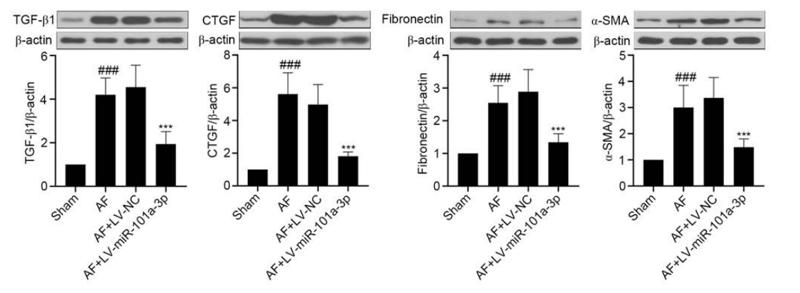
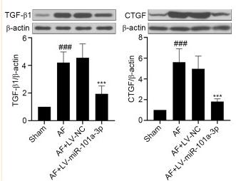
 Western blot analysis for α-smooth muscle actin (α-SMA), collagen I, transforming growth factor (TGF)-β1 and connective tissue growth factor (CTGF). The relative expression of (B) α-SMA, (C) collagen I, (D) TGF-β1 and (E) CTGF. Data were shown as mean value ± SD (n=6). ***P<0.001, versus Control group. &&P<0.01, &&&P<0.001, versus NC agomir group.")
Control Products

Related Downloads
Protocols
Product Info
*The optimal dilutions should be determined by the end user. For optimal experimental results, antibody reuse is not recommended.
*Tips:
WB: For western blot detection of denatured protein samples. IHC: For immunohistochemical detection of paraffin sections (IHC-p) or frozen sections (IHC-f) of tissue samples. IF/ICC: For immunofluorescence detection of cell samples. ELISA(peptide): For ELISA detection of antigenic peptide.
Cite Format: Affinity Biosciences Cat# DF7091, RRID:AB_2839046.
Fold/Unfold
CCN 2; CCN family member 2; CCN2; Connective tissue growth factor; Ctgf; CTGF_HUMAN; Hcs 24; Hcs24; Hypertrophic chondrocyte specific protein 24; Hypertrophic chondrocyte-specific gene product 24; Hypertrophic chondrocyte-specific protein 24; IBP-8; IGF-binding protein 8; IGFBP-8; IGFBP8; Insulin like Growth Factor Binding Protein 8; Insulin-like growth factor-binding protein 8; MGC102839; NOV 2; NOV2;
Immunogens
A synthesized peptide derived from human CTGF, corresponding to a region within the internal amino acids.
Expressed in bone marrow and thymic cells. Also expressed one of two Wilms tumors tested.
- P29279 CCN2_HUMAN:
- Protein BLAST With
- NCBI/
- ExPASy/
- Uniprot
MTAASMGPVRVAFVVLLALCSRPAVGQNCSGPCRCPDEPAPRCPAGVSLVLDGCGCCRVCAKQLGELCTERDPCDPHKGLFCHFGSPANRKIGVCTAKDGAPCIFGGTVYRSGESFQSSCKYQCTCLDGAVGCMPLCSMDVRLPSPDCPFPRRVKLPGKCCEEWVCDEPKDQTVVGPALAAYRLEDTFGPDPTMIRANCLVQTTEWSACSKTCGMGISTRVTNDNASCRLEKQSRLCMVRPCEADLEENIKKGKKCIRTPKISKPIKFELSGCTSMKTYRAKFCGVCTDGRCCTPHRTTTLPVEFKCPDGEVMKKNMMFIKTCACHYNCPGDNDIFESLYYRKMYGDMA
Predictions
Score>80(red) has high confidence and is suggested to be used for WB detection. *The prediction model is mainly based on the alignment of immunogen sequences, the results are for reference only, not as the basis of quality assurance.
High(score>80) Medium(80>score>50) Low(score<50) No confidence
Research Backgrounds
Major connective tissue mitoattractant secreted by vascular endothelial cells. Promotes proliferation and differentiation of chondrocytes. Mediates heparin- and divalent cation-dependent cell adhesion in many cell types including fibroblasts, myofibroblasts, endothelial and epithelial cells. Enhances fibroblast growth factor-induced DNA synthesis.
Secreted>Extracellular space>Extracellular matrix. Secreted.
Expressed in bone marrow and thymic cells. Also expressed one of two Wilms tumors tested.
Belongs to the CCN family.
Research Fields
· Environmental Information Processing > Signal transduction > Apelin signaling pathway. (View pathway)
· Environmental Information Processing > Signal transduction > Hippo signaling pathway. (View pathway)
References

Application: WB Species: rat Sample:

Application: WB Species: Mouse Sample:

Application: WB Species: human Sample:
Application: WB Species: Rat Sample: tumor tissues
Application: WB Species: rat Sample: atrial tissues of heart
Application: WB Species: Rat Sample: atrial tissues
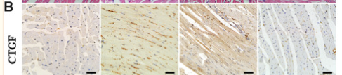
Application: IHC Species: Rat Sample: myocardium
Application: WB Species: Rat Sample: myocardium
Restrictive clause
Affinity Biosciences tests all products strictly. Citations are provided as a resource for additional applications that have not been validated by Affinity Biosciences. Please choose the appropriate format for each application and consult Materials and Methods sections for additional details about the use of any product in these publications.
For Research Use Only.
Not for use in diagnostic or therapeutic procedures. Not for resale. Not for distribution without written consent. Affinity Biosciences will not be held responsible for patent infringement or other violations that may occur with the use of our products. Affinity Biosciences, Affinity Biosciences Logo and all other trademarks are the property of Affinity Biosciences LTD.