 and mouse anti-beta tubulin Ab(T0023 1:200) for 1 hour at 37°C. An AlexaFluor594 conjugated goat anti-rabbit IgG(H+L) Ab(Red) and an AlexaFluor488 conjugated goat anti-mouse IgG(H+L) Ab(Green) were used as the secondary antibody.
The nuclear counter stain is DAPI(blue).")
| Product: | GRM1 Antibody |
| Catalog: | DF7120 |
| Description: | Rabbit polyclonal antibody to GRM1 |
| Application: | WB IHC IF/ICC |
| Cited expt.: | WB |
| Reactivity: | Human, Mouse, Rat |
| Prediction: | Pig, Horse, Sheep, Dog |
| Mol.Wt.: | 132kDa(Observed); 132kD(Calculated). |
| Uniprot: | Q13255 |
| RRID: | AB_2839074 |
Control Products

Related Downloads
Protocols
Product Info
*The optimal dilutions should be determined by the end user. For optimal experimental results, antibody reuse is not recommended.
*Tips:
WB: For western blot detection of denatured protein samples. IHC: For immunohistochemical detection of paraffin sections (IHC-p) or frozen sections (IHC-f) of tissue samples. IF/ICC: For immunofluorescence detection of cell samples. ELISA(peptide): For ELISA detection of antigenic peptide.
Cite Format: Affinity Biosciences Cat# DF7120, RRID:AB_2839074.
Fold/Unfold
Glutamate receptor metabotropic 1; glutamate receptor, metabotropic 1; GPRC1A; GRM1; GRM1-Alpha; GRM1_HUMAN; GRM1A; Metabotropic glutamate receptor 1; mGlu1; mGluR1; MGLUR1-Alpha; MGLUR1A; SCAR13;
Immunogens
A synthesized peptide derived from human GRM1, corresponding to a region within the internal amino acids.
- Q13255 GRM1_HUMAN:
- Protein BLAST With
- NCBI/
- ExPASy/
- Uniprot
MVGLLLFFFPAIFLEVSLLPRSPGRKVLLAGASSQRSVARMDGDVIIGALFSVHHQPPAEKVPERKCGEIREQYGIQRVEAMFHTLDKINADPVLLPNITLGSEIRDSCWHSSVALEQSIEFIRDSLISIRDEKDGINRCLPDGQSLPPGRTKKPIAGVIGPGSSSVAIQVQNLLQLFDIPQIAYSATSIDLSDKTLYKYFLRVVPSDTLQARAMLDIVKRYNWTYVSAVHTEGNYGESGMDAFKELAAQEGLCIAHSDKIYSNAGEKSFDRLLRKLRERLPKARVVVCFCEGMTVRGLLSAMRRLGVVGEFSLIGSDGWADRDEVIEGYEVEANGGITIKLQSPEVRSFDDYFLKLRLDTNTRNPWFPEFWQHRFQCRLPGHLLENPNFKRICTGNESLEENYVQDSKMGFVINAIYAMAHGLQNMHHALCPGHVGLCDAMKPIDGSKLLDFLIKSSFIGVSGEEVWFDEKGDAPGRYDIMNLQYTEANRYDYVHVGTWHEGVLNIDDYKIQMNKSGVVRSVCSEPCLKGQIKVIRKGEVSCCWICTACKENEYVQDEFTCKACDLGWWPNADLTGCEPIPVRYLEWSNIESIIAIAFSCLGILVTLFVTLIFVLYRDTPVVKSSSRELCYIILAGIFLGYVCPFTLIAKPTTTSCYLQRLLVGLSSAMCYSALVTKTNRIARILAGSKKKICTRKPRFMSAWAQVIIASILISVQLTLVVTLIIMEPPMPILSYPSIKEVYLICNTSNLGVVAPLGYNGLLIMSCTYYAFKTRNVPANFNEAKYIAFTMYTTCIIWLAFVPIYFGSNYKIITTCFAVSLSVTVALGCMFTPKMYIIIAKPERNVRSAFTTSDVVRMHVGDGKLPCRSNTFLNIFRRKKAGAGNANSNGKSVSWSEPGGGQVPKGQHMWHRLSVHVKTNETACNQTAVIKPLTKSYQGSGKSLTFSDTSTKTLYNVEEEEDAQPIRFSPPGSPSMVVHRRVPSAATTPPLPSHLTAEETPLFLAEPALPKGLPPPLQQQQQPPPQQKSLMDQLQGVVSNFSTAIPDFHAVLAGPGGPGNGLRSLYPPPPPPQHLQMLPLQLSTFGEELVSPPADDDDDSERFKLLQEYVYEHEREGNTEEDELEEEEEDLQAASKLTPDDSPALTPPSPFRDSVASGSSVPSSPVSESVLCTPPNVSYASVILRDYKQSSSTL
Predictions
Score>80(red) has high confidence and is suggested to be used for WB detection. *The prediction model is mainly based on the alignment of immunogen sequences, the results are for reference only, not as the basis of quality assurance.
High(score>80) Medium(80>score>50) Low(score<50) No confidence
Research Backgrounds
G-protein coupled receptor for glutamate. Ligand binding causes a conformation change that triggers signaling via guanine nucleotide-binding proteins (G proteins) and modulates the activity of down-stream effectors. Signaling activates a phosphatidylinositol-calcium second messenger system. May participate in the central action of glutamate in the CNS, such as long-term potentiation in the hippocampus and long-term depression in the cerebellum. May function in the light response in the retina (By similarity).
Cell membrane>Multi-pass membrane protein.
Detected in brain.
Belongs to the G-protein coupled receptor 3 family.
Research Fields
· Cellular Processes > Cellular community - eukaryotes > Gap junction. (View pathway)
· Environmental Information Processing > Signal transduction > Calcium signaling pathway. (View pathway)
· Environmental Information Processing > Signal transduction > FoxO signaling pathway. (View pathway)
· Environmental Information Processing > Signal transduction > Phospholipase D signaling pathway. (View pathway)
· Environmental Information Processing > Signaling molecules and interaction > Neuroactive ligand-receptor interaction.
· Organismal Systems > Nervous system > Long-term potentiation.
· Organismal Systems > Nervous system > Retrograde endocannabinoid signaling. (View pathway)
· Organismal Systems > Nervous system > Glutamatergic synapse.
· Organismal Systems > Nervous system > Long-term depression.
· Organismal Systems > Sensory system > Taste transduction.
· Organismal Systems > Endocrine system > Estrogen signaling pathway. (View pathway)
References

Application: WB Species: Rat Sample: hippocampus
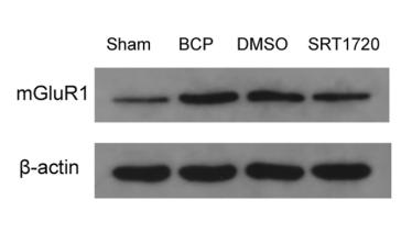
Application: WB Species: mouse Sample: spinal cord
Restrictive clause
Affinity Biosciences tests all products strictly. Citations are provided as a resource for additional applications that have not been validated by Affinity Biosciences. Please choose the appropriate format for each application and consult Materials and Methods sections for additional details about the use of any product in these publications.
For Research Use Only.
Not for use in diagnostic or therapeutic procedures. Not for resale. Not for distribution without written consent. Affinity Biosciences will not be held responsible for patent infringement or other violations that may occur with the use of our products. Affinity Biosciences, Affinity Biosciences Logo and all other trademarks are the property of Affinity Biosciences LTD.