

| Product: | SLC3A2 Antibody |
| Catalog: | DF7468 |
| Description: | Rabbit polyclonal antibody to SLC3A2 |
| Application: | WB IHC |
| Cited expt.: | WB |
| Reactivity: | Human, Rat |
| Prediction: | Pig, Bovine, Horse, Sheep, Rabbit, Dog |
| Mol.Wt.: | 67kDa(Observed); 68kD(Calculated). |
| Uniprot: | P08195 |
| RRID: | AB_2839405 |
Control Products

Related Downloads
Protocols
Product Info
*The optimal dilutions should be determined by the end user. For optimal experimental results, antibody reuse is not recommended.
*Tips:
WB: For western blot detection of denatured protein samples. IHC: For immunohistochemical detection of paraffin sections (IHC-p) or frozen sections (IHC-f) of tissue samples. IF/ICC: For immunofluorescence detection of cell samples. ELISA(peptide): For ELISA detection of antigenic peptide.
Cite Format: Affinity Biosciences Cat# DF7468, RRID:AB_2839405.
Fold/Unfold
4F2; 4F2 cell surface antigen heavy chain; 4F2 cell-surface antigen heavy chain; 4F2 heavy chain; 4F2 heavy chain antigen; 4F2_HUMAN; 4F2hc; 4T2HC; Antigen defined by monoclonal antibody 4F2 heavy chain; Antigen identified by monoclonal antibodies 4F2 TRA1.10 TROP4 and T43; CD 98; CD98; CD98 antigen; CD98 heavy chain; CD98HC; Heavy chain; Lymphocyte activation antigen 4F2 large subunit; MDU 1; MDU1; Monoclonal antibody 44D7; NACAE; Slc3a2; Solute carrier family 3 (activators of dibasic and neutral amino acid transport) member 2; Solute carrier family 3 member 2;
Immunogens
A synthesized peptide derived from human SLC3A2, corresponding to a region within the internal amino acids.
Expressed ubiquitously in all tissues tested with highest levels detected in kidney, placenta and testis and weakest level in thymus. During gestation, expression in the placenta was significantly stronger at full-term than at the mid-trimester stage. Expressed in HUVECS and at low levels in resting peripheral blood T-lymphocytes and quiescent fibroblasts. Also expressed in fetal liver and in the astrocytic process of primary astrocytic gliomas. Expressed in retinal endothelial cells and in the intestinal epithelial cell line C2BBe1.
- P08195 4F2_HUMAN:
- Protein BLAST With
- NCBI/
- ExPASy/
- Uniprot
MELQPPEASIAVVSIPRQLPGSHSEAGVQGLSAGDDSELGSHCVAQTGLELLASGDPLPSASQNAEMIETGSDCVTQAGLQLLASSDPPALASKNAEVTGTMSQDTEVDMKEVELNELEPEKQPMNAASGAAMSLAGAEKNGLVKIKVAEDEAEAAAAAKFTGLSKEELLKVAGSPGWVRTRWALLLLFWLGWLGMLAGAVVIIVRAPRCRELPAQKWWHTGALYRIGDLQAFQGHGAGNLAGLKGRLDYLSSLKVKGLVLGPIHKNQKDDVAQTDLLQIDPNFGSKEDFDSLLQSAKKKSIRVILDLTPNYRGENSWFSTQVDTVATKVKDALEFWLQAGVDGFQVRDIENLKDASSFLAEWQNITKGFSEDRLLIAGTNSSDLQQILSLLESNKDLLLTSSYLSDSGSTGEHTKSLVTQYLNATGNRWCSWSLSQARLLTSFLPAQLLRLYQLMLFTLPGTPVFSYGDEIGLDAAALPGQPMEAPVMLWDESSFPDIPGAVSANMTVKGQSEDPGSLLSLFRRLSDQRSKERSLLHGDFHAFSAGPGLFSYIRHWDQNERFLVVLNFGDVGLSAGLQASDLPASASLPAKADLLLSTQPGREEGSPLELERLKLEPHEGLLLRFPYAA
Predictions
Score>80(red) has high confidence and is suggested to be used for WB detection. *The prediction model is mainly based on the alignment of immunogen sequences, the results are for reference only, not as the basis of quality assurance.
High(score>80) Medium(80>score>50) Low(score<50) No confidence
Research Backgrounds
Component of several heterodimeric amino acid transporter complexes. The precise substrate specificity depends on the other subunit in the heterodimer. The heterodimer with SLC3A2 functions as sodium-independent, high-affinity transporter that mediates uptake of large neutral amino acids such as phenylalanine, tyrosine, L-DOPA, leucine, histidine, methionine and tryptophan. The complexes with SLC7A6 and SLC7A7 mediate uptake of dibasic amino acids. The complexes function as amino acid exchangers. Required for targeting of SLC7A5 and SLC7A8 to the plasma membrane and for channel activity. Plays a role in nitric oxide synthesis in human umbilical vein endothelial cells (HUVECs) via transport of L-arginine. The heterodimer with SLC7A5/LAT1 may play a role in the transport of L-DOPA across the blood-brain barrier (By similarity). May mediate blood-to-retina L-leucine transport across the inner blood-retinal barrier (By similarity). The heterodimer with SLC7A5/LAT1 can mediate the transport of thyroid hormones triiodothyronine (T3) and thyroxine (T4) across the cell membrane. When associated with SLC7A5 or SLC7A8, involved in the cellular activity of small molecular weight nitrosothiols, via the stereoselective transport of L-nitrosocysteine (L-CNSO) across the transmembrane. The heterodimer with SLC7A5 is involved in the uptake of toxic methylmercury (MeHg) when administered as the L-cysteine or D,L-homocysteine complexes. Together with ICAM1, regulates the transport activity SLC7A8 in polarized intestinal cells, by generating and delivering intracellular signals. When associated with LAPTM4B, the heterodimer formed by SLC3A2 and SLC7A5 is recruited to lysosomes to promote leucine uptake into these organelles, and thereby mediates mTORC1 activation.
Phosphorylation on Ser-406; Ser-408 or Ser-410 and on Ser-527 or Ser-531 by ecto-protein kinases favors heterotypic cell-cell interactions.
Apical cell membrane. Cell membrane>Single-pass type II membrane protein. Cell junction. Lysosome membrane. Melanosome.
Note: Identified by mass spectrometry in melanosome fractions from stage I to stage IV (PubMed:17081065). Localized at the plasma membrane when associated with SLC7A5 or SLC7A8 (PubMed:9751058, PubMed:11311135). Localized to the apical membrane of placental syncytiotrophoblastic cells (PubMed:11742812). Recruited to lysosomes by LAPTM4B (PubMed:25998567). Located selectively at cell-cell adhesion sites (By similarity). Colocalized with SLC7A8/LAT2 at the basolateral membrane of kidney proximal tubules and small intestine epithelia. Expressed in both luminal and abluminal membranes of brain capillary endothelial cells (By similarity).
Expressed ubiquitously in all tissues tested with highest levels detected in kidney, placenta and testis and weakest level in thymus. During gestation, expression in the placenta was significantly stronger at full-term than at the mid-trimester stage. Expressed in HUVECS and at low levels in resting peripheral blood T-lymphocytes and quiescent fibroblasts. Also expressed in fetal liver and in the astrocytic process of primary astrocytic gliomas. Expressed in retinal endothelial cells and in the intestinal epithelial cell line C2BBe1.
Belongs to the SLC3A transporter family.
Research Fields
· Cellular Processes > Cell growth and death > Ferroptosis. (View pathway)
· Environmental Information Processing > Signal transduction > mTOR signaling pathway. (View pathway)
· Organismal Systems > Digestive system > Protein digestion and absorption.
References
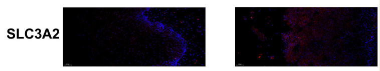
Application: IF/ICC Species: Human Sample: ESCC

Application: IF/ICC Species: human Sample: breast cancer

Application: WB Species: human Sample: LO2 cells
Restrictive clause
Affinity Biosciences tests all products strictly. Citations are provided as a resource for additional applications that have not been validated by Affinity Biosciences. Please choose the appropriate format for each application and consult Materials and Methods sections for additional details about the use of any product in these publications.
For Research Use Only.
Not for use in diagnostic or therapeutic procedures. Not for resale. Not for distribution without written consent. Affinity Biosciences will not be held responsible for patent infringement or other violations that may occur with the use of our products. Affinity Biosciences, Affinity Biosciences Logo and all other trademarks are the property of Affinity Biosciences LTD.