 Antibody.
Lane 1: 293 cells(heat-shock treatment), blocked with antigen-specific peptides,
Lane 2: 293 cells(heat-shock treatment),
Lane 3: A549 cells(serum starvation treatment).")
 by IF/ICC. The samples were fixed with PFA and permeabilized in 0.1% Triton X-100,then blocked in 10% serum for 45 minutes at 25°C. Samples were then incubated with primary Ab(DF7576) and mouse anti-beta tubulin Ab(T0023) for 1 hour at 37°C. An AlexaFluor594 conjugated goat anti-rabbit IgG(H+L) Ab(Red) and an AlexaFluor488 conjugated goat anti-mouse IgG(H+L) Ab(Green) were used as the secondary antibody.
The nuclear counter stain is DAPI(blue).")


 the ER stress markers GRP 78, caspase 12, and CHOP protein expressions; (b–d) the ratios of p-PERK, p-eIF2α/eIF2α, and p-IRE 1/IRE 1 in the left ventricle")
 ISRIB (100 nM), the specific PERKinhibitor, inhibited eIF2 phosphorylation and reversed β-catenin inhibition induced by BA.")
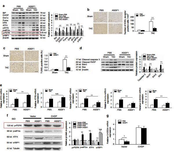
 Western blot analyses of global protein, PERK, p‐PERK, eIf2α, p‐eIf2α, ATF4, GRP78, and CHOP levels in mouse hearts. Data are expressed as mean ± SD, n = 3. One‐way ANOVA was used for evaluating significant differences in the mean values. ATF4, activating transcription factor 4; eIF2α, eukaryotic initiation factor‐2α; ERS, endoplasmic reticulum stress; GAPDH, glyceraldehyde 3‐phosphate dehydrogenase; GRP78, 78‐kDa glucose‐regulated protein; PERK, protein endoplasmic reticulum kinase; SHAM, transverse aortic constriction without clip placement; TAC, transverse aortic constriction; TE, TAC with aerobic exercise; TC, TAC with choline treatment; TEC, TAC with exercise and choline treatment. *p < .05, **p < .01")
. b Nuclear translocation of ATF6. HeLa cells were infected
with NDV or mock-infected for 16 h, subjected to immunostaining with ATF6 and NP antibody. c–e Pharmacological inhibition of UPR suppresses
virus proliferation and reduces apoptosis. HeLa cells were pretreated with 4-PBA (2.5 mM) or PBS (control) for 2 h, infected with NDV, and maintained
with 2.5 mM 4-PBA during infection. Mock infection group without 4-PBA treatment was included as control. Cells were harvested at 16 and 20 h.p.i.,
analyzed with western blot (c) and TUNEL assay (d). In parallel, the culture medium was subjected to TCID50 assay, to measure the released progeny
virus (e). The intensities of indicated protein bands were determined, normalized to eIF2α, IRE1α, or β-actin, respectively, and shown as fold change of
4-PBA (+:−). The protein bands intensities in NDV-infected cells with PBS treatment were set as 1. Western blot, Immunofluorescence, and TUNEL
assay are representative of three independent experiments. Virus titer represents means ± SD of three independent determinations. *p < 0.05.
**p < 0.01.")
 A and B, (b1) A and B) after 72 h culture.
Changes in the TUNEL index in SWFs ((a1) C and D, (b1) C and D) after coculture with GCs. Green: apoptotic cells. BrdU was used to
label the cells ((a1) E and F, (b1) E and F) that were proliferating (red). After coculture of the D580-SYF-GCs with D280-SWFs, ER stress
marker GRP78 ((a1) G and H) was increased in the GCs. After coculture of the D580-SWFs with D280-SYF-GCs, the ER marker GRP78
((b1) G and H) was reduced in both GCs and TCs. Scale bar: 20 μm. Western blot and grey analysis of BCL2, p-PERK, ATF4, caspase12,
ASK1, and GRP78 (a2) expression in D280-SWFs with D580-SYF-GCs in coculture. Western blot and grey analysis of BCL2, p-PERK,
ATF4, caspase12, ASK1, and GRP78 (b2) expression in D580-SWFs with D280-SYF-GCs in coculture. By qRT-PCR analysis, CALR,
PERK, GRP78, and CHOP mRNAs (a3) increased in D280-SWFs (coculture with D580-SYF-GCs). The mRNAs of CALR, PERK, GRP78,
and CHOP (b3) decreased in D580-SWFs (coculture with D280-SYF-GCs). Values were the mean ± SEM of three experiments. Different
lowercase letters indicated significant difference (p < 0:05).")

via suppression of activated ER stress. NRCMs were pretreated with 5 mM 4-PBA (an inhibitor of ERS) or 10 ng/mL tunicamycin (Tm, an ERS
inducer) for 2 h and then exposed to glucose (33 mM) in the presence or absence of ZNS (3 μM) for 24 h. a–b Representative and quantitative
images showing the protein expression of ERS markers, including GRP78, XBP-1s, ATF6, p-PERK, PERK, ATF4, CHOP, and Hrd1.
c Immunofluorescence staining of cardiomyocytes with phalloidin (red) and cell nuclei with DAPI (blue), Scale bar = 50 μm. d Quantitative
analysis of cell surface area by ImageJ software. e–f Representative Western blotting and analysis of Bax and Bcl-2 expression. g–h
Representative and quantitative images of GRP78, ATF6, p-PERK, PERK, ATF4, and CHOP expression. All values are the fold changes normalized
to their control group. The results are presented as the means ± SEM (n = 6). *P < 0.05, **P < 0.01 vs. Con; #P < 0.05, ##P < 0.01 vs. HG; $P < 0.05,
$$P < 0.01 vs. HG + ZNS.")
 in the presence of RANKL (100 ng/mL) for
4 days, and DCFH-DA probe was used to detect intracellular ROS levels. Scale bar = 200 μm. b, c RANKL (100 ng/mL) and NAC were used to stimulate
BMMs, after 4 days, the total proteins were extracted and western blot was conducted to test the expression of PERK and its phosphorylation level. d, e BMMs were treated with NAC and PERK activator CCT020312 in the presence of RANKL (100 ng/mL) for 4 days, total proteins were extracted and
Western Blot was performed to examine the expression of osteoclast-related proteins NFATc1, c-fos and Autophagy-related proteins Beclin1, LC3B. f
The schematic model of the hypothesized mechanism by which PERK inhibition affects osteoclast differentiation. Densitometric analysis of an
immunoblot from three independent experiments; *p < 0.05, **p < 0.01.")
 Gene Set
Enrichment Analysis (GSEA) of UPR/ER stress pathway signatures in the Melatonin (2 mM) plus Lapatinib (1 μM) -treated HCC1954 cells versus Lapatinib alone
treated cells. Drug treatment time: 24 h. NES, FDR q values and p values of the correlation was shown. (B) Heatmap showing the expression of UPR/ER stress pathway
genes in the HCC1954 cells treated with Melatonin and Lapatinib as single-agents or in combination. Row Z-score value is shown. (C) Quantitative RT-PCR analysis of
the expression of ATF4, ATF6, PERKA and IRE1 in HCC1954 and MDA-MB-453 cells subjected to drug treatment as indicated. ACTB was used as an endogenous
control. HCC1954: 2 mM Melatonin, 1 μM Lapatinib; MDA-MB-453: 2 mM Melatonin, 2 μM Lapatinib. (D) Western Blot analysis of the protein levels in the HER2-
positive breast cancer cells subjected to drug treatment as indicated. HCC1954 and MDA-MB-453 cells were treated as in (C); MCF7/HER2: 1 mM Melatonin, 1 μM
Lapatinib. Vinculin was used as a loading control. (E–F) Cell viability was measured by crystal violet assay for the HCC1954 treated with Lapatinib at different
concentrations and with or without BFA (0.1 μM). (E) 96-well format; (F) 24-well format. (G) Western Blot analysis of the protein levels in the HCC1954 cells treated
with Lapatinib (1.0 μM) and BFA (0.1 μM), either alone or in combination. Data was shown as Mean ± S.D. *p < 0.05, **p < 0.01, ***p < 0.001 (Student’s t-test).")
 Tumor tissues from each group were processed for the proteins p‑PERK, PERK, p‑eIF2α, eIF2α, ATF4 and Lcn2 detection.")
 Immunoblots of PERK, p-PERK, eIF2α, p-eIF2α, ATF4 and CHOP.")
 Representative immunoreactive bands of GRP78, PERK, p-PERK, IRE1α, p-IRE1α, and ATF6")
 Representative blots and histograms showing (B) p‑PERK/PERK ratio; (C) p‑eIF2α/eIF2α ratio; (D) ATF4 expression; and (E) CHOP expression.")

 Protein expression of the UPR markers Bip, ATF6, p50-ATF6, p-IRE1α, IRE1α, p-PERK, and PERK was measured after reprogramming by western blotting. β-tubulin expression was used as an equal loading control. Representative blotting shows the protein levels of these proteins (Supplementary Figure 1). (B–H) The expression of Bip, ATF6, p50-ATF6, p-IRE1α, IRE1α, p-PERK, and PERK in WT iPSCs, COL4A5 missense mutant iPSCs, COL4A5 splicing iPSCs and COL4A5 frameshifted mutant iPSCs were quantified by densitometric analysis. The protein levels of ER chaperone protein Bip, three ER transducers (ATF6, IRE1α, and PERK) and their activated forms (p50-ATF6, p-IRE1α, and p-PERK) were similar between the WT and mutant samples examined. Data are means ± SEM. n = 3, (WT, control; MM, missense mutation; SM, splicing mutation; FM, frameshift mutation).")

 Detection of protein expression levels of caspase-3, cleaved-caspase-3, bax and bcl-2 in BEAS-2B and 16-HBE cells by Western blot. (B) Detection of protein expression levels of GRP78, PERK, p-PERK, eIF2α, p-eIF2α, and CHOP in BEAS-2B and 16-HBE cells by Western blot. Results are presented by three independent experiments (n = 3). Values are presented as means ± SD. #P < 0.05, ##P < 0.01 and ###P < 0.001 compared with control group; *P < 0.05, **P < 0.01 and ***P < 0.001 compared with CSE group.")
 and 10 μmol/L MLT (except NC and Ox groups). (a) RT-PCR histograms of mRNA levels of DDIT3, GRP78, ATF6, and XBP1s relative to GAPDH. (b) Western blot and corresponding histograms of DDIT3, GRP78, ATF6, PERK, p-PERK, IRE1, p-IRE1, and XBP1s protein expression relative to GAPDH. Data are presented as the mean ± SEM from three experiments. *P < 0.05, **P < 0.01, ***P < 0.001, ****P < 0.0001 versus the NC group; #P < 0.05, ##P < 0.01, ###P < 0.001, ####P < 0.0001 versus the Ox group")
 Protein expression levels of CHOP, ATF6, p‑PERK, PERK, p‑IRE1,IRE1 and β‑actin in H9C2 cells treated with LPS, TA and TA + LPS, as determined by western blot analysis.")
 Protein expression levels of CHOP, ATF6, p-PERK, PERK, p-IRE1, IRE1 and β-actin in H9C2 cells treated with LPS, TA and TA + LPS, as determined by western blot analysis. Semi-quantification of the protein expression levels of (B) CHOP and ATF6, and (C) p-PERK, PERK and p-IRE1/IRE1 in the LPS, TA and TA + LPS groups. Data are presented as the mean ± SEM (n=3). *P<0.05 and **P<0.01 vs. control group; #P<0.05 vs. LPS group. TA, tannic acid; LPS, lipopolysaccharide; CHOP, C/EBP-homologous protein; ATF6, activating transcription factor 6; p, phosphorylated; PERK, protein kinase-like endoplasmic reticulum kinase; IRE1, inositol-requiring enzyme 1.")

B. Effect of G-1 treatment on CHOP expression in colitis (The figure above was the
original one, and the figure below was the statistical one, n=4, ***P<0.001,
**P<0.01).
C. Effect of G-1 treatment on PERK activity in colitis (The figure above was the
original one, and the figure below was the statistical one, n=4, *P <0.05).
D. Effect of G-1 treatment on IRE1αactivity in colitis (The figure above was the
original one, and the figure below was the statistical one, n=4, **P<0.01, ***P
<0.001).
This article has not been copyedited and formatted. The final version may differ from this version.
JPET Fast Forward. Published on December 14, 2020 as DOI: 10.1124/jpet.120.000216
at ASPET Journals on December 16, 2020 jpet.aspetjournals.org Downloaded from
38
E. Effect of G-1 treatment on ATF6 expression in colitis (The figure above was the
original one, and the figure below was the statistical one, n=4, **P<0.01, ***P
<0.001).
The mice were divided into three groups: control, DSS treatment group, DSS plus G-1
treatment. Data were expressed as mean±SEM. Statistical analyses were performed
using One-Way ANOVA followed by Student-Newman-Keuls Method.")
 ER stress‐related proteins (p‐eIF2α, eIF2α, ATF4 and CHOP) and (B) UPR signal transduction molecules (p‐PERK, PERK, p‐IRE1α, IRE1α and sXBP1) in HepG2 cells after administration of BMS‐303141. ATF4p‐eIF2α, eIF2α were activated 3 h post‐treatment; CHOP was activated 8 h post‐treatment. (* P < .05, ** P < .01 and *** P < .001, compared with control group) (C) Western blot analysis of protein expression after ATF4 knockdown. (D) Annexin V‐FITC/PI double staining was performed to determine the apoptosis rate of HepG2 cells after ATF4 knockdown via flow cytometry. (* P < .05, ** P < .01 and *** P < .001, compared with con siRNA group). All experiments were repeated 3 times")
 Representative blots of SIRT1, GRP78, p‑PERK, p‑eIF2α, CHOP and caspase‑12. Semiquantitative analysis of (B) SIRT1, (C) GRP78, (D) p‑PERK, (E) p‑eIF2α, (F) CHOP and (G) caspase‑12.")
 Protein expression levels of CHOP, ATF6, p-PERK, PERK, p-IRE1, IRE1 and β-actin in H9C2 cells treated with LPS, TA and TA + LPS, as determined by western blot analysis. Semi-quantification of the protein expression levels of (B) CHOP and ATF6, and (C) p-PERK, PERK and p-IRE1/IRE1 in the LPS, TA and TA + LPS groups. Data are presented as the mean ± SEM (n=3). *P<0.05 and **P<0.01 vs. control group; #P<0.05 vs. LPS group. TA, tannic acid; LPS, lipopolysaccharide; CHOP, C/EBP-homologous protein; ATF6, activating transcription factor 6; p, phosphorylated; PERK, protein kinase-like endoplasmic reticulum kinase; IRE1, inositol-requiring enzyme 1.")
 Representative blots of SIRT1, GRP78, p-PERK, p-eIF2α, CHOP and caspase-12. Semiquantitative analysis of (B) SIRT1, (C) GRP78, (D) p-PERK, (E) p-eIF2α, (F) CHOP and (G) caspase-12. (H) The mRNA levels of caspase-12 in the myocardium. Data are presented as the mean ± standard deviation. n=3. ##P<0.01 vs. Sham. *P<0.05 and **P<0.01 vs. MI/R. IOE, Inonotus obliquus extract; SIRT1, NAD-dependent protein deacetylase sirtuin-1; GRP78, glucose-regulated protein 78; PERK, protein kinase R-like endoplasmic reticulum kinase; eIF2α, eukaryotic translation initiation factor 2 subunit α; CHOP, C/EBP homologous protein; p, phosphorylated; MI/R, myocardial ischemia/reperfusion.")

 The effect of PPRV on target proteins was analyzed by Western blot probed with specific antibodies. (B) Relative expression of PPRV in Vero cells over time was analyzed by Western blot probed with a mouse monoclonal anti-PPRV N. (C,D) Densitometry quantification of p-PERK and p-eIF2α was normalized to β-actin and fold change was measured within each time point compared with mock-infected by ImageJ analysis. Statistical analysis was performed using GraphPad Prism 8.0 software. Shown are representative immunoblots and the error bars represent the mean ± SD from three independent experiments. The asterisks represent the statistically significant difference between mock and infected cells (*P < 0.5, **P < 0.01, ***P < 0.001).")
 MCP5 podocytes were treated either with 10% serum from control C57BL/KsJ dm/m mice or with 10% serum from C57BL/KsJ dm/dm DN mice for 24 h. Western blot image showing activation of PERK-eIF2α-ATF4 in ER stress signaling pathway and increased apoptosis-related molecule cleaved caspase-3 in podocytes treated with serum from DN mice as compared to control mice. (B) Densitometric quantification of protein expression from Figure 1(A). (**p < 0.01, *p < 0.05).")
 Levels of p-PERK, p-EIF 2α, p-NF-κB, and p-IRE 1α in MLE-12 cells, measured by Western blotting. Data are presented as the mean ± SD; n = 3 per group; (b) SA-β-gal staining; (c) coexpression of PCNA and p21 in MLE-12 cells. Bar = 50 μm; (d) levels of p-ATM, p-ATR, p-p53, p21, and p16 in MLE-12 cells measured by Western blotting. Data are presented as the mean ± SD; n = 3 per group.")
 Immunohistochemistry analysis of GRP78 (original magnification ×200). (c, d) Proteins of hepatic tissues were determined by western blotting for the determination of GRP78, p-PERK, p-EIF2S1, and CHOP and β-actin expression. Relative protein abundance was semiquantified by densitometry (n = 4). Data were expressed as mean ± standard deviation (SD) values. ∗P < 0.05, ∗∗P < 0.01, and ∗∗∗P < 0.001 versus the sham group; #P < 0.05, ##P < 0.01, and ###P < 0.001 versus the I/R group; NS: no significance.")
 Western blotting and quantification of ERS-elevated protein expression [p-PERK (A,B), p-IRE1α (A,C), GRP78 (A,D), ATF-6 (A,E), CHOP (A,F)] in kidney of mice from each group (n = 3). Protein expression was normalized to the expression of β-actin. (G,H) Representative images of immunohistochemical staining of CHOP in kidney of mice from each group (n = 3). Scale bar = 20 μm. Data are presented as mean ± SEM; *p < 0.05, **p < 0.01, ***p < 0.001 vs. db/m control; # p < 0.05, ## p < 0.01, ### p < 0.001 vs. db/db mice.")
 The expression of BiP/p-PERK/p-eIF2α/ATF4/CHOP were measured by western blot under MHO7 treatment in MDA-MB-231 cells.")
 IS promotes the expression of the proapoptotic transcription factor CHOP. Cells were harvested after 24 h of treatment. Data were from at least three independent experiments. 4-PBA: 4-phyenylbutyric acid.")
 HUVEC cells in a six-well plate were transfected with control siRNA (NC siRNA) or NGBR siRNA for 24 h in serum-free medium, followed by switching the cells into complete medium to culture for another 24 h. After treatment with rosiglitazone (10 μM) for 12 h, the cells were treated with tunicamycin (0.5 μg/ml) with or without rosiglitazone for another 12 h. Expression of CHOP, BIP, NGBR p-PERK, p-IRE1α and c-ATF6 protein was determined by Western blot (A, B). Expression of CHOP, BIP and NGBR mRNA was determined by qPCR (C). Values were expressed as means ± SD. *p < 0.05; **p < 0.01; ns, not significant (n = 3). (D) liver total proteins were collected from Figure 4. Expression of BIP and CHOP in mouse liver was determined by Western blot. Values were expressed as means ± SD. *p < 0.05; **p < 0.01; ns, not significant (n = 3).")
 Expression of p-PERK in NR8383 cells treated with SiO2 at different doses observed by immunofluorescence (IF) staining (scale bar = 50 µm). (B) Expression of p-IRE-1α in NR8383 cells treated with SiO2 at different doses observed by immunohistochemistry (IHC) staining (scale bar = 50 µm). (C) Intracellular ROS production in NR8383 cells treated with SiO2 at different doses observed by 2,7-dichlorodihydrofluorescein diacetate (DCFH-DA) staining (scale bar = 100 µm). (D) Protein expression of p-PERK and p-IRE-1α in NR8383 cells treated with SiO2 at different doses measured by Western blotting. * Compared with control group, p < 0.05. Data are presented as the mean ± SD, n = 4 per group.")
 Expression of p-PERK in NR8383 cells treated with SiO2 at different doses observed by immunofluorescence (IF) staining (scale bar = 50 µm). (B) Expression of p-IRE-1α in NR8383 cells treated with SiO2 at different doses observed by immunohistochemistry (IHC) staining (scale bar = 50 µm). (C) Intracellular ROS production in NR8383 cells treated with SiO2 at different doses observed by 2,7-dichlorodihydrofluorescein diacetate (DCFH-DA) staining (scale bar = 100 µm). (D) Protein expression of p-PERK and p-IRE-1α in NR8383 cells treated with SiO2 at different doses measured by Western blotting. * Compared with control group, p < 0.05. Data are presented as the mean ± SD, n = 4 per group.")
 and thapsigargin (1 μM) for 6 h. (A–E) Total mRNA was extracted, and real-time PCR was used to measure the mRNA expression levels of Bip (A), Chop (B), IRE1α (C), ATF6 (D), ATF4 (E). (F) The expression of BIP, IRE1α, p-PERK, PERK, p-eIF2α, eIF2α, and ATF4 was analysed by western blotting. GAPDH was used as a loading control. The data are presented for at least three independent experiments. The data are presented as the mean ± SD. *P < 0.05; **P < 0.01 and ****P < 0.0001.")
 H2S content was determined by Methylene blue method in the LAA of patients with SR or AF (n = 6 in each group). (B) Representative Western blotting and relative densitometry analysis of CSE, CBS, 3-MST, PDK-4, LDHA, PDH, ATF-4, CHOP, caspase-12 and GAPDH in the LAA of patients with SR or AF (n = 6 in each group). (C) Western blotting was used to analyze the protein levels of p-PERK and t-PERK in the LAA of patients with SR or AF (n = 6 in each group). (D) Western blotting was used to analyze the protein levels of p-elf2α and t-elf2α in the LAA of patients with SR or AF (n = 6 in each group). (E) Lactate acid was determined by kit analysis in the LAA of patients with SR or AF (n = 6 in each group). Blue staining with representative Masson staining (scale bar = 50 μm) and quantification of atrial fibrosis (n = 6 in each group, with ≥40 fields in each group) were used in the (F) AF group and (G) SR group. (H) Western blotting was used to analyze the protein levels of MMP-9 and GAPDH in the LAA of patients with SR or AF (n = 6 in each group). Representative RT-PCR and relative densitometry analysis of (I) collagen Iα and (J) collagen IIIα in the LAA of patients with SR or AF (n = 6 in each group). Left atrial function was measured by color Doppler echocardiography (K) LAESVI, (L) LAEF and (M) LAFI (n = 6 in each group) in the patients with SR or AF during the echocardiographic studies. **p < 0.01 VS AF. A Student’s t-test was used for each of these comparisons between AF and SR groups.")
 The levels of endoplasmic reticulum stress-related proteins, as detected using Western blotting (final concentration of IL-10 was 50 ng/ml). ##p < 0.01, compared with shCtrl+M0; ∗p < 0.05, ∗∗p < 0.01, compared with shACLY+M0.")
 The protein levels of endoplasmic reticulum stress-related proteins in decidua of patients with the RSA and the HC groups, as measured using immunohistochemistry (n = 5). ∗∗p < 0.01, compared with the HC group. IOD: Integrated Optical Density. Scale bar: 200x = 50 μm, 400x = 20 μm.")
 NB4 and NB4-R1 cells were treated with different concentrations of CBG for 24 h, western blotting detects the PERK-eIF2α-ATF4 expression. (C, D) RT-PCR was used to measure the CHOP expression. Results are expressed as mean ± SD (n = 3). Compared with solvent group, *P < 0.05, **P < 0.01, ***P < 0.001. (E, F) NB4 and NB4-R1 cells were incubated with 60 nM CBG, and immunofluorescence was performed to observe CHOP protein. Results are expressed as mean ± SD (n = 3).")
 Representative Western blot band of GRP 78 activation in the lung tissues. (b) Representative Western blot band of p-PERK activation in the lung tissues. (c) Representative Western blot band of p-eIF 2α activation in the lung tissues. (d) Representative Western blot band of IκB activation in the lung tissues. (e) Representative Western blot band of p-NF-κB p65 activation in the lung tissues. Data are expressed as mean ± SEM (n = 6). ##P < 0.01 vs. control group, *P < 0.05, **P < 0.01 versus MCT group. MCT: monocrotaline, 18β-GA: 18β-glycyrrhetinic acid, SEM: Standard error of the mean.")
 Expression of p-PERK in silicosis mice measured by IF staining (scale bar = 50 µm). (B–E) Expression levels of p-PERK, p-eIF-2α, and p-IRE-1α in the lungs of mice measured by Western blot. Data are presented as the mean ± SD, n = 3 per group.")
 Expression of p-PERK in silicosis mice measured by IF staining (scale bar = 50 µm). (B–E) Expression levels of p-PERK, p-eIF-2α, and p-IRE-1α in the lungs of mice measured by Western blot. Data are presented as the mean ± SD, n = 3 per group.")
 stress and its effects on cytotoxic T lymphocyte (CTL) functions. RNA-seq was used to detect differentially expressed genes in hepatocarcinoma-infiltrating CTLs. (A) Gene ontology (GO) Enrichment Plot of upregulated genes. (B) GO Enrichment Plot of downregulated genes. (C) The protein expression levels of GRP78, PERK, and P-PERK in CTLs were detected by western blots. (D) Morphological changes of the ER were observed by transmission electron microscopy. (E) Protein levels of PD-1 and TIM-3 in CTLs cultured with 4-PBA were analyzed using western blots. The secreted levels of (F) Granzyme B and (G) perforin in CTLs cultured with 4-PBA were assessed by enzyme-linked immunosorbent assays (ELISAs).")
 The TMAO contents in serum. The mRNA levels of (B) PERK, (C) FoxO1, (D) Bim(E) PUMA(F) TNFSF10. (G) Representative images of western blot bands are shown. (H) The protein levels of phosphorylated PERK (p-PERK), (I)The protein levels of phosphorylated FoxO1 (p-FoxO1), (J) The protein levels of phosphorylated Bim (p-Bim), (K) The protein levels of PUMA, (L) The protein levels of TNFSF10. ## P")
 Protein expression levels of CHOP, ATF6, p-PERK, PERK, p-IRE1, IRE1 and β-actin in H9C2 cells treated with LPS, TA and TA + LPS, as determined by western blot analysis. Semi-quantification of the protein expression levels of (B) CHOP and ATF6, and (C) p-PERK, PERK and p-IRE1/IRE1 in the LPS, TA and TA + LPS groups. Data are presented as the mean ± SEM (n=3). *P")
 for 0–12 h. (A) The representative western blotting images of p-PERK, p-eIF2α, as well as ATF4 expressions in HUVECs. (B-D) Quantification for p-PERK, p-eIF2α, and ATF4. All experimental data were expressed as mean ± SEM. (n = 3 per group). Compared with the untreated group,")
 Effect of G-1 treatment on GRP78 expression in colitis (the figure above was the original one, and the figure below was the statistical one; n = 4, **P < 0.01). (B) Effect of G-1 treatment on CHOP expression in colitis (the figure above was the original one, and the figure below was the statistical one; n = 4, ***P < 0.001, **P < 0.01). (C) Effect of G-1 treatment on PERK activity in colitis (the figure above was the original one, and the figure below was the statistical one; n = 4, *P < 0.05). (D) Effect of G-1 treatment on IRE1α activity in colitis (the figure above was the original one, and the figure below was the statistical one; n = 4, **P < 0.01, ***P < 0.001). (E) Effect of G-1 treatment on ATF6 expression in colitis (the figure above was the original one, and the figure below was the statistical one; n = 4, **P < 0.01, ***P < 0.001). The mice were divided into three groups: control, DSS treatment group, and DSS + G-1 treatment. Data are expressed as means ± S.E.M. Statistical analyses were performed using one-way ANOVA followed by the Student-Newman-Keuls method.")
 Western blot showing changes in the protein expression of GRP78, IRE1α, p-IRE1α, PERK, p-PERK and ATF6 in the lung tissue of each group. (B-E) Quantifying GRP78, IRE1α, p-IRE1α, PERK, p-PERK and ATF6 protein expression by densitometry. (F-H) Immunofluorescence of p-IRE1α, p-PERK and ATF6 in each group’s lung alveolar/airway epithelial cells (×200). Data represent the mean ± SD.")
 Western blot showing changes in the protein expression of GRP78, IRE1α, p-IRE1α, PERK, p-PERK and ATF6 in the lung tissue of each group. (B-E) Quantifying GRP78, IRE1α, p-IRE1α, PERK, p-PERK and ATF6 protein expression by densitometry. (F-H) Immunofluorescence of p-IRE1α, p-PERK and ATF6 in each group’s lung alveolar/airway epithelial cells (×200). Data represent the mean ± SD.")
. Western blot results of ER stress-related proteins (BIP, p-PERK, PERK, ATF6, CHOP) were analyed through Western blot on APP-SH-SY5Y cell line (c, d, e). Immunofluorescence expression of ATF6 and p-PERK on APP-SH-SY5Y cell line (f). Data are presented as mean ± standard deviation.")
 Expression of PERK pathway (GRP78, PERK, p-PERK, eIF2α, p-eIF2α, ATF4 and CHOP) in rat testis; TM3 and TM4 cells were co-treated with MEHP and SS-31 for 24 h. (B) The level of MitoROS was detected by MitoSOX Red (scale bar= 20 µm); (C) The change of MMP was detected by JC-1 (scale bar= 20 µm); (D) Expression and quantification of ER stress-related proteins (GRP78, PERK, p-PERK and CHOP) in TM3 and TM4 cells. All data are presented as mean ± SEM. * P")
 The protein expressions of p-PERK, PERK, pIREα, IREα, and cleaved ATF6 were determined in triplicate by using Western blot analysis. n = 3. (B,C) A confocal image provided by immunofluorescence determined the expression levels of ATF4, XBP1, and ATF6 in bEECs. Scale bars: 50 μm and 10 μm (bottom, n = 2). (D) RT-qPCR analysis of ATF4, CHOP, and GRP78 mRNA expression normalized to the expression of GAPDH. n = 4. The data are presented as the mean ± SEM. Experiments were repeated n times with duplicate biological replicates. *p < 0.05; **p < 0.01; ***p < 0.001.")
 The protein expression of GRP78, PERK, IRE1, ATF6 and CHOP. (B-F) Statistical analysis of the protein expression (n = 3, mean ± SD. *p")
 CTRP9 mRNA and protein expression levels. (B) ATF6a mRNA and protein expression levels. (C) mRNA and protein expression levels of CRP78. (D) Expression levels of IRE1a mRNA and p-IRE1a protein. (E) Expression levels of PERK mRNA and p-PERK protein. (F) SREBP1c mRNA and protein expression levels. (G) SREBP2 mRNA and protein expression levels. The experiment was performed in triplicate, and the representative images are shown. *P")
 CTRP9 mRNA and protein expression levels. (B) ATF6a mRNA and protein expression levels. (C) mRNA and protein expression levels of CRP78. (D) Expression levels of IRE1a mRNA and p-IRE1a protein. (E) Expression levels of PERK mRNA and p-PERK protein. (F) SREBP1c mRNA and protein expression levels. (G) SREBP2 mRNA and protein expression levels. The experiment was performed in triplicate, and the representative images are shown. *P")
 or VLA (1.0, 0.5, or 0.1 mM) for 24 h. The treatment of the inhibitor group was as follows, HepG2 cells were pretreated with AKT inhibitor LY294002 (10 µM) for 30 min, and then treated with FFA or VLA (1.0 mM) for 24 h. (A) Western analysis determined the expression of AKT, p-AKT, PERK, and p-PERK. (B and C) AKT, p-AKT, PERK, and p-PERK protein level was normalized by β-actin. Values of (B) and (C) were expressed as the mean ± SD (n = 3). * p < 0.05.")
 The DEGs between renal I/R injury and sham mice (GSE212678). The dotted line indicates the threshold for DEGs, with blue and red dots representing genes with low and high expression in renal I/R injury mice, respectively. (B) GO and KEGG enrichment analyses of DEGs. (C) Representative images of WB assays are shown. (D) The relative protein expression level of ATF6 was determined via normalization to that of GAPDH, and the Sham group was set as the baseline (value of 1). (E–G) The relative phosphorylation levels of PERK, IRE1α, and eIF2α were detected by measuring the ratio of phosphorylated to total proteins, and the Sham group was set as the baseline (value of 1). (H) The relative levels of expression of ERS-mediated apoptosis proteins (ATF4, CHOP, and DR5) in kidney tissues were evaluated and normalized to those of GAPDH, and the Sham group was set as the baseline (value of 1). (I) Relative fluorescence intensity of CHOP (red). (J) Quantification of the TUNEL staining results. (K) CHOP expression in renal tissues was visualized by conducting IF staining (×400, scale bar: 40 μm). (L) Representative TUNEL staining (green) of renal tissues (×400, scale bar: 40 μm). The data are presented as the mean ± SD and evaluated by conducting one-way ANOVA and the Bonferroni correction for multiple comparisons, n = 5 for each group; *P < 0.05, **P < 0.01, and ***P < 0.001.")
 and Huh7 (b) cells following different SHB plasmid transfections.")
 from control subjects, MDA5- DM and MDA5 + DM patients for 24 h (A-D), or 48 h (E-G). Expressions of protein were measured by Western blotting (A-F). Immunofluorescence staining was performed to show ER stress (G). Immunofluorescence staining was performed to reveal protein localisation of PERK and collagen I (Col-I) (H). Control subject, n = 6; MDA5- DM, n = 6; MDA5 + DM, n = 12. P values were calculated using One-way ANOVA followed by Bonferroni’s test.")
 Analysis of ER stress proteins in chondrocytes after treatment with 10 ng/ml IL-1β at designated time points. (B) Left panel: Light microscopy images of chondrocytes treated with 10 ng/ml IL-1β after CHX pretreatment. Arrows point to cytoplasmic vacuoles (scale bar: 20 μm). Right panel: at least 100 cells were visually examined to determine the proportion of cells showing vacuoles. (C) Fluorescence and bright-field images of DsRed-ER chondrocytes treated with 10 ng/ml IL-1β for 24 h, with or without CHX pretreatment (5 μM for 4 h) (scale bar: 20 μm). (D) Protein levels were analyzed after pretreating cells with CHX for 4 h, followed by 10 ng/ml IL-1β exposure for 12 or 24 h. (E) Western blot and quantification of Alix levels in chondrocytes treated with 10 ng/ml IL-1β at specified time points. (F–G) Western blot and quantification of proteins in Alix-overexpressing chondrocytes treated with 10 ng/ml IL-1β for 24 h. (H) Left panel: Representative phase-contrast images of cells overexpressing Alix after 24 h exposure to 10 ng/ml IL-1β. Arrows showing cytoplasmic vacuoles (scale bar: 20 μm). Right panel: at least 100 cells were visually examined to determine the proportion of cells showing vacuoles. (I) Cell viability of chondrocytes overexpressing Alix exposed to 10 ng/ml IL-1β for 48 h. Data are shown as mean ± SD (n = 3).")
 on the expression of ATF4, CHOP, and p-PERK in Huh7 cells. B The gray value results of the western blotting experiment in Huh7 cells. C Flow cytometry demonstrated the effect of ISRIB combined with lenvatinib on CRT translocation to the membranes of HCC cells. D The HMGB1 levels in cell supernatant after treatments of ISRIB combined with lenvatinib in Huh7 cells. E The ATP level was analyzed after lenvatinib or ISRIB treatment in the cell supernatant of Huh7 cells. Each experiment is conducted in triplicate, and the represented data are standard deviation (SD) (±) mean for each experiment.")
 (B) Quantification of hepatocytes displaying ER dilation. For each biological replicate (n = 4 per group), ten non-overlapping fields of view were analyzed, with ten hepatocytes randomly selected per field (total 40 hepatocytes per group). C Representative images of the western blotting results for p-PERK, PERK, p-IRE1, IRE1 and CHOP in the liver tissues; Actin was used as a loading control. D The bar graphs show quantification of the indicated proteins. (n = 4). Data are expressed as the mean ± SD and analyzed by two-way ANOVA (B and D). *P")
 Schematic diagram of ER stress-induced ICD caused by lactate accumulation. B) Western blot analysis showing the expression levels of SERCA2, PERK, p-PERK, eIF2α, and p-eIF2α ARTICLE IN PRESS in H22 cells following treatment with PBS, As³⁺, As⁵⁺, GD NPs, and GD-A NPs. C) The Ca2+-ATPase activity was assessed following the different treatment conditions (n=3). D) Western blot analysis showing the expression levels of CHOP, ATF4, GRP78 and SERCA2 in H22 cells following treatment with PBS, As³⁺, As⁵⁺, GD NPs, and GD-A NPs. E-F) Average fluorescence intensity of HMGB1 and CRT quantification in H22 cells after treatment with different groups (n=3). G) Results of extracellular ATP concentration measurements in H22 cells treated with PBS, As³⁺, As⁵⁺, GD NPs, and GD-A NPs (n=3). H) CLSM images of CRT expression in H22 cells treated with PBS, As³⁺, As⁵⁺, GD NPs, and GD-A NPs, scale bar: 20 μm. I) CLSM images of HMGB1 expression in H22 cells after treatment with PBS, As³⁺, As⁵⁺, GD NPs, and GD-A NPs, scale bar: 20 μm. J) Schematic diagram of H22 cell culture medium co-incubated with GD-A NPs and BMDCs for 24 h to induce maturation. K-M) Flow cytometric evaluation of CD80⁺/CD86⁺ expression in CD11c⁺ BMDCs following treatment with PBS, As³⁺, As⁵⁺, GD NPs, or GD-A NPs. (n=3). Results are expressed as mean ± SD; differences from the control group are denoted by * p")
. Western blotting and quantification of p-AKT, AKT, GRP78, p-PERK, PERK, p-eIF2α, eIF2α, and CHOP (n=3). (G−J). mRNA levels of GRP78, PERK, eIF2α, and CHOP (n=3).")
| Product: | Phospho-PERK (Thr982) Antibody |
| Catalog: | DF7576 |
| Description: | Rabbit polyclonal antibody to Phospho-PERK (Thr982) |
| Application: | WB IHC IF/ICC |
| Cited expt.: | WB, IHC, IF/ICC |
| Reactivity: | Human, Mouse, Rat |
| Prediction: | Pig, Bovine, Horse, Sheep, Rabbit, Dog, Chicken, Xenopus |
| Mol.Wt.: | 125~180kDa(Observed); 125kD(Calculated). |
| Uniprot: | Q9NZJ5 |
| RRID: | AB_2833024 |
Control Products

Related Downloads
Protocols
Product Info
*The optimal dilutions should be determined by the end user. For optimal experimental results, antibody reuse is not recommended.
*Tips:
WB: For western blot detection of denatured protein samples. IHC: For immunohistochemical detection of paraffin sections (IHC-p) or frozen sections (IHC-f) of tissue samples. IF/ICC: For immunofluorescence detection of cell samples. ELISA(peptide): For ELISA detection of antigenic peptide.
Cite Format: Affinity Biosciences Cat# DF7576, RRID:AB_2833024.
Fold/Unfold
DKFZp781H1925; E2AK3_HUMAN; EC 2.7.11.1; Eif2ak3; Eukaryotic translation initiation factor 2 alpha kinase 3; Eukaryotic translation initiation factor 2-alpha kinase 3; Heme regulated EIF2 alpha kinase; HRI; HsPEK; Pancreatic eIF2 alpha kinase; Pancreatic eIF2-alpha kinase; PEK; PRKR like endoplasmic reticulum kinase; PRKR-like endoplasmic reticulum kinase; WRS;
Immunogens
A synthesized peptide derived from human PERK around the phosphorylation site of Thr982.
- Q9NZJ5 E2AK3_HUMAN:
- Protein BLAST With
- NCBI/
- ExPASy/
- Uniprot
MERAISPGLLVRALLLLLLLLGLAARTVAAGRARGLPAPTAEAAFGLGAAAAPTSATRVPAAGAVAAAEVTVEDAEALPAAAGEQEPRGPEPDDETELRPRGRSLVIISTLDGRIAALDPENHGKKQWDLDVGSGSLVSSSLSKPEVFGNKMIIPSLDGALFQWDQDRESMETVPFTVESLLESSYKFGDDVVLVGGKSLTTYGLSAYSGKVRYICSALGCRQWDSDEMEQEEDILLLQRTQKTVRAVGPRSGNEKWNFSVGHFELRYIPDMETRAGFIESTFKPNENTEESKIISDVEEQEAAIMDIVIKVSVADWKVMAFSKKGGHLEWEYQFCTPIASAWLLKDGKVIPISLFDDTSYTSNDDVLEDEEDIVEAARGATENSVYLGMYRGQLYLQSSVRISEKFPSSPKALESVTNENAIIPLPTIKWKPLIHSPSRTPVLVGSDEFDKCLSNDKFSHEEYSNGALSILQYPYDNGYYLPYYKRERNKRSTQITVRFLDNPHYNKNIRKKDPVLLLHWWKEIVATILFCIIATTFIVRRLFHPHPHRQRKESETQCQTENKYDSVSGEANDSSWNDIKNSGYISRYLTDFEPIQCLGRGGFGVVFEAKNKVDDCNYAIKRIRLPNRELAREKVMREVKALAKLEHPGIVRYFNAWLEAPPEKWQEKMDEIWLKDESTDWPLSSPSPMDAPSVKIRRMDPFATKEHIEIIAPSPQRSRSFSVGISCDQTSSSESQFSPLEFSGMDHEDISESVDAAYNLQDSCLTDCDVEDGTMDGNDEGHSFELCPSEASPYVRSRERTSSSIVFEDSGCDNASSKEEPKTNRLHIGNHCANKLTAFKPTSSKSSSEATLSISPPRPTTLSLDLTKNTTEKLQPSSPKVYLYIQMQLCRKENLKDWMNGRCTIEERERSVCLHIFLQIAEAVEFLHSKGLMHRDLKPSNIFFTMDDVVKVGDFGLVTAMDQDEEEQTVLTPMPAYARHTGQVGTKLYMSPEQIHGNSYSHKVDIFSLGLILFELLYPFSTQMERVRTLTDVRNLKFPPLFTQKYPCEYVMVQDMLSPSPMERPEAINIIENAVFEDLDFPGKTVLRQRSRSLSSSGTKHSRQSNNSHSPLPSN
Predictions
Score>80(red) has high confidence and is suggested to be used for WB detection. *The prediction model is mainly based on the alignment of immunogen sequences, the results are for reference only, not as the basis of quality assurance.
High(score>80) Medium(80>score>50) Low(score<50) No confidence
Research Backgrounds
Metabolic-stress sensing protein kinase that phosphorylates the alpha subunit of eukaryotic translation initiation factor 2 (eIF-2-alpha/EIF2S1) on 'Ser-52' during the unfolded protein response (UPR) and in response to low amino acid availability. Converts phosphorylated eIF-2-alpha/EIF2S1 either in a global protein synthesis inhibitor, leading to a reduced overall utilization of amino acids, or to a translation initiation activator of specific mRNAs, such as the transcriptional activator ATF4, and hence allowing ATF4-mediated reprogramming of amino acid biosynthetic gene expression to alleviate nutrient depletion. Serves as a critical effector of unfolded protein response (UPR)-induced G1 growth arrest due to the loss of cyclin-D1 (CCND1). Involved in control of mitochondrial morphology and function.
Oligomerization of the N-terminal ER luminal domain by ER stress promotes PERK trans-autophosphorylation of the C-terminal cytoplasmic kinase domain at multiple residues including Thr-982 on the kinase activation loop (By similarity). Autophosphorylated. Phosphorylated at Tyr-619 following endoplasmic reticulum stress, leading to activate its tyrosine-protein kinase activity. Dephosphorylated by PTPN1/TP1B, leading to inactivate its enzyme activity.
N-glycosylated.
ADP-ribosylated by PARP16 upon ER stress, which increases kinase activity.
Endoplasmic reticulum membrane>Single-pass type I membrane protein.
Ubiquitous. A high level expression is seen in secretory tissues.
The lumenal domain senses perturbations in protein folding in the ER, probably through reversible interaction with HSPA5/BIP.
Belongs to the protein kinase superfamily. Ser/Thr protein kinase family. GCN2 subfamily.
Research Fields
· Cellular Processes > Transport and catabolism > Autophagy - animal. (View pathway)
· Cellular Processes > Cell growth and death > Apoptosis. (View pathway)
· Genetic Information Processing > Folding, sorting and degradation > Protein processing in endoplasmic reticulum. (View pathway)
· Human Diseases > Endocrine and metabolic diseases > Non-alcoholic fatty liver disease (NAFLD).
· Human Diseases > Neurodegenerative diseases > Alzheimer's disease.
· Human Diseases > Infectious diseases: Viral > Hepatitis C.
· Human Diseases > Infectious diseases: Viral > Measles.
· Human Diseases > Infectious diseases: Viral > Influenza A.
· Human Diseases > Infectious diseases: Viral > Herpes simplex infection.
· Human Diseases > Infectious diseases: Viral > Epstein-Barr virus infection.
References
Application: WB Species: human Sample: fibroblasts
Application: WB Species: rat Sample:
Application: WB Species: mouse Sample: liver

Application: WB Species: mouse Sample: CD8+T cells
Restrictive clause
Affinity Biosciences tests all products strictly. Citations are provided as a resource for additional applications that have not been validated by Affinity Biosciences. Please choose the appropriate format for each application and consult Materials and Methods sections for additional details about the use of any product in these publications.
For Research Use Only.
Not for use in diagnostic or therapeutic procedures. Not for resale. Not for distribution without written consent. Affinity Biosciences will not be held responsible for patent infringement or other violations that may occur with the use of our products. Affinity Biosciences, Affinity Biosciences Logo and all other trademarks are the property of Affinity Biosciences LTD.