, using Phospho-IRE1 (Ser724) Antibody at 1/1000 dilution.")


 and mouse anti-beta tubulin Ab(T0023 1:200) for 1 hour at 37°C. An AlexaFluor594 conjugated goat anti-rabbit IgG(H+L) Ab(Red) and an AlexaFluor488 conjugated goat anti-mouse IgG(H+L) Ab(Green) were used as the secondary antibody.
The nuclear counter stain is DAPI(blue).")
 and mouse anti-beta tubulin Ab(T0023 1:200) for 1 hour at 37°C. An AlexaFluor594 conjugated goat anti-rabbit IgG(H+L) Ab(Red) and an AlexaFluor488 conjugated goat anti-mouse IgG(H+L) Ab(Green) were used as the secondary antibody.
The nuclear counter stain is DAPI(blue).")

 the ER stress markers GRP 78, caspase 12, and CHOP protein expressions; (b–d) the ratios of p-PERK, p-eIF2α/eIF2α, and p-IRE 1/IRE 1 in the left ventricle")
 Western blot analysis of
p‐IRE1α, IRE1α, XBP1s, p‐PERK, PERK, p‐eIF2α, eIF2α, and CHOP proteins in the kidney tissues of mice. (b) Apoptosis in renal cortex was
visualized by TUNEL immunohistochemical staining. Data are presented as mean ± SD (n = 6). CHOP, C/EBP homologous protein; eIF2α,
eukaryotic initiation factor 2 α; IRE1α, inositol‐requiring transmembrane kinase/endoribonuclease 1α; TUNEL, terminal deoxynucleotidyl
transferase dUTP nick end labeling; XBP1, X‐box binding protein 1")
 Hepatic
stellate cells‐T6 were treated with 0–4 μM NaAsO2 for 24 hr. (a) The level of IRE1α phosphorylation in cells was monitored through western
blot. (b) The expression level of NOX4 and p22phox in cells was monitored through western blot. (c) The expression level of Collagen‐1 and
α‐SMA in cells was monitored through western blot. Values are mean ± standard deviation, and n = 3. *p < .05, **p < .01 compared to the control
group. Collagen‐1, collagen type 1; GAPDH, glyceraldehyde‐3‐phosphate dehydrogenase; NOX4, nicotinamide adenine dinucleotide phosphate
oxidase 4; p‐IRE1α, phospho‐inositol‐requiring enzyme 1α; α‐SMA, α‐smooth muscle actin")
 MIN6-PC and MIN6-MKP5 cellswere exposed to GP for 24 h, after which the activation of caspase-3, -9, -8, and the expression of PARP-1 were assessed by western blotting (A). Relative Bcl-2/Bax expression was determined by real-time PCR (B).(C) MIN6 cells infected with Ad-MKP5 or Ad-GFP were exposed to GP for 9 h or 12 h, and the expression of MKP-5 and the activation of IRE-1α, eIF-2α and caspase-12 were assessed via western blotting.")
 Western blot analysis of p-IRE1α, IRE1α, and sXBP1 proteins in HK2 cells transfected with miR-185-5p mimics or scrambled RNA (NC) after TGF-β1 treatment.")

 HT-22 cells were exposed to 0, 0.25, 0.5, 1.0 and 2.0 mM METH for 24 h or 0, 1 h, 3
h, 6 h, 12 h and 24 h of 1mM METH. Western blotting analysis for PERK and p-PERK expression
levels (n = 3). (B) HT-22 cells were treated with GSK-2656157(500 nM) for 2 h followed by
METH (1 mM) treatment for 24 h. Western blotting analysis for p-eIF2α, ATF4, CHOP, cleavedcaspase-3 and cleaved-GSDME-NT expression levels (n = 3). Levels of LDH release (n = 4) and
SYTOX green acid positive cells (n = 6) were determined by cell death. Data were expressed as
the mean ± SEM. * p < 0.05, ** p < 0.01, ***p < 0.001.
J")
 and 10 μmol/L MLT (except NC and Ox groups). (a) RT-PCR histograms of mRNA levels of DDIT3, GRP78, ATF6, and XBP1s relative to GAPDH. (b) Western blot and corresponding histograms of DDIT3, GRP78, ATF6, PERK, p-PERK, IRE1, p-IRE1, and XBP1s protein expression relative to GAPDH. Data are presented as the mean ± SEM from three experiments. *P < 0.05, **P < 0.01, ***P < 0.001, ****P < 0.0001 versus the NC group; #P < 0.05, ##P < 0.01, ###P < 0.001, ####P < 0.0001 versus the Ox group")
 The protein levels of p-PERK, PERK, p-IRE1a, IRE1a, and ATF6 were determined by Western blot. (GeI) The protein secretion levels of IL-6,
TNF-a and IL-18 in the cell culture supernatant were determined by ELISA. All data are presented as mean ± SEM (n ¼ 3e4; *P < 0.05; **P < 0.01; ***P < 0.001; ****P < 0.0001).")
 Protein expression levels of CHOP, ATF6, p‑PERK, PERK, p‑IRE1,IRE1 and β‑actin in H9C2 cells treated with LPS, TA and TA + LPS, as determined by western blot analysis.")
 Effects of allicin on AA-induced ERS proteins levels in Kupffer cells. (B) Effects of
allicin on AA-induced ERS proteins levels in SD rats liver. GRP78, CHOP, p-IRE1α, p-ASK, XBP-1s and TRAF2 proteins expression were tested by Western-blotting
analysis. The values are presented as means ± of SD (n = 3). *P < 0.05, **P < 0.01, versus the control group; #P < 0.05, ##P < 0.01, versus the model group.")
 Protein expression levels of CHOP, ATF6, p-PERK, PERK, p-IRE1, IRE1 and β-actin in H9C2 cells treated with LPS, TA and TA + LPS, as determined by western blot analysis. Semi-quantification of the protein expression levels of (B) CHOP and ATF6, and (C) p-PERK, PERK and p-IRE1/IRE1 in the LPS, TA and TA + LPS groups. Data are presented as the mean ± SEM (n=3). *P<0.05 and **P<0.01 vs. control group; #P<0.05 vs. LPS group. TA, tannic acid; LPS, lipopolysaccharide; CHOP, C/EBP-homologous protein; ATF6, activating transcription factor 6; p, phosphorylated; PERK, protein kinase-like endoplasmic reticulum kinase; IRE1, inositol-requiring enzyme 1.")
 ER stress‐related proteins (p‐eIF2α, eIF2α, ATF4 and CHOP) and (B) UPR signal transduction molecules (p‐PERK, PERK, p‐IRE1α, IRE1α and sXBP1) in HepG2 cells after administration of BMS‐303141. ATF4p‐eIF2α, eIF2α were activated 3 h post‐treatment; CHOP was activated 8 h post‐treatment. (* P < .05, ** P < .01 and *** P < .001, compared with control group) (C) Western blot analysis of protein expression after ATF4 knockdown. (D) Annexin V‐FITC/PI double staining was performed to determine the apoptosis rate of HepG2 cells after ATF4 knockdown via flow cytometry. (* P < .05, ** P < .01 and *** P < .001, compared with con siRNA group). All experiments were repeated 3 times")
 The expression levels of p-PERK, p-elf2α, and ATF-4 were increased in the hippocampus after 2 weeks of IH exposure (n = 4). The expression of p-IRE1 (b) and p-ATF-6 (c) was not altered(n = 4). ∗P < 0:05; ∗∗P < 0:01; ns: not significant.")
 Representative images of the
western blot analyses from hepatic Bip, IRE1, phospho-IRE1, and NLRP3 expression. (B) The semi-quantitative analysis of Bip, and (C and D) representative images of the IHC analyses from hepatic phospho-IRE1 and NLRP3 expression (samples from 3 rats in each group were analyzed). Values are
expressed as means ± SEM (n = 3); ###P < 0.001, versus control; ***P < 0.001, versus HF-HCC.")
 The expression levels of p-PERK, p-elf2α, and ATF-4 were increased in the hippocampus after 2 weeks of IH exposure (n = 4). The expression of p-IRE1 (b) and p-ATF-6 (c) was not altered (n = 4). ∗P < 0.05; ∗∗P < 0.01; ns: not significant.")
 The expression levels of p-PERK, p-elf2α, and ATF-4 were increased in the hippocampus after 2 weeks of IH exposure (n = 4). The expression of p-IRE1 (b) and p-ATF-6 (c) was not altered (n = 4). ∗P < 0.05; ∗∗P < 0.01; ns: not significant.")
 HUVEC cells in a six-well plate were transfected with control siRNA (NC siRNA) or NGBR siRNA for 24 h in serum-free medium, followed by switching the cells into complete medium to culture for another 24 h. After treatment with rosiglitazone (10 μM) for 12 h, the cells were treated with tunicamycin (0.5 μg/ml) with or without rosiglitazone for another 12 h. Expression of CHOP, BIP, NGBR p-PERK, p-IRE1α and c-ATF6 protein was determined by Western blot (A, B). Expression of CHOP, BIP and NGBR mRNA was determined by qPCR (C). Values were expressed as means ± SD. *p < 0.05; **p < 0.01; ns, not significant (n = 3). (D) liver total proteins were collected from Figure 4. Expression of BIP and CHOP in mouse liver was determined by Western blot. Values were expressed as means ± SD. *p < 0.05; **p < 0.01; ns, not significant (n = 3).")

 in CD4+ T cells were quantified by western blotting and showed as the relative expression values of β-actin, which was used as a loading control to normalize protein levels. For highlighting the activation level of p-Akt and p-JNK in this signaling pathway, the ratio of p-Akt to Akt (p/t Akt) and p-JNK to JNK (p/t JNK) were used for statistics. Data are shown as Mean ± SD (n = 6). Statistically significant differences were determined by one-way analysis of variance (ANOVA) followed by Dunn’s/Tukey’s test. *P < 0.05, **P < 0.01, ***P < 0.001, ****P < 0.0001")
 Western blot analyzed the expression of PERK, p-PERK, GRP78, ATF6, ATF4, IRE1α, p-IRE1α. n = 4 per group. (B) CTRP1 affected the interaction between PERK and GRP78 after CIRI. n = 3 per group. ****p < 0.0001, ***p < 0.001, **p < 0.01, *p < 0.05 vs. sham group, ####p < 0.0001, ###p < 0.001, ## p < 0.01, #p < 0.05 vs. MCAO/R + LV-NC group.")
 The levels of endoplasmic reticulum stress-related proteins, as detected using Western blotting (final concentration of IL-10 was 50 ng/ml). ##p < 0.01, compared with shCtrl+M0; ∗p < 0.05, ∗∗p < 0.01, compared with shACLY+M0.")
 The protein levels of endoplasmic reticulum stress-related proteins in decidua of patients with the RSA and the HC groups, as measured using immunohistochemistry (n = 5). ∗∗p < 0.01, compared with the HC group. IOD: Integrated Optical Density. Scale bar: 200x = 50 μm, 400x = 20 μm.")
 or high-fat diet (10% fat, HFD) for 12 weeks. (A) polyacrylamide gel electrophoresis (SDS-PAGE) of MUC2, GPR 78, IRE1, p-IRE1 and occludin in intestinal tissue or isolated goblet cells. (B) Quantitative western blot analysis of MUC2, GPR 78, IRE1, p-IRE1 and occludin in intestinal tissue or isolated goblet cells. (C) Immunohistochemical analysis for the distribution of MUC2 in the intestinal tissue. Goblet cells are marked with red arrows. (D,E) The ratio of spliced to unspliced XBP1 mRNA in ntestinal tissue (D) or isolated goblet cells (E). (F–H) Immunofluorescence analysis for the relocalization of GPR 78, p-IRE1 and occludin. The nucleus is stained by DAPA (blue), goblet cells is stained by MUC2 (red) and GPR 78 (F), p-IRE1 (G) and occludin (H) is stained by corresponding antibody (green). Data are shown as mean ± SEM. N = 4. Two-tailed unpaired Student's t-test was used. *P < 0.05, **P < 0.01.")
 or high-fat diet (10% fat, HFD) for 12 weeks. (A) polyacrylamide gel electrophoresis (SDS-PAGE) of MUC2, GPR 78, IRE1, p-IRE1 and occludin in intestinal tissue or isolated goblet cells. (B) Quantitative western blot analysis of MUC2, GPR 78, IRE1, p-IRE1 and occludin in intestinal tissue or isolated goblet cells. (C) Immunohistochemical analysis for the distribution of MUC2 in the intestinal tissue. Goblet cells are marked with red arrows. (D,E) The ratio of spliced to unspliced XBP1 mRNA in ntestinal tissue (D) or isolated goblet cells (E). (F–H) Immunofluorescence analysis for the relocalization of GPR 78, p-IRE1 and occludin. The nucleus is stained by DAPA (blue), goblet cells is stained by MUC2 (red) and GPR 78 (F), p-IRE1 (G) and occludin (H) is stained by corresponding antibody (green). Data are shown as mean ± SEM. N = 4. Two-tailed unpaired Student's t-test was used. *P < 0.05, **P < 0.01.")
 Protein expression levels of CHOP, ATF6, p-PERK, PERK, p-IRE1, IRE1 and β-actin in H9C2 cells treated with LPS, TA and TA + LPS, as determined by western blot analysis. Semi-quantification of the protein expression levels of (B) CHOP and ATF6, and (C) p-PERK, PERK and p-IRE1/IRE1 in the LPS, TA and TA + LPS groups. Data are presented as the mean ± SEM (n=3). *P")
 The relative protein expression levels of GRP78 and CHOP are depicted as the ratio of each to β-actin. The relative p-IRE1α protein expression levels are presented as the ratio of p-IRE1α to IRE1α; β-actin blots were used as the loading control. (B) The same method was used to indicate the relative expression levels of NOD1 and ATF6 proteins. (C) Relative p-PERK protein expression levels. Data are presented as the mean ± SD (n=3 repeats per group). *P")
 Western blot showing changes in the protein expression of GRP78, IRE1α, p-IRE1α, PERK, p-PERK and ATF6 in the lung tissue of each group. (B-E) Quantifying GRP78, IRE1α, p-IRE1α, PERK, p-PERK and ATF6 protein expression by densitometry. (F-H) Immunofluorescence of p-IRE1α, p-PERK and ATF6 in each group’s lung alveolar/airway epithelial cells (×200). Data represent the mean ± SD.")
 Western blot showing changes in the protein expression of GRP78, IRE1α, p-IRE1α, PERK, p-PERK and ATF6 in the lung tissue of each group. (B-E) Quantifying GRP78, IRE1α, p-IRE1α, PERK, p-PERK and ATF6 protein expression by densitometry. (F-H) Immunofluorescence of p-IRE1α, p-PERK and ATF6 in each group’s lung alveolar/airway epithelial cells (×200). Data represent the mean ± SD.")
 Western blots for p-PERK (Ser1096), p-IRE1 (Ser724), ATF6 and GRP78. Quantification of (B) p-PERK (Ser1096), (C) p-IRE1 (Ser724), (D) ATF6, and (E) GRP78 normalized to β-actin. Results are represented as the mean ± SD. The data are the means of three replicates with two fishes per replicate (n = 3). Mean values within the same row with different superscripts are significantly different (P < 0.05). p-PERK (Ser1096) = protein kinase R-like endoplasmic reticulum kinase (phospho-Ser1096); p-IRE1 (Ser724) = inositol-requiring enzyme 1 (phospho-Ser724); ATF6 = activating transcription factor 6; GRP78 = glucose regulatory protein 78.")
 The protein expressions of p-PERK, PERK, pIREα, IREα, and cleaved ATF6 were determined in triplicate by using Western blot analysis. n = 3. (B,C) A confocal image provided by immunofluorescence determined the expression levels of ATF4, XBP1, and ATF6 in bEECs. Scale bars: 50 μm and 10 μm (bottom, n = 2). (D) RT-qPCR analysis of ATF4, CHOP, and GRP78 mRNA expression normalized to the expression of GAPDH. n = 4. The data are presented as the mean ± SEM. Experiments were repeated n times with duplicate biological replicates. *p < 0.05; **p < 0.01; ***p < 0.001.")
 The protein expression of GRP78, PERK, IRE1, ATF6 and CHOP. (B-F) Statistical analysis of the protein expression (n = 3, mean ± SD. *p")
: mRNA expression in liver samples; (B): protein expression in liver samples. NC: Normal control group, HFD: HFD group, HFD + KP: HFD + KP group. For (A,B), Quantification of mRNA and protein level expression was normalized to β-actin levels, except protein phosphorylation level expression of PERK, eIF2α, IRE1α expression was normalized to their prototypes. Compared with group NC, *, p < 0.05, **, p < 0.01; Compared with group HFD, △, p < 0.05, △△, p < 0.01.")
 CTRP9 mRNA and protein expression levels. (B) ATF6a mRNA and protein expression levels. (C) mRNA and protein expression levels of CRP78. (D) Expression levels of IRE1a mRNA and p-IRE1a protein. (E) Expression levels of PERK mRNA and p-PERK protein. (F) SREBP1c mRNA and protein expression levels. (G) SREBP2 mRNA and protein expression levels. The experiment was performed in triplicate, and the representative images are shown. *P")
 CTRP9 mRNA and protein expression levels. (B) ATF6a mRNA and protein expression levels. (C) mRNA and protein expression levels of CRP78. (D) Expression levels of IRE1a mRNA and p-IRE1a protein. (E) Expression levels of PERK mRNA and p-PERK protein. (F) SREBP1c mRNA and protein expression levels. (G) SREBP2 mRNA and protein expression levels. The experiment was performed in triplicate, and the representative images are shown. *P")
, Xbp1s (B) and c-MYC (C) expression in cardiac tissues in different groups. (D) Western blot analysis of IRE1α, p-IRE1α, Xbp1s and c-MYC in cardiac tissues in different groups. Statistical analysis of protein level of IRE1α (E), p-IRE1α (F), Xbp1s (G) and c-MYC (H) in different groups. GAPDH is used as a loading control. Data are shown as the mean ± SD (n = 3). Significance: *p")
 The representative images of cardiac oxidative fluorescence intensity assessed by hydroethidine (DHE) staining (magnification ×200, scale bars = 75 μm); (B) The quantitation of relative average DHE fluorescence intensity (n = 6); (C) NADPH oxidative activity (n = 6); (D) The representative WB bands of GRP78 and CHOP; The quantitation protein level of GRP78 (E) and CHOP (F), (n = 6); The representative WB images of p-IRE1 and IRE1 (G); The ratio of p-IRE1/IRE1 (H), (n = 6) *p < 0.05.")
 The protein bands of pIRE1α, IRE1α, and Nox4. (B,C) Quantification of the p-IRE1α/IRE1α and Nox4 protein levels at various time points (n = 4). (D,E) Quantification of the Nox4 mRNA and miR-25-3p expression levels at various time points (n = 4). Data are the mean ± SD. *p")
 The DEGs between renal I/R injury and sham mice (GSE212678). The dotted line indicates the threshold for DEGs, with blue and red dots representing genes with low and high expression in renal I/R injury mice, respectively. (B) GO and KEGG enrichment analyses of DEGs. (C) Representative images of WB assays are shown. (D) The relative protein expression level of ATF6 was determined via normalization to that of GAPDH, and the Sham group was set as the baseline (value of 1). (E–G) The relative phosphorylation levels of PERK, IRE1α, and eIF2α were detected by measuring the ratio of phosphorylated to total proteins, and the Sham group was set as the baseline (value of 1). (H) The relative levels of expression of ERS-mediated apoptosis proteins (ATF4, CHOP, and DR5) in kidney tissues were evaluated and normalized to those of GAPDH, and the Sham group was set as the baseline (value of 1). (I) Relative fluorescence intensity of CHOP (red). (J) Quantification of the TUNEL staining results. (K) CHOP expression in renal tissues was visualized by conducting IF staining (×400, scale bar: 40 μm). (L) Representative TUNEL staining (green) of renal tissues (×400, scale bar: 40 μm). The data are presented as the mean ± SD and evaluated by conducting one-way ANOVA and the Bonferroni correction for multiple comparisons, n = 5 for each group; *P < 0.05, **P < 0.01, and ***P < 0.001.")
 GO analysis indicating that ER stress pathways significantly changed in the AS-treated group. (B) Real-time PCR analysis detecting the mRNA levels of IRE1 in MDA-MB-231 cells or 4T1 cells. (C) The mRNA level of IRE1 in MDA-MB-231 cells and 4T1 cells transfected with si-IRE1 was detected by real-time PCR. (D) Western blotting detecting the levels of total and phosphorylated IRE1, AKT, and p38 after treatment with AS with or without IRE1 knockdown. (E,F) Quantification of C. (G) Real-time PCR analysis detecting the mRNA levels of Ddit3 in 4T1 cells treated with AS with or without IRE1 knockdown. (H) Real-time PCR analysis detecting the mRNA levels of Ddit3 in 4T1 cells treated with AS in the absence or presence of SC79. AS, astragalin.")
 Morphologic changes of the ER in ENCs of the colonic myenteric plexus of AP rats under TEM. The structures marked with red arrows are ER (n = 6). (B) PPI network between key signaling pathways of ERS and PI3K/Akt signaling pathway, nNOS in the STRING database. (C) Molecular docking diagram of toyocamycin with IRE1α. (D-E) H&E staining and histopathological scores of pancreatic tissues from control and AP rats sampled 24 h after modeling (n = 6). (F-G) WB analysis showing nNOS, p-nNOSSer1417, IRE1α, p-IRE1α, XBP1s, PI3K, p-PI3K, Akt, p-Akt expression in colonic tissues from different groups of rats, and the results were quantified (n = 6). (Hsingle bondI) WB analysis showing IRE1α, p-IRE1α, and XBP1s expression in colonic tissues from different groups of rats, and the results were quantified (n = 6). (J-K) Recording and quantitative analysis of contractile activity of colonic smooth muscle strips from different groups of rats (n = 6). (L-M) Effects of different drug treatments on the contractile activity of isolated rat colonic smooth muscle strips and quantitative analysis of the results (n = 6). (N) NO concentration in the colon of rats from each group (n = 6). (O) Quantification of the phosphorylation level of nNOS at Ser1417 in colonic tissues from different groups of rats (n = 6). (P-Q) Immunohistochemical localization and quantitative analysis of p-nNOSSer1417 in the colonic myenteric plexus of rats in each group (n = 6). *p < 0.05; **p < 0.01; ***p < 0.001; ****p < 0.0001; ns represents no statistical significance. (For interpretation of the references to colour in this figure legend, the reader is referred to the web version of this article.)")

P<0.05,2)P<0.01;A7DG干预组与OX组比较")
 on proliferation and IRE1 signaling in 12Z cells (a) xCELLigence analysis of 12Z cells individually and in combination with NAC, IFN-ɣ, Anta. Blocking IFN-ɣ signaling reduced IFN-ɣ and IFN-ɣ+NAC mediated antiproliferative effects (b–j) Ki-67 (green) and p-IRE1 (red) immunofluorescence labeling of 12Z cells. NAC, IFN-ɣ, and IFN-ɣ+NAC treatments decreased Ki-67 labeling compared to Anta including groups, significantly while p-IRE1 expression was significantly higher in IFN-ɣ-treated cells compared to the IFN-ɣ+Anta and IFN-ɣ+NAC + Anta groups. NAC treatment also increased p-IRE1 levels compared to the IFN-ɣ+Anta and IFN-ɣ+NAC + Anta groups. DAPI (blue) Scale bars: 75 μm (630x). (n = 3) Statistical differences between groups are indicated by bars and are significant at *p")
 Western blot analysis of ER stress sensors in MCF-7 MT/R1KO (A) and MDA-MB-231 shCtrl/shR1 (B) breast cancer cells. (C,D) Western blot analysis of ER stress sensors in MCF-7 MT/R1KO (C) and MDA-MB-231 shCtrl/shR1 (D) breast cancer cells treated with thapsigargin (TG) (2 μM). Representative images (left) and data summaries (right) from three experiments are shown. Data are presented as the mean ± SD, n = 3.")
 (B) Quantification of hepatocytes displaying ER dilation. For each biological replicate (n = 4 per group), ten non-overlapping fields of view were analyzed, with ten hepatocytes randomly selected per field (total 40 hepatocytes per group). C Representative images of the western blotting results for p-PERK, PERK, p-IRE1, IRE1 and CHOP in the liver tissues; Actin was used as a loading control. D The bar graphs show quantification of the indicated proteins. (n = 4). Data are expressed as the mean ± SD and analyzed by two-way ANOVA (B and D). *P")
 The bar graph presents the top 30 enriched pathways identified by KEGG analysis. Notably, the “Response to endoplasmic reticulum stress” pathway was highly enriched in A2780/DNAJC5 cells. (b) Volcano plot illustrating differentially expressed genes in transcriptome sequencing of A2780 vs. A2780/DNAJC5 cells. A total of 25 genes were upregulated and 63 genes were downregulated, with significance criteria of p adj 0.5. The differential expression of HSPA5 (BiP) was particularly significant (p = 2.25 × 10⁻²⁰⁶). (c,d) Western blot analysis of BiP and the three branches—PERK, IRE1α, and ATF6—in A2780 and A2780/DNAJC5 cells. (e) Western blot verification of BiP-IRE1α-XBP1 branch protein expression in siDNAJC5-transfected A2780/DDP cells compared to untreated A2780/DDP cells. (f–h) IP analysis detected the interaction between DNAJC5 and BiP in A2780/DNAJC5 cells, A2780/DNAJC5 cells treated with 3.5 μM DDP for 12 h, and A2780/DDP cells; (i,j) Western blot analysis of autophagy-related proteins expression in A2780/DNAJC5 and siDNAJC5-transfected A2780/DDP cells. (k) Determination of IC50 values in A2780/DNAJC5 cells following autophagy inhibition with chloroquine using CCK-8 assay. Cells were treated with 10µM chloroquine for 8 h, followed by incubation with different concentrations of DDP for 72 h before absorbance measurement.")
 on the ER-stress pathway and ER-phagy pathway in MAC-T cells induced with LPS (10 μg/mL for 12 h). (A) The mRNA expression of ER-stress factors XBP1, HSPA5, and ATF4. (B) The protein expression of ER-stress genes p-PERK, PERK, p-IRE1α, IRE1α, ATF6, GRP78, and CHOP, and quantification of p-PERK /PERK, pIRE1α /IRE1α, ATF6, GRP78 CHOP and β-actin. (C) The mRNA expression of ER-phagy genes FAM134B, TEX264, SQSTM1, and MAP1LC3B. (D) The protein expression of FAM134B, ATL3, SEC62, TEX264, p62, LC3B, and β-actin (left), and quantification of FAM134B, ATL3, SEC62, TEX264, p62 and LC3B (right). Data are presented as the mean ± SD (n = 3). Different lowercase letters in graphs denote significant differences (P < 0.05, Tukey's test).")
| Product: | Phospho-IRE1 (Ser724) Antibody |
| Catalog: | AF7150 |
| Description: | Rabbit polyclonal antibody to Phospho-IRE1 (Ser724) |
| Application: | WB IHC IF/ICC |
| Cited expt.: | WB, IHC, IF/ICC |
| Reactivity: | Human, Mouse, Rat |
| Prediction: | Pig, Bovine, Horse, Sheep, Rabbit, Dog, Chicken, Xenopus |
| Mol.Wt.: | 110kDa(Observed); 110kD(Calculated). |
| Uniprot: | O75460 |
| RRID: | AB_2843590 |
Control Products

Related Downloads
Protocols
Product Info
*The optimal dilutions should be determined by the end user. For optimal experimental results, antibody reuse is not recommended.
*Tips:
WB: For western blot detection of denatured protein samples. IHC: For immunohistochemical detection of paraffin sections (IHC-p) or frozen sections (IHC-f) of tissue samples. IF/ICC: For immunofluorescence detection of cell samples. ELISA(peptide): For ELISA detection of antigenic peptide.
Cite Format: Affinity Biosciences Cat# AF7150, RRID:AB_2843590.
Fold/Unfold
Endoplasmic reticulum (ER) to nucleus signalling 1; Endoplasmic reticulum to nucleus signaling 1; Endoplasmic reticulum-to-nucleus signaling 1; Endoribonuclease; ER to nucleus signaling 1; ERN 1; Ern1; ERN1_HUMAN; hIRE 1p; hIRE1p; Inositol requiring 1; Inositol requiring 1, S. cerevisiae, homolog of; Inositol requiring enzyme 1, S. cerevisiae, homolog of; Inositol requiring protein 1; inositol-requiring enzyme 1; Inositol-requiring protein 1; IRE 1; IRE 1a; IRE 1P; Ire1 alpha; Ire1-alpha; IRE1a; Ire1alpha; IRE1P; MGC163277; MGC163279; Protein kinase/endoribonuclease; RGD1559716; Serine/threonine protein kinase/endoribonuclease IRE1; Endoplasmic reticulum (ER) to nucleus signalling 1; Endoplasmic reticulum to nucleus signaling 1; Endoplasmic reticulum-to-nucleus signaling 1; Endoribonuclease; ER to nucleus signaling 1; ERN 1; Ern1; ERN1_HUMAN; hIRE 1p; hIRE1p; Inositol requiring 1; Inositol requiring 1, S. cerevisiae, homolog of; Inositol requiring enzyme 1, S. cerevisiae, homolog of; Inositol requiring protein 1; inositol-requiring enzyme 1; Inositol-requiring protein 1; IRE 1; IRE 1a; IRE 1P; Ire1 alpha; Ire1-alpha; IRE1a; Ire1alpha; IRE1P; MGC163277; MGC163279; Protein kinase/endoribonuclease; RGD1559716; Serine/threonine protein kinase/endoribonuclease IRE1;
Immunogens
A synthesized peptide derived from human IRE1 around the phosphorylation site of phospho S724.
- O75460 ERN1_HUMAN:
- Protein BLAST With
- NCBI/
- ExPASy/
- Uniprot
MPARRLLLLLTLLLPGLGIFGSTSTVTLPETLLFVSTLDGSLHAVSKRTGSIKWTLKEDPVLQVPTHVEEPAFLPDPNDGSLYTLGSKNNEGLTKLPFTIPELVQASPCRSSDGILYMGKKQDIWYVIDLLTGEKQQTLSSAFADSLCPSTSLLYLGRTEYTITMYDTKTRELRWNATYFDYAASLPEDDVDYKMSHFVSNGDGLVVTVDSESGDVLWIQNYASPVVAFYVWQREGLRKVMHINVAVETLRYLTFMSGEVGRITKWKYPFPKETEAKSKLTPTLYVGKYSTSLYASPSMVHEGVAVVPRGSTLPLLEGPQTDGVTIGDKGECVITPSTDVKFDPGLKSKNKLNYLRNYWLLIGHHETPLSASTKMLERFPNNLPKHRENVIPADSEKKSFEEVINLVDQTSENAPTTVSRDVEEKPAHAPARPEAPVDSMLKDMATIILSTFLLIGWVAFIITYPLSMHQQQQLQHQQFQKELEKIQLLQQQQQQLPFHPPGDTAQDGELLDTSGPYSESSGTSSPSTSPRASNHSLCSGSSASKAGSSPSLEQDDGDEETSVVIVGKISFCPKDVLGHGAEGTIVYRGMFDNRDVAVKRILPECFSFADREVQLLRESDEHPNVIRYFCTEKDRQFQYIAIELCAATLQEYVEQKDFAHLGLEPITLLQQTTSGLAHLHSLNIVHRDLKPHNILISMPNAHGKIKAMISDFGLCKKLAVGRHSFSRRSGVPGTEGWIAPEMLSEDCKENPTYTVDIFSAGCVFYYVISEGSHPFGKSLQRQANILLGACSLDCLHPEKHEDVIARELIEKMIAMDPQKRPSAKHVLKHPFFWSLEKQLQFFQDVSDRIEKESLDGPIVKQLERGGRAVVKMDWRENITVPLQTDLRKFRTYKGGSVRDLLRAMRNKKHHYRELPAEVRETLGSLPDDFVCYFTSRFPHLLAHTYRAMELCSHERLFQPYYFHEPPEPQPPVTPDAL
Predictions
Score>80(red) has high confidence and is suggested to be used for WB detection. *The prediction model is mainly based on the alignment of immunogen sequences, the results are for reference only, not as the basis of quality assurance.
High(score>80) Medium(80>score>50) Low(score<50) No confidence
Research Backgrounds
Serine/threonine-protein kinase and endoribonuclease that acts as a key sensor for the endoplasmic reticulum unfolded protein response (UPR). In unstressed cells, the endoplasmic reticulum luminal domain is maintained in its inactive monomeric state by binding to the endoplasmic reticulum chaperone HSPA5/BiP. Accumulation of misfolded protein in the endoplasmic reticulum causes release of HSPA5/BiP, allowing the luminal domain to homodimerize, promoting autophosphorylation of the kinase domain and subsequent activation of the endoribonuclease activity. The endoribonuclease activity is specific for XBP1 mRNA and excises 26 nucleotides from XBP1 mRNA. The resulting spliced transcript of XBP1 encodes a transcriptional activator protein that up-regulates expression of UPR target genes. Acts as an upstream signal for ER stress-induced GORASP2-mediated unconventional (ER/Golgi-independent) trafficking of CFTR to cell membrane by modulating the expression and localization of SEC16A.
Autophosphorylated following homodimerization. Autophosphorylation promotes activation of the endoribonuclease domain.
ADP-ribosylated by PARP16 upon ER stress, which increases both kinase and endonuclease activities.
Endoplasmic reticulum membrane>Single-pass type I membrane protein.
Ubiquitously expressed. High levels observed in pancreatic tissue.
Belongs to the protein kinase superfamily. Ser/Thr protein kinase family.
Research Fields
· Cellular Processes > Transport and catabolism > Autophagy - animal. (View pathway)
· Cellular Processes > Cell growth and death > Apoptosis. (View pathway)
· Genetic Information Processing > Folding, sorting and degradation > Protein processing in endoplasmic reticulum. (View pathway)
· Human Diseases > Endocrine and metabolic diseases > Non-alcoholic fatty liver disease (NAFLD).
· Human Diseases > Neurodegenerative diseases > Alzheimer's disease.
References

Application: WB Species: human Sample: Molm13 cell

Application: WB Species: Human Sample: LN229 and U251 cells

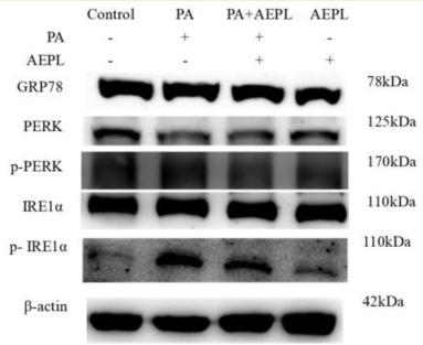
Application: WB Species: Mouse Sample:
Application: WB Species: Human Sample: HepG2 Cells
Restrictive clause
Affinity Biosciences tests all products strictly. Citations are provided as a resource for additional applications that have not been validated by Affinity Biosciences. Please choose the appropriate format for each application and consult Materials and Methods sections for additional details about the use of any product in these publications.
For Research Use Only.
Not for use in diagnostic or therapeutic procedures. Not for resale. Not for distribution without written consent. Affinity Biosciences will not be held responsible for patent infringement or other violations that may occur with the use of our products. Affinity Biosciences, Affinity Biosciences Logo and all other trademarks are the property of Affinity Biosciences LTD.