 Antibody. The lane on the left was treated with blocking peptide.")
 Antibody. The lane on the left was treated with blocking peptide.")


| Product: | Phospho-VASP (Ser157) Antibody |
| Catalog: | AF3337 |
| Description: | Rabbit polyclonal antibody to Phospho-VASP (Ser157) |
| Application: | WB IHC IF/ICC |
| Cited expt.: | WB |
| Reactivity: | Human, Mouse, Rat |
| Prediction: | Pig, Bovine, Sheep, Dog |
| Mol.Wt.: | 46kDa(Observed); 40kD(Calculated). |
| Uniprot: | P50552 |
| RRID: | AB_2834752 |
Control Products

Product Info
*The optimal dilutions should be determined by the end user. For optimal experimental results, antibody reuse is not recommended.
*Tips:
WB: For western blot detection of denatured protein samples. IHC: For immunohistochemical detection of paraffin sections (IHC-p) or frozen sections (IHC-f) of tissue samples. IF/ICC: For immunofluorescence detection of cell samples. ELISA(peptide): For ELISA detection of antigenic peptide.
Cite Format: Affinity Biosciences Cat# AF3337, RRID:AB_2834752.
Fold/Unfold
Vasodilator stimulated phosphoprotein; Vasodilator-stimulated phosphoprotein; VASP; VASP_HUMAN;
Immunogens
A synthesized peptide derived from human VASP around the phosphorylation site of Ser157.
- P50552 VASP_HUMAN:
- Protein BLAST With
- NCBI/
- ExPASy/
- Uniprot
MSETVICSSRATVMLYDDGNKRWLPAGTGPQAFSRVQIYHNPTANSFRVVGRKMQPDQQVVINCAIVRGVKYNQATPNFHQWRDARQVWGLNFGSKEDAAQFAAGMASALEALEGGGPPPPPALPTWSVPNGPSPEEVEQQKRQQPGPSEHIERRVSNAGGPPAPPAGGPPPPPGPPPPPGPPPPPGLPPSGVPAAAHGAGGGPPPAPPLPAAQGPGGGGAGAPGLAAAIAGAKLRKVSKQEEASGGPTAPKAESGRSGGGGLMEEMNAMLARRRKATQVGEKTPKDESANQEEPEARVPAQSESVRRPWEKNSTTLPRMKSSSSVTTSETQPCTPSSSDYSDLQRVKQELLEEVKKELQKVKEEIIEAFVQELRKRGSP
Predictions
Score>80(red) has high confidence and is suggested to be used for WB detection. *The prediction model is mainly based on the alignment of immunogen sequences, the results are for reference only, not as the basis of quality assurance.
High(score>80) Medium(80>score>50) Low(score<50) No confidence
Research Backgrounds
Ena/VASP proteins are actin-associated proteins involved in a range of processes dependent on cytoskeleton remodeling and cell polarity such as axon guidance, lamellipodial and filopodial dynamics, platelet activation and cell migration. VASP promotes actin filament elongation. It protects the barbed end of growing actin filaments against capping and increases the rate of actin polymerization in the presence of capping protein. VASP stimulates actin filament elongation by promoting the transfer of profilin-bound actin monomers onto the barbed end of growing actin filaments. Plays a role in actin-based mobility of Listeria monocytogenes in host cells. Regulates actin dynamics in platelets and plays an important role in regulating platelet aggregation.
Major substrate for cAMP-dependent (PKA) and cGMP-dependent protein kinase (PKG) in platelets. The preferred site for PKA is Ser-157, the preferred site for PKG/PRKG1, Ser-239. In ADP-activated platelets, phosphorylation by PKA or PKG on Ser-157 leads to fibrinogen receptor inhibition. Phosphorylation on Thr-278 requires prior phosphorylation on Ser-157 and Ser-239. In response to phorbol ester (PMA) stimulation, phosphorylated by PKC/PRKCA. In response to thrombin, phosphorylated by both PKC and ROCK1. Phosphorylation at Thr-278 by AMPK does not require prior phosphorylation at Ser-157 or Ser-239. Phosphorylation at Ser-157 by PKA is required for localization to the tight junctions in epithelial cells. Phosphorylation modulates F-actin binding, actin filament elongation and platelet activation. Phosphorylation at Ser-322 by AMPK also alters actin filament binding. Carbon monoxide (CO) promotes phosphorylation at Ser-157, while nitric oxide (NO) promotes phosphorylation at Ser-157, but also at Ser-239. Response to NO and CO is blunted in platelets from diabetic patients, and VASP is not phosphorylated efficiently at Ser-157 and Ser-239.
Cytoplasm. Cytoplasm>Cytoskeleton. Cell junction>Focal adhesion. Cell junction>Tight junction. Cell projection>Lamellipodium membrane. Cell projection>Filopodium membrane.
Note: Targeted to stress fibers and focal adhesions through interaction with a number of proteins including MRL family members. Localizes to the plasma membrane in protruding lamellipodia and filopodial tips. Stimulation by thrombin or PMA, also translocates VASP to focal adhesions. Localized along the sides of actin filaments throughout the peripheral cytoplasm under basal conditions. In pre-apoptotic cells, colocalizes with MEFV in large specks (pyroptosomes).
Highly expressed in platelets.
The EVH2 domain is comprised of 3 regions. Block A is a thymosin-like domain required for G-actin binding. The KLKR motif within this block is essential for the G-actin binding and for actin polymerization. Block B is required for F-actin binding and subcellular location, and Block C for tetramerization.
The WH1 domain mediates interaction with XIRP1.
Belongs to the Ena/VASP family.
Research Fields
· Cellular Processes > Cellular community - eukaryotes > Focal adhesion. (View pathway)
· Cellular Processes > Cellular community - eukaryotes > Tight junction. (View pathway)
· Environmental Information Processing > Signal transduction > Rap1 signaling pathway. (View pathway)
· Environmental Information Processing > Signal transduction > cGMP-PKG signaling pathway. (View pathway)
· Organismal Systems > Immune system > Platelet activation. (View pathway)
· Organismal Systems > Immune system > Fc gamma R-mediated phagocytosis. (View pathway)
· Organismal Systems > Immune system > Leukocyte transendothelial migration. (View pathway)
References
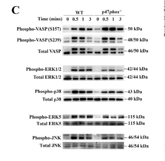
Application: WB Species: Human Sample: platelets
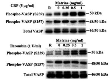
Application: WB Species: Human Sample:
Restrictive clause
Affinity Biosciences tests all products strictly. Citations are provided as a resource for additional applications that have not been validated by Affinity Biosciences. Please choose the appropriate format for each application and consult Materials and Methods sections for additional details about the use of any product in these publications.
For Research Use Only.
Not for use in diagnostic or therapeutic procedures. Not for resale. Not for distribution without written consent. Affinity Biosciences will not be held responsible for patent infringement or other violations that may occur with the use of our products. Affinity Biosciences, Affinity Biosciences Logo and all other trademarks are the property of Affinity Biosciences LTD.