Product: Phospho-Trk B (Tyr706) Antibody
Catalog: AF3461
Description: Rabbit polyclonal antibody to Phospho-Trk B (Tyr706)
Application: WB IHC IF/ICC
Reactivity: Human, Mouse, Rat
Prediction: Horse, Sheep, Rabbit, Chicken
Mol.Wt.: 90~145kDa; 92kD(Calculated).
Uniprot: Q16620
RRID: AB_2834899

View similar products>>

   Size Price Inventory
 100ul $280 In stock
 200ul $350 In stock

Lead Time: Same day delivery

For pricing and ordering contact:
Local distributors

Product Info

Source:
Rabbit
Application:
WB 1:500-1:2000, IHC 1:50-1:200, IF/ICC 1:100-1:500
*The optimal dilutions should be determined by the end user.
*Tips:

WB: For western blot detection of denatured protein samples. IHC: For immunohistochemical detection of paraffin sections (IHC-p) or frozen sections (IHC-f) of tissue samples. IF/ICC: For immunofluorescence detection of cell samples. ELISA(peptide): For ELISA detection of antigenic peptide.

Reactivity:
Human,Mouse,Rat
Prediction:
Horse(100%), Sheep(100%), Rabbit(100%), Chicken(100%)
Clonality:
Polyclonal
Specificity:
Phospho-Trk B (Tyr706) Antibody detects endogenous levels of Trk B only when phosphorylated at Tyrosine 706.
RRID:
AB_2834899
Cite Format: Affinity Biosciences Cat# AF3461, RRID:AB_2834899.
Conjugate:
Unconjugated.
Purification:
The antibody is from purified rabbit serum by affinity purification via sequential chromatography on phospho-peptide and non-phospho-peptide affinity columns.
Storage:
Rabbit IgG in phosphate buffered saline , pH 7.4, 150mM NaCl, 0.02% sodium azide and 50% glycerol. Store at -20 °C. Stable for 12 months from date of receipt.
Alias:

Fold/Unfold

AI848316; BDNF tropomyosine receptor kinase B; BDNF/NT 3 growth factors receptor; BDNF/NT-3 growth factors receptor; Brain derived neurotrophic factor receptor; C030027L06Rik; EC 2.7.10.1; GP145 TrkB; GP145-TrkB; GP145-TrkB/GP95-TrkB; GP95 TrkB; Neurotrophic receptor tyrosine kinase 2; Neurotrophic tyrosine kinase receptor type 2; Neurotrophin receptor tyrosine kinase type 2; NTRK 2; Ntrk2; NTRK2_HUMAN; Obesity, hyperphagia, and developmental delay, included; RATTRKB1; Tkrb; Trk B; Trk-B; TRKB; TrkB tyrosine kinase; TRKB1; Tropomyosin related kinase B; tyrosine kinase receptor B; Tyrosine receptor kinase B;

Immunogens

Immunogen:
Uniprot:
Gene(ID):
Expression:
Q16620 NTRK2_HUMAN:

Isoform TrkB is expressed in the central and peripheral nervous system. In the central nervous system (CNS), expression is observed in the cerebral cortex, hippocampus, thalamus, choroid plexus, granular layer of the cerebellum, brain stem, and spinal cord. In the peripheral nervous system, it is expressed in many cranial ganglia, the ophthalmic nerve, the vestibular system, multiple facial structures, the submaxillary glands, and dorsal root ganglia. Isoform TrkB-T1 is mainly expressed in the brain but also detected in other tissues including pancreas, kidney and heart. Isoform TrkB-T-Shc is predominantly expressed in the brain.

Description:
This gene encodes a member of the neurotrophic tyrosine receptor kinase (NTRK) family. This kinase is a membrane-bound receptor that, upon neurotrophin binding, phosphorylates itself and members of the MAPK pathway. Signalling through this kinase leads to cell differentiation.
Sequence:
MSSWIRWHGPAMARLWGFCWLVVGFWRAAFACPTSCKCSASRIWCSDPSPGIVAFPRLEPNSVDPENITEIFIANQKRLEIINEDDVEAYVGLRNLTIVDSGLKFVAHKAFLKNSNLQHINFTRNKLTSLSRKHFRHLDLSELILVGNPFTCSCDIMWIKTLQEAKSSPDTQDLYCLNESSKNIPLANLQIPNCGLPSANLAAPNLTVEEGKSITLSCSVAGDPVPNMYWDVGNLVSKHMNETSHTQGSLRITNISSDDSGKQISCVAENLVGEDQDSVNLTVHFAPTITFLESPTSDHHWCIPFTVKGNPKPALQWFYNGAILNESKYICTKIHVTNHTEYHGCLQLDNPTHMNNGDYTLIAKNEYGKDEKQISAHFMGWPGIDDGANPNYPDVIYEDYGTAANDIGDTTNRSNEIPSTDVTDKTGREHLSVYAVVVIASVVGFCLLVMLFLLKLARHSKFGMKGPASVISNDDDSASPLHHISNGSNTPSSSEGGPDAVIIGMTKIPVIENPQYFGITNSQLKPDTFVQHIKRHNIVLKRELGEGAFGKVFLAECYNLCPEQDKILVAVKTLKDASDNARKDFHREAELLTNLQHEHIVKFYGVCVEGDPLIMVFEYMKHGDLNKFLRAHGPDAVLMAEGNPPTELTQSQMLHIAQQIAAGMVYLASQHFVHRDLATRNCLVGENLLVKIGDFGMSRDVYSTDYYRVGGHTMLPIRWMPPESIMYRKFTTESDVWSLGVVLWEIFTYGKQPWYQLSNNEVIECITQGRVLQRPRTCPQEVYELMLGCWQREPHMRKNIKGIHTLLQNLAKASPVYLDILG

Predictions

Predictions:

Score>80(red) has high confidence and is suggested to be used for WB detection. *The prediction model is mainly based on the alignment of immunogen sequences, the results are for reference only, not as the basis of quality assurance.

Species
Results
Score
Horse
100
Sheep
100
Chicken
100
Rabbit
100
Pig
0
Bovine
0
Dog
0
Xenopus
0
Zebrafish
0
Model Confidence:
High(score>80) Medium(80>score>50) Low(score<50) No confidence

PTMs - Q16620 As Substrate

Site PTM Type Enzyme
N67 N-Glycosylation
N95 N-Glycosylation
N121 N-Glycosylation
S141 Phosphorylation
T151 Phosphorylation
S153 Phosphorylation
T161 Phosphorylation
N178 N-Glycosylation
N205 N-Glycosylation
N241 N-Glycosylation
T253 Phosphorylation
N254 N-Glycosylation
S256 Phosphorylation
S257 Phosphorylation
S260 Phosphorylation
N280 N-Glycosylation
N338 N-Glycosylation
N412 N-Glycosylation
K461 Acetylation
S479 Phosphorylation
Y490 Phosphorylation
T506 Phosphorylation
Y516 Phosphorylation Q16620 (NTRK2)
Y558 Phosphorylation
T573 Phosphorylation
Y670 Phosphorylation
Y674 Phosphorylation
Y675 Phosphorylation
S698 Phosphorylation
Y702 Phosphorylation
Y706 Phosphorylation
Y707 Phosphorylation
Y785 Phosphorylation
Y817 Phosphorylation

PTMs - Q16620 As Enzyme

Substrate Site Source
O43559 (FRS3) Y417 Uniprot
O43559 (FRS3) Y455 Uniprot
P22001 (KCNA3) Y161 Uniprot
P22001 (KCNA3) Y162 Uniprot
P22001 (KCNA3) Y163 Uniprot
P22001 (KCNA3) Y499 Uniprot
Q00535 (CDK5) Y15 Uniprot
Q13009 (TIAM1) Y829 Uniprot
Q16620 (NTRK2) Y516 Uniprot
Q16620-4 (NTRK2) Y532 Uniprot
Q16620-1 (NTRK2) Y702 Uniprot
Q16620-1 (NTRK2) Y706 Uniprot
Q16620-1 (NTRK2) Y707 Uniprot
Q16620-4 (NTRK2) Y718 Uniprot
Q16620-4 (NTRK2) Y722 Uniprot
Q16620-4 (NTRK2) Y723 Uniprot
Q16620-1 (NTRK2) Y817 Uniprot
Q16620-4 (NTRK2) Y833 Uniprot
Q92529-2 (SHC3) Y218 Uniprot
Q92529-2 (SHC3) Y219 Uniprot
Q92529-2 (SHC3) Y256 Uniprot
Q92529-2 (SHC3) Y257 Uniprot
Q92529-2 (SHC3) Y283 Uniprot
Q92529-2 (SHC3) Y301 Uniprot

Research Backgrounds

Function:

Receptor tyrosine kinase involved in the development and the maturation of the central and the peripheral nervous systems through regulation of neuron survival, proliferation, migration, differentiation, and synapse formation and plasticity (By similarity). Receptor for BDNF/brain-derived neurotrophic factor and NTF4/neurotrophin-4. Alternatively can also bind NTF3/neurotrophin-3 which is less efficient in activating the receptor but regulates neuron survival through NTRK2. Upon ligand-binding, undergoes homodimerization, autophosphorylation and activation. Recruits, phosphorylates and/or activates several downstream effectors including SHC1, FRS2, SH2B1, SH2B2 and PLCG1 that regulate distinct overlapping signaling cascades. Through SHC1, FRS2, SH2B1, SH2B2 activates the GRB2-Ras-MAPK cascade that regulates for instance neuronal differentiation including neurite outgrowth. Through the same effectors controls the Ras-PI3 kinase-AKT1 signaling cascade that mainly regulates growth and survival. Through PLCG1 and the downstream protein kinase C-regulated pathways controls synaptic plasticity. Thereby, plays a role in learning and memory by regulating both short term synaptic function and long-term potentiation. PLCG1 also leads to NF-Kappa-B activation and the transcription of genes involved in cell survival. Hence, it is able to suppress anoikis, the apoptosis resulting from loss of cell-matrix interactions. May also play a role in neutrophin-dependent calcium signaling in glial cells and mediate communication between neurons and glia.

PTMs:

Phosphorylated. Undergoes ligand-mediated autophosphorylation that is required for interaction with SHC1 and PLCG1 and other downstream effectors. Isoform TrkB-T-Shc is not phosphorylated.

Ubiquitinated. Undergoes polyubiquitination upon activation; regulated by NGFR. Ubiquitination regulates the internalization of the receptor (By similarity).

Subcellular Location:

Cell membrane>Single-pass type I membrane protein. Endosome membrane>Single-pass type I membrane protein. Early endosome membrane. Cell projection>Axon. Cell projection>Dendrite. Cytoplasm>Perinuclear region. Cell junction>Synapse>Postsynaptic density.
Note: Internalized to endosomes upon ligand-binding.

Extracellular region or secreted Cytosol Plasma membrane Cytoskeleton Lysosome Endosome Peroxisome ER Golgi apparatus Nucleus Mitochondrion Manual annotation Automatic computational assertionSubcellular location
Tissue Specificity:

Isoform TrkB is expressed in the central and peripheral nervous system. In the central nervous system (CNS), expression is observed in the cerebral cortex, hippocampus, thalamus, choroid plexus, granular layer of the cerebellum, brain stem, and spinal cord. In the peripheral nervous system, it is expressed in many cranial ganglia, the ophthalmic nerve, the vestibular system, multiple facial structures, the submaxillary glands, and dorsal root ganglia. Isoform TrkB-T1 is mainly expressed in the brain but also detected in other tissues including pancreas, kidney and heart. Isoform TrkB-T-Shc is predominantly expressed in the brain.

Subunit Structure:

Exists in a dynamic equilibrium between monomeric (low affinity) and dimeric (high affinity) structures. Interacts (phosphorylated upon activation by BDNF) with SHC1; mediates SHC1 phosphorylation and activation. Interacts (phosphorylated upon activation by BDNF) with PLCG1 and/or PLCG2; mediates PLCG1 phosphorylation and activation. Interacts with SH2B1 and SH2B2. Interacts with NGFR; may regulate the ligand specificity of the receptor (By similarity). Interacts with SORCS2; this interaction is important for normal targeting to post-synaptic densities in response to high-frequency stimulation (By similarity). Interacts (phosphorylated upon ligand-binding) with SH2D1A; regulates NTRK2. Interacts with SQSTM1 and KIDINS220 (By similarity). Interacts (phosphorylated upon ligand-binding) with FRS2; activates the MAPK signaling pathway. Interacts with APPL1 (By similarity). Interacts with MAPK8IP3/JIP3 and KLC1; interaction with KLC1 is mediated by MAPK8IP3/JIP3 (By similarity). Interacts with SORL1; this interaction facilitates NTRK2 trafficking between synaptic plasma membranes, postsynaptic densities and cell soma, hence positively regulates BDNF signaling (By similarity).

Family&Domains:

Belongs to the protein kinase superfamily. Tyr protein kinase family. Insulin receptor subfamily.

Research Fields

· Environmental Information Processing > Signal transduction > MAPK signaling pathway.   (View pathway)

· Environmental Information Processing > Signal transduction > Ras signaling pathway.   (View pathway)

· Environmental Information Processing > Signal transduction > PI3K-Akt signaling pathway.   (View pathway)

· Human Diseases > Substance dependence > Alcoholism.

· Organismal Systems > Nervous system > Neurotrophin signaling pathway.   (View pathway)

References

1). Purkinje-cell-specific MeCP2 deficiency leads to motor deficits and autistic-like behavior due to aberrations in PTP1B-TrkB-SK signaling. Cell reports, 2023 (PubMed: 38100348) [IF=8.8]

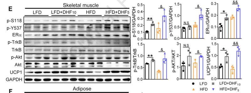
2). Crosstalk between the muscular estrogen receptor α and BDNF/TrkB signaling alleviates metabolic syndrome via 7, 8-dihydroxyflavone in female mice. Molecular Metabolism, 2021 (PubMed: 33352311) [IF=8.1]

Application: WB    Species: mouse    Sample: skeletal muscle and adipose

Figure 3.| Effect of 7,8-DHF treatment on gene and protein levels of ERα and ERβ in both skeletal muscle and adipose tissues.(E)–(F) Western blotting analysis of skeletal muscle and adipose tissues isolated from mice that have been treated with 7,8-DHF for 24 weeks. Protein levels of ERα and its two phosphorylated forms p-S118 and p-Y537, the phosphorylation of TrkB and two key energy metabolism-related proteins, AKT and UCP1 were measured. Representative immunoblots and quantification are shown (n = 3 per group. *p <0.05, **p < 0.01, N.S: not significant, LFD control vs. LFD+DHF10, Student’s t test; &p < 0.05, &&p < 0.01, N.S: not significant, HFD control vs. HFD+DHF5, Student’s t test).

3). SIRT3 alleviates mitochondrial dysfunction induced by recurrent low glucose and improves the supportive function of astrocytes to neurons. Free Radical Biology and Medicine, 2022 (PubMed: 36306990) [IF=7.4]

4). Kaempferol-3-O-sophoroside (PCS-1) contributes to modulation of depressive-like behaviour in C57BL/6J mice by activating AMPK. British journal of pharmacology, 2023 (PubMed: 37949672) [IF=7.3]

5). TAT-Ngn2 Enhances Cognitive Function Recovery and Regulates Caspase-Dependent and Mitochondrial Apoptotic Pathways After Experimental Stroke. Frontiers in Cellular Neuroscience, 2018 (PubMed: 30618628) [IF=5.3]

6). Brain-derived neurotrophic factor-TrkB signaling in the medial prefrontal cortex plays a role in the anhedonia-like phenotype after spared nerve injury. EUROPEAN ARCHIVES OF PSYCHIATRY AND CLINICAL NEUROSCIENCE, 2020 (PubMed: 29882089) [IF=4.7]

Application: WB    Species: Rat    Sample: mPFC,NAc and hippocampus tissue

Fig. 4 Levels of p-TrkB and TrkB in selected tissues among the sham, anhedonia susceptible, and unsusceptible groups. a p-TrkB/TrkB ratio (F(2,15) = 6.994, P = 0.027) and TrkB (F(2,15) = 1.565, P = 0.241) levels in the mPFC. b p-TrkB/TrkB ratio (F(2,15) = 10.551, P = 0.002) and TrkB (F2,15=1.729, P = 0.211) levels in the NAc; c p-TrkB/TrkB ratio (F(2,15) = 8.163, P = 0.018) and TrkB (F(2,15) = 1.67, P = 0.221) levels in the hippocampus. d p-TrkB/TrkB ratio (F(2,15) = 24.098, P < 0.001) and TrkB (F(2,15) = 2.376, P = 0.127) levels in the L2–5 spinal cord. e p-TrkB/TrkB ratio (F(2,15) = 9.747, P = 0.002) and TrkB (F(2,15) = 2.133, P = 0.153) levels in the muscle. f p-TrkB/TrkB ratio (F(2,15) = 11.089, P = 0.001) and TrkB (F(2,15) = 0.13, P = 0.879) levels in the liver. Data are shown as mean ± S.E.M. (n = 6). *P < 0.05, **P < 0.01 or ***P < 0.001. mPFC medial prefrontal cortex, NAc nucleus accumbens, N.S. not significant, p-TrkB phosphorylation-tropomyosin-receptor kinase B

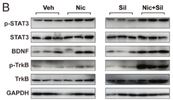
7). Nicotine exposure exacerbates silica-induced pulmonary fibrosis via STAT3-BDNF-TrkB-mediated epithelial-mesenchymal transition in alveolar type II cells. Food and Chemical Toxicology, 2023 (PubMed: 36868510) [IF=4.3]

Application: WB    Species: Mouse    Sample: lung tissue

Fig. 5. Nicotine accelerates silica-induced EMT through activation of STAT3-BDNF-TrkB and its downstream signalling axis. A) Representative image of IHC staining for BDNF (n = 3); Orange arrowheads indicate BDNF-positive cells present in the lung tissue; Scale bar = 50 μm; B) Relative protein levels of p-STAT3, STAT3, BDNF, p-TrkB and TrkB revealed by western blot and their quantification (n = 3); C) p-AKT, AKT, Twist and Snail expression in the lungs of each group was analysed by western blot and quantified (n = 3); D) Average optical density (AOD) values of BDNF; Data are expressed as mean ± SEM; Statistical significance was determined by one-way ANOVA. *p < 0.05, **p < 0.01, ***p < 0.001, ****p < 0.0001. (For interpretation of the references to colour in this figure legend, the reader is referred to the Web version of this article.)

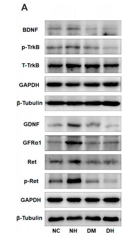
8). Recurrent non-severe hypoglycemia aggravates cognitive decline in diabetes and induces mitochondrial dysfunction in cultured astrocytes. Molecular and Cellular Endocrinology, 2021 (PubMed: 33545179) [IF=4.1]

Application: WB    Species: mice    Sample: cortex tissue

Fig. 3. Recurrent non-severe hypoglycemia (RH) inhibits brain-derived neurotrophic factor (BDNF) and glial cell derived neurotrophic factor (GDNF) signaling in diabetic mice. (A) The expression of BDNF/tropomyosin (t)-receptor (r)-kinase (k) B (TrkB) signaling and GDNF/GDNF family receptor alpha-1 (GFRa1)/receptor tyrosine kinase (Ret) signaling in mice cerebral cortex, measured using western blotting. (B–G) Relative protein levels of BDNF, TrkB, p-TrkB, GDNF, GFRa1, Ret, and p-Ret (Tyr1062), as determined by ImageJ. *P < 0.05 vs. normal control (NC), **P < 0.01 vs. NC, #P < 0.05 vs. diabetes mellitus (DM), ##P < 0.05 vs. DM.

9). PirB Overexpression Exacerbates Neuronal Apoptosis by Inhibiting TrkB and mTOR Phosphorylation After Oxygen and Glucose Deprivation Injury. CELLULAR AND MOLECULAR NEUROBIOLOGY, 2017 (PubMed: 27443384) [IF=4.0]

Application: WB    Species: mouse    Sample:


10). PGC-1α-FNDC5-BDNF signaling pathway in skeletal muscle confers resilience to stress in mice subjected to chronic social defeat. PSYCHOPHARMACOLOGY, 2018 (PubMed: 30259075) [IF=3.4]

Load more

Restrictive clause


Affinity Biosciences tests all products strictly. Citations are provided as a resource for additional applications that have not been validated by Affinity Biosciences. Please choose the appropriate format for each application and consult Materials and Methods sections for additional details about the use of any product in these publications.

For Research Use Only.
Not for use in diagnostic or therapeutic procedures. Not for resale. Not for distribution without written consent. Affinity Biosciences will not be held responsible for patent infringement or other violations that may occur with the use of our products. Affinity Biosciences, Affinity Biosciences Logo and all other trademarks are the property of Affinity Biosciences LTD.