 RT-qPCR was performed to examine the mRNA level of TrkA, TrkB, and TrkC. (B, D, and F) The upper panels represent results of the Western blots. The lower panels show the semi-quantitative analysis of TrkA, TrkB, and TrkC after normalization to the corresponding β-actin. (*P<0.05, **P<0.01 vs control group; #P<0.05, ##P<0.01, ###P<0.001 vs PI, one-way ANOVA followed by Bonferroni’s multiple comparison post hoc test, n=3–4 rats per group). Each value represents mean±SEM. PI+OXY, postoperative oxycodone group; OXY+PI, preoperative oxycodone group.")
 inhibits brain-derived neurotrophic factor (BDNF) and glial cell derived neurotrophic factor (GDNF) signaling in
diabetic mice. (A) The expression of BDNF/tropomyosin (t)-receptor (r)-kinase (k) B (TrkB) signaling and GDNF/GDNF family receptor alpha-1 (GFRa1)/receptor
tyrosine kinase (Ret) signaling in mice cerebral cortex, measured using western blotting. (B–G) Relative protein levels of BDNF, TrkB, p-TrkB, GDNF, GFRa1, Ret, and
p-Ret (Tyr1062), as determined by ImageJ. *P < 0.05 vs. normal control (NC), **P < 0.01 vs. NC, #P < 0.05 vs. diabetes mellitus (DM), ##P < 0.05 vs. DM.")

brain-derived neurotrophic factor (BDNF) and tropomyosin-related
kinase B (TrkB) in morphine-induced conditioned place preference (CPP). a The mRNA expression of BDNF. F(2,6)=6.716. b The
mRNA expression of TrkB. F(2,6)=7.553. c The representative immunoblots. d Graphic representation of relative expression of BDNF to
β-actin. F(2,6)=9.132. e Graphic representation of relative expression of TrkB to β-actin. F(2,6)=12.13. f Immunohistochemical staining
for BDNF and TrkB expression. Figures were magnifed by ×200. g
Comparison of the percentage of BDNF-positive cells. F(2,6)=8.633.
h Comparison of the percentage of TrkB-positive cells. F(2,6)=9.624.
Data are presented as the mean±SEM, n=3. One-way ANOVA with
a post hoc Newman–Keuls test. *:vs the con group, P<0.05; **:vs
the con group, P<0.001; #:vs the mod group, P<0.05 of TrkB to β-actin. F(2,6)=12.13. f Immunohistochemical staining
for BDNF and TrkB expression. Figures were magnifed by ×200. g
Comparison of the percentage of BDNF-positive cells. F(2,6)=8.633.
h Comparison of the percentage of TrkB-positive cells. F(2,6)=9.624.
Data are presented as the mean±SEM, n=3. One-way ANOVA with
a post hoc Newman–Keuls test. *:vs the con group, P<0.05; **:vs
the con group, P<0.001; #:vs the mod group, P<0.05")
 Representative image of IHC staining for BDNF (n = 3); Orange arrowheads indicate BDNF-positive cells present in the lung tissue; Scale bar = 50 μm; B) Relative protein levels of p-STAT3, STAT3, BDNF, p-TrkB and TrkB revealed by western blot and their quantification (n = 3); C) p-AKT, AKT, Twist and Snail expression in the lungs of each group was analysed by western blot and quantified (n = 3); D) Average optical density (AOD) values of BDNF; Data are expressed as mean ± SEM; Statistical significance was determined by one-way ANOVA. *p < 0.05, **p < 0.01, ***p < 0.001, ****p < 0.0001. (For interpretation of the references to colour in this figure legend, the reader is referred to the Web version of this article.)")
| Product: | Trk B Antibody |
| Catalog: | AF6461 |
| Description: | Rabbit polyclonal antibody to Trk B |
| Application: | WB IHC IF/ICC |
| Cited expt.: | WB |
| Reactivity: | Human, Mouse, Rat |
| Prediction: | Horse, Sheep, Rabbit, Chicken |
| Mol.Wt.: | 145kDa(Observed); 92kD(Calculated). |
| Uniprot: | Q16620 |
| RRID: | AB_2835281 |
Control Products

Related Downloads
Protocols
Product Info
*The optimal dilutions should be determined by the end user. For optimal experimental results, antibody reuse is not recommended.
*Tips:
WB: For western blot detection of denatured protein samples. IHC: For immunohistochemical detection of paraffin sections (IHC-p) or frozen sections (IHC-f) of tissue samples. IF/ICC: For immunofluorescence detection of cell samples. ELISA(peptide): For ELISA detection of antigenic peptide.
Cite Format: Affinity Biosciences Cat# AF6461, RRID:AB_2835281.
Fold/Unfold
AI848316; BDNF tropomyosine receptor kinase B; BDNF/NT 3 growth factors receptor; BDNF/NT-3 growth factors receptor; Brain derived neurotrophic factor receptor; C030027L06Rik; EC 2.7.10.1; GP145 TrkB; GP145-TrkB; GP145-TrkB/GP95-TrkB; GP95 TrkB; Neurotrophic receptor tyrosine kinase 2; Neurotrophic tyrosine kinase receptor type 2; Neurotrophin receptor tyrosine kinase type 2; NTRK 2; Ntrk2; NTRK2_HUMAN; Obesity, hyperphagia, and developmental delay, included; RATTRKB1; Tkrb; Trk B; Trk-B; TRKB; TrkB tyrosine kinase; TRKB1; Tropomyosin related kinase B; tyrosine kinase receptor B; Tyrosine receptor kinase B;
Immunogens
A synthesized peptide derived from human Trk B, corresponding to a region within C-terminal amino acids.
Isoform TrkB is expressed in the central and peripheral nervous system. In the central nervous system (CNS), expression is observed in the cerebral cortex, hippocampus, thalamus, choroid plexus, granular layer of the cerebellum, brain stem, and spinal cord. In the peripheral nervous system, it is expressed in many cranial ganglia, the ophthalmic nerve, the vestibular system, multiple facial structures, the submaxillary glands, and dorsal root ganglia. Isoform TrkB-T1 is mainly expressed in the brain but also detected in other tissues including pancreas, kidney and heart. Isoform TrkB-T-Shc is predominantly expressed in the brain.
- Q16620 NTRK2_HUMAN:
- Protein BLAST With
- NCBI/
- ExPASy/
- Uniprot
MSSWIRWHGPAMARLWGFCWLVVGFWRAAFACPTSCKCSASRIWCSDPSPGIVAFPRLEPNSVDPENITEIFIANQKRLEIINEDDVEAYVGLRNLTIVDSGLKFVAHKAFLKNSNLQHINFTRNKLTSLSRKHFRHLDLSELILVGNPFTCSCDIMWIKTLQEAKSSPDTQDLYCLNESSKNIPLANLQIPNCGLPSANLAAPNLTVEEGKSITLSCSVAGDPVPNMYWDVGNLVSKHMNETSHTQGSLRITNISSDDSGKQISCVAENLVGEDQDSVNLTVHFAPTITFLESPTSDHHWCIPFTVKGNPKPALQWFYNGAILNESKYICTKIHVTNHTEYHGCLQLDNPTHMNNGDYTLIAKNEYGKDEKQISAHFMGWPGIDDGANPNYPDVIYEDYGTAANDIGDTTNRSNEIPSTDVTDKTGREHLSVYAVVVIASVVGFCLLVMLFLLKLARHSKFGMKGPASVISNDDDSASPLHHISNGSNTPSSSEGGPDAVIIGMTKIPVIENPQYFGITNSQLKPDTFVQHIKRHNIVLKRELGEGAFGKVFLAECYNLCPEQDKILVAVKTLKDASDNARKDFHREAELLTNLQHEHIVKFYGVCVEGDPLIMVFEYMKHGDLNKFLRAHGPDAVLMAEGNPPTELTQSQMLHIAQQIAAGMVYLASQHFVHRDLATRNCLVGENLLVKIGDFGMSRDVYSTDYYRVGGHTMLPIRWMPPESIMYRKFTTESDVWSLGVVLWEIFTYGKQPWYQLSNNEVIECITQGRVLQRPRTCPQEVYELMLGCWQREPHMRKNIKGIHTLLQNLAKASPVYLDILG
Predictions
Score>80(red) has high confidence and is suggested to be used for WB detection. *The prediction model is mainly based on the alignment of immunogen sequences, the results are for reference only, not as the basis of quality assurance.
High(score>80) Medium(80>score>50) Low(score<50) No confidence
Research Backgrounds
Receptor tyrosine kinase involved in the development and the maturation of the central and the peripheral nervous systems through regulation of neuron survival, proliferation, migration, differentiation, and synapse formation and plasticity (By similarity). Receptor for BDNF/brain-derived neurotrophic factor and NTF4/neurotrophin-4. Alternatively can also bind NTF3/neurotrophin-3 which is less efficient in activating the receptor but regulates neuron survival through NTRK2. Upon ligand-binding, undergoes homodimerization, autophosphorylation and activation. Recruits, phosphorylates and/or activates several downstream effectors including SHC1, FRS2, SH2B1, SH2B2 and PLCG1 that regulate distinct overlapping signaling cascades. Through SHC1, FRS2, SH2B1, SH2B2 activates the GRB2-Ras-MAPK cascade that regulates for instance neuronal differentiation including neurite outgrowth. Through the same effectors controls the Ras-PI3 kinase-AKT1 signaling cascade that mainly regulates growth and survival. Through PLCG1 and the downstream protein kinase C-regulated pathways controls synaptic plasticity. Thereby, plays a role in learning and memory by regulating both short term synaptic function and long-term potentiation. PLCG1 also leads to NF-Kappa-B activation and the transcription of genes involved in cell survival. Hence, it is able to suppress anoikis, the apoptosis resulting from loss of cell-matrix interactions. May also play a role in neutrophin-dependent calcium signaling in glial cells and mediate communication between neurons and glia.
Phosphorylated. Undergoes ligand-mediated autophosphorylation that is required for interaction with SHC1 and PLCG1 and other downstream effectors. Isoform TrkB-T-Shc is not phosphorylated.
Ubiquitinated. Undergoes polyubiquitination upon activation; regulated by NGFR. Ubiquitination regulates the internalization of the receptor (By similarity).
Cell membrane>Single-pass type I membrane protein. Endosome membrane>Single-pass type I membrane protein. Early endosome membrane. Cell projection>Axon. Cell projection>Dendrite. Cytoplasm>Perinuclear region. Cell junction>Synapse>Postsynaptic density.
Note: Internalized to endosomes upon ligand-binding.
Isoform TrkB is expressed in the central and peripheral nervous system. In the central nervous system (CNS), expression is observed in the cerebral cortex, hippocampus, thalamus, choroid plexus, granular layer of the cerebellum, brain stem, and spinal cord. In the peripheral nervous system, it is expressed in many cranial ganglia, the ophthalmic nerve, the vestibular system, multiple facial structures, the submaxillary glands, and dorsal root ganglia. Isoform TrkB-T1 is mainly expressed in the brain but also detected in other tissues including pancreas, kidney and heart. Isoform TrkB-T-Shc is predominantly expressed in the brain.
Belongs to the protein kinase superfamily. Tyr protein kinase family. Insulin receptor subfamily.
Research Fields
· Environmental Information Processing > Signal transduction > MAPK signaling pathway. (View pathway)
· Environmental Information Processing > Signal transduction > Ras signaling pathway. (View pathway)
· Environmental Information Processing > Signal transduction > PI3K-Akt signaling pathway. (View pathway)
· Human Diseases > Substance dependence > Alcoholism.
· Organismal Systems > Nervous system > Neurotrophin signaling pathway. (View pathway)
References
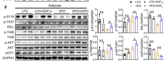
Application: WB Species: mouse Sample: skeletal muscle and adipose
Application: WB Species: rat Sample: gastrocnemius muscles
Restrictive clause
Affinity Biosciences tests all products strictly. Citations are provided as a resource for additional applications that have not been validated by Affinity Biosciences. Please choose the appropriate format for each application and consult Materials and Methods sections for additional details about the use of any product in these publications.
For Research Use Only.
Not for use in diagnostic or therapeutic procedures. Not for resale. Not for distribution without written consent. Affinity Biosciences will not be held responsible for patent infringement or other violations that may occur with the use of our products. Affinity Biosciences, Affinity Biosciences Logo and all other trademarks are the property of Affinity Biosciences LTD.