


 The impact of different treatment groups on copper ion transport. (B,C) FCM was used to analyze the expression of JC-1 in different treatment groups. (D,E) Flow cytometry was used to analyze the expression of LPO in different treatment groups. (F) Fluorescence microscopy is used to detect changes in ROS and JC-1 probe levels in different treatment groups. (G,H) WB was used to verify the protein expression of cuproptosis across different treatment groups. (I) Representative Bio-TEM images of WT-Huh7 cells and LenR HCC cells before and after treatment with DSF@CuO. (G1: corn oil, G2: DSF, G3: CuO NPs, G4: Len@CuO, and G5: DSF@CuO). Data are presented as mean ± SD and are representative of three independent experiments. ** p < 0.01, *** p < 0.001.")
 Representative images of AR staining of mice from each group, scale bar = 400 μm and 100 μm respectively. (B) Representative images of von kossa staining of mice from each group, scale bar = 400 μm and 100 μm respectively. (C) Representative images of ALP staining of mice from each group, scale bar = 400 μm and 100 μm respectively. (D) Representative images of p16 staining of mice from each group, scale bar = 400 μm and 100 μm respectively. (E) Quantification of positive area of Alizarin red staining, n = 6. (F) Quantification of positive area of von kossa staining, n = 6. (G) Quantification of the relative expression of ALP, n = 6. (H) Quantification of the relative expression of p16, n = 6.")
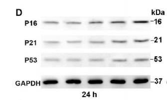
 mRNA relative expression of SDHB; (B) mRNA relative expression of ULK1; (C) mRNA relative expression of CCS; (D) Protein relative expressions of SDHB and CCS; (E) protein relative expressions of ULK1, CDKN2A, and CMC1 *p")
 Immunofluorescence double staining showed the location and expression of CD47 in skin tissues of each group. Fibroblasts were labeled using Vimentin (green) and nuclei were stained with DAPI (blue). CD47 was labeled as red. The quantification of CD47/Vimentin double-positive cells was shown in the right side. Scale bar, 50 μm. (B) Images of SA-β-gal staining and quantification of the SA-β-gal-positive rate in UVA-irradiated fibroblasts. Scale bar, 200 μm. (C) The protein expression levels of CD47, p53, p21, and p16 in control and UVA-irradiated fibroblasts were detected by Western blotting. (D) Immunofluorescence detection of CD47 protein expression in control and UVA-irradiated fibroblasts. The red marker indicates cells expressing CD47 and the blue marker indicates DAPI-stained nuclei. Scale bar, 50 μm. Data are the means ± SD from three independent experiments. *p")
 Triple immunofluorescence staining for CD47 (deep red), Vimentin (green) and p21 (red) in skin tissues of sun-protected and sun-exposed groups. Blue: DAPI nuclear counterstaining. Bar graphs represent the number of CD47, p16, and Vimentin triple-positive cells. Scale bar, 50 μm. (B) Double immunofluorescence staining for CD68 (green) and SIRPα (red) in skin tissues of each group. Blue: DAPI nuclear counterstaining. Bar graphs represent the number of CD68/SIRPα double-positive cells. Data are the means ± SD from three independent experiments. ***p")
 Expression levels of group was imaged at 0 and 12 hours using a microscope. (K) The recover areas were calculated by ImageJ software in different groups at different time point. (L) Cell migration assay results of ES-2 cells in the control group and CuET treated group (0.5 μg/mL). (M) Calculate the degree of cell migration in the control group and CuET treated group (0.5 μg/mL) using ImageJ software. (N, O) Tumor size (N) and volume (O) measurements in the CDX model following CuET treatment (5mg/kg and 15mg/kg). (P) Body weights of mice across different treatment groups. n = 3, independent experiments (D, E, F, G, H, K, L and M). Data presented as mean ± S.D. Statistical significance was assessed using Student's unpaired t-test. *p < 0.05, **p < 0.01, **p < 0.001.")
| Product: | CDKN2A/p16INK4a Antibody |
| Catalog: | AF5484 |
| Description: | Rabbit polyclonal antibody to CDKN2A/p16INK4a |
| Application: | WB IF/ICC |
| Cited expt.: | WB, IF/ICC |
| Reactivity: | Human, Mouse |
| Prediction: | Pig, Bovine, Horse, Rabbit |
| Mol.Wt.: | 16 kDa(Observed); 17kD(Calculated). |
| Uniprot: | P42771 |
| RRID: | AB_2837964 |
Control Products

Product Info
*The optimal dilutions should be determined by the end user. For optimal experimental results, antibody reuse is not recommended.
*Tips:
WB: For western blot detection of denatured protein samples. IHC: For immunohistochemical detection of paraffin sections (IHC-p) or frozen sections (IHC-f) of tissue samples. IF/ICC: For immunofluorescence detection of cell samples. ELISA(peptide): For ELISA detection of antigenic peptide.
Cite Format: Affinity Biosciences Cat# AF5484, RRID:AB_2837964.
Fold/Unfold
Cyclin-dependent kinase inhibitor 2A;Cyclin-dependent kinase 4 inhibitor A;CDK4I;Multiple tumor suppressor 1;MTS-1;p16-INK4a;p16-INK4;p16INK4A;CDKN2A;CDKN2;MTS1;
Immunogens
A synthesized peptide derived from human CDKN2A/p16INK4a, corresponding to a region within N-terminal amino acids.
Widely expressed but not detected in brain or skeletal muscle. Isoform 3 is pancreas-specific.
- P42771 CDN2A_HUMAN:
- Protein BLAST With
- NCBI/
- ExPASy/
- Uniprot
MEPAAGSSMEPSADWLATAAARGRVEEVRALLEAGALPNAPNSYGRRPIQVMMMGSARVAELLLLHGAEPNCADPATLTRPVHDAAREGFLDTLVVLHRAGARLDVRDAWGRLPVDLAEELGHRDVARYLRAAAGGTRGSNHARIDAAEGPSDIPD
Predictions
Score>80(red) has high confidence and is suggested to be used for WB detection. *The prediction model is mainly based on the alignment of immunogen sequences, the results are for reference only, not as the basis of quality assurance.
High(score>80) Medium(80>score>50) Low(score<50) No confidence
Research Backgrounds
Acts as a negative regulator of the proliferation of normal cells by interacting strongly with CDK4 and CDK6. This inhibits their ability to interact with cyclins D and to phosphorylate the retinoblastoma protein.
Phosphorylation seems to increase interaction with CDK4.
Cytoplasm. Nucleus.
Widely expressed but not detected in brain or skeletal muscle. Isoform 3 is pancreas-specific.
Belongs to the CDKN2 cyclin-dependent kinase inhibitor family.
Research Fields
· Cellular Processes > Cell growth and death > Cell cycle. (View pathway)
· Cellular Processes > Cell growth and death > p53 signaling pathway. (View pathway)
· Cellular Processes > Cell growth and death > Cellular senescence. (View pathway)
· Human Diseases > Drug resistance: Antineoplastic > Endocrine resistance.
· Human Diseases > Drug resistance: Antineoplastic > Platinum drug resistance.
· Human Diseases > Infectious diseases: Viral > HTLV-I infection.
· Human Diseases > Cancers: Overview > Pathways in cancer. (View pathway)
· Human Diseases > Cancers: Overview > Viral carcinogenesis.
· Human Diseases > Cancers: Overview > MicroRNAs in cancer.
· Human Diseases > Cancers: Specific types > Pancreatic cancer. (View pathway)
· Human Diseases > Cancers: Specific types > Glioma. (View pathway)
· Human Diseases > Cancers: Specific types > Melanoma. (View pathway)
· Human Diseases > Cancers: Specific types > Bladder cancer. (View pathway)
· Human Diseases > Cancers: Specific types > Chronic myeloid leukemia. (View pathway)
· Human Diseases > Cancers: Specific types > Non-small cell lung cancer. (View pathway)
· Human Diseases > Cancers: Specific types > Hepatocellular carcinoma. (View pathway)
References

Application: IF/ICC Species: Mouse Sample:

Application: IHC Species: human Sample: VICs

Application: WB Species: goat Sample: NPCs
Application: WB Species: mice Sample: bone marrow mesenchymal stem (BMSCs)

Application: IHC Species: Rat Sample:

Application: IF/ICC Species: Rat Sample:

Application: WB Species: Mouse Sample:

Application: IF/ICC Species: Mouse Sample:
Restrictive clause
Affinity Biosciences tests all products strictly. Citations are provided as a resource for additional applications that have not been validated by Affinity Biosciences. Please choose the appropriate format for each application and consult Materials and Methods sections for additional details about the use of any product in these publications.
For Research Use Only.
Not for use in diagnostic or therapeutic procedures. Not for resale. Not for distribution without written consent. Affinity Biosciences will not be held responsible for patent infringement or other violations that may occur with the use of our products. Affinity Biosciences, Affinity Biosciences Logo and all other trademarks are the property of Affinity Biosciences LTD.