Product: APG5L/ATG5 Antibody
Catalog: DF6010
Description: Rabbit polyclonal antibody to APG5L/ATG5
Application: WB IHC
Reactivity: Human, Mouse, Rat
Prediction: Pig, Zebrafish, Bovine, Horse, Sheep, Rabbit, Dog, Chicken, Xenopus
Mol.Wt.: 35kD,56kD(complex); 32kD(Calculated).
Uniprot: Q9H1Y0
RRID: AB_2837987

View similar products>>

   Size Price Inventory
 100ul $280 In stock
 200ul $350 In stock

Lead Time: Same day delivery

For pricing and ordering contact:
Local distributors

Product Info

Source:
Rabbit
Application:
WB 1:500-1:2000, IHC 1:50-1:200
*The optimal dilutions should be determined by the end user.
*Tips:

WB: For western blot detection of denatured protein samples. IHC: For immunohistochemical detection of paraffin sections (IHC-p) or frozen sections (IHC-f) of tissue samples. IF/ICC: For immunofluorescence detection of cell samples. ELISA(peptide): For ELISA detection of antigenic peptide.

Reactivity:
Human,Mouse,Rat
Prediction:
Pig(100%), Zebrafish(93%), Bovine(100%), Horse(100%), Sheep(100%), Rabbit(100%), Dog(100%), Chicken(93%), Xenopus(100%)
Clonality:
Polyclonal
Specificity:
APG5L/ATG5 Antibody detects endogenous levels of total APG5L/ATG5.
RRID:
AB_2837987
Cite Format: Affinity Biosciences Cat# DF6010, RRID:AB_2837987.
Conjugate:
Unconjugated.
Purification:
The antiserum was purified by peptide affinity chromatography using SulfoLink™ Coupling Resin (Thermo Fisher Scientific).
Storage:
Rabbit IgG in phosphate buffered saline , pH 7.4, 150mM NaCl, 0.02% sodium azide and 50% glycerol. Store at -20 °C. Stable for 12 months from date of receipt.
Alias:

Fold/Unfold

APG 5; APG 5L; APG5; APG5 autophagy 5 like; APG5 like; APG5-like; APG5L; Apoptosis specific protein; Apoptosis-specific protein; ASP; ATG 5; Atg5; ATG5 autophagy related 5 homolog; ATG5_HUMAN; Autophagy protein 5; Autophagy related 5; hAPG5; Homolog of S Cerevisiae autophagy 5; OTTHUMP00000040507;

Immunogens

Immunogen:
Uniprot:
Gene(ID):
Expression:
Q9H1Y0 ATG5_HUMAN:

Ubiquitous. The mRNA is present at similar levels in viable and apoptotic cells, whereas the protein is dramatically highly expressed in apoptotic cells.

Description:
Autophagy is a catabolic process for the autophagosomic-lysosomal degradation of bulk cytoplasmic contents (1,2). Autophagy is generally activated by conditions of nutrient deprivation but has also been associated with a number of physiological processes including development, differentiation, neurodegeneration, infection, and cancer (3). The molecular machinery of autophagy was largely discovered in yeast and referred to as autophagy-related (Atg) genes. Formation of the autophagosome involves a ubiquitin-like conjugation system in which Atg12 is covalently bound to Atg5 and targeted to autophagosome vesicles (4-6). This conjugation reaction is mediated by the ubiquitin E1-like enzyme Atg7 and the E2-like enzyme Atg10 (7,8).
Sequence:
MTDDKDVLRDVWFGRIPTCFTLYQDEITEREAEPYYLLLPRVSYLTLVTDKVKKHFQKVMRQEDISEIWFEYEGTPLKWHYPIGLLFDLLASSSALPWNITVHFKSFPEKDLLHCPSKDAIEAHFMSCMKEADALKHKSQVINEMQKKDHKQLWMGLQNDRFDQFWAINRKLMEYPAEENGFRYIPFRIYQTTTERPFIQKLFRPVAADGQLHTLGDLLKEVCPSAIDPEDGEKKNQVMIHGIEPMLETPLQWLSEHLSYPDNFLHISIIPQPTD

Predictions

Predictions:

Score>80(red) has high confidence and is suggested to be used for WB detection. *The prediction model is mainly based on the alignment of immunogen sequences, the results are for reference only, not as the basis of quality assurance.

Species
Results
Score
Pig
100
Horse
100
Bovine
100
Sheep
100
Dog
100
Xenopus
100
Rabbit
100
Zebrafish
93
Chicken
93
Model Confidence:
High(score>80) Medium(80>score>50) Low(score<50) No confidence

PTMs - Q9H1Y0 As Substrate

Site PTM Type Enzyme
M1 Acetylation
K5 Ubiquitination
Y35 Phosphorylation
Y36 Phosphorylation
T75 Phosphorylation
Y81 Phosphorylation
T101 Phosphorylation
K138 Ubiquitination
K147 Ubiquitination
K171 Ubiquitination
K220 Ubiquitination

Research Backgrounds

Function:

Involved in autophagic vesicle formation. Conjugation with ATG12, through a ubiquitin-like conjugating system involving ATG7 as an E1-like activating enzyme and ATG10 as an E2-like conjugating enzyme, is essential for its function. The ATG12-ATG5 conjugate acts as an E3-like enzyme which is required for lipidation of ATG8 family proteins and their association to the vesicle membranes. Involved in mitochondrial quality control after oxidative damage, and in subsequent cellular longevity. Plays a critical role in multiple aspects of lymphocyte development and is essential for both B and T lymphocyte survival and proliferation. Required for optimal processing and presentation of antigens for MHC II. Involved in the maintenance of axon morphology and membrane structures, as well as in normal adipocyte differentiation. Promotes primary ciliogenesis through removal of OFD1 from centriolar satellites and degradation of IFT20 via the autophagic pathway.

May play an important role in the apoptotic process, possibly within the modified cytoskeleton. Its expression is a relatively late event in the apoptotic process, occurring downstream of caspase activity. Plays a crucial role in IFN-gamma-induced autophagic cell death by interacting with FADD.

(Microbial infection) May act as a proviral factor. In association with ATG12, negatively regulates the innate antiviral immune response by impairing the type I IFN production pathway upon vesicular stomatitis virus (VSV) infection. Required for the translation of incoming hepatitis C virus (HCV) RNA and, thereby, for initiation of HCV replication, but not required once infection is established.

PTMs:

Conjugated to ATG12; which is essential for autophagy, but is not required for association with isolation membrane.

Acetylated by EP300.

Subcellular Location:

Cytoplasm. Preautophagosomal structure membrane>Peripheral membrane protein.
Note: Colocalizes with nonmuscle actin. The conjugate detaches from the membrane immediately before or after autophagosome formation is completed (By similarity). Localizes also to discrete punctae along the ciliary axoneme and to the base of the ciliary axoneme.

Extracellular region or secreted Cytosol Plasma membrane Cytoskeleton Lysosome Endosome Peroxisome ER Golgi apparatus Nucleus Mitochondrion Manual annotation Automatic computational assertionSubcellular location
Tissue Specificity:

Ubiquitous. The mRNA is present at similar levels in viable and apoptotic cells, whereas the protein is dramatically highly expressed in apoptotic cells.

Subunit Structure:

Forms a conjugate with ATG12. The ATG5-ATG12 conjugate forms a complex with several units of ATG16L. Interacts with TECPR1; the interaction is direct and does not take place when ATG16L is associated with the ATG5-ATG12 conjugate. Interacts with DHX58/RIG-1, IFIH1/MDA5 and MAVS/IPS-1 in monomeric form as well as in ATG12-ATG5 conjugate form. The interaction with MAVS is further enhanced upon vesicular stomatitis virus (VSV) infection. Interacts with ATG3. Interacts with ATG7 and ATG10 (By similarity). Interacts with FADD. Interacts with Bassoon/BSN; this interaction is important for the regulation of presynaptic autophagy (By similarity).

(Microbial infection) Interacts transiently interacts with hepatitis C virus (HCV) protein NS5B during HCV infection.

Family&Domains:

Belongs to the ATG5 family.

Research Fields

· Cellular Processes > Transport and catabolism > Autophagy - other.   (View pathway)

· Cellular Processes > Transport and catabolism > Autophagy - animal.   (View pathway)

· Cellular Processes > Cell growth and death > Ferroptosis.   (View pathway)

· Human Diseases > Infectious diseases: Bacterial > Shigellosis.

· Organismal Systems > Aging > Longevity regulating pathway.   (View pathway)

· Organismal Systems > Aging > Longevity regulating pathway - multiple species.   (View pathway)

· Organismal Systems > Immune system > NOD-like receptor signaling pathway.   (View pathway)

· Organismal Systems > Immune system > RIG-I-like receptor signaling pathway.   (View pathway)

References

1). d-Borneol enhances cisplatin sensitivity via autophagy dependent EMT signaling and NCOA4-mediated ferritinophagy. PHYTOMEDICINE (PubMed: 36030746) [IF=7.9]

2). Chrysin induces autophagy-dependent ferroptosis to increase chemosensitivity to gemcitabine by targeting CBR1 in pancreatic cancer cells. Biochemical Pharmacology (PubMed: 34673014) [IF=5.8]

3). Respiratory Syncytial Virus Replication Is Promoted by Autophagy-Mediated Inhibition of Apoptosis. Journal of Virology (PubMed: 29386287) [IF=5.4]

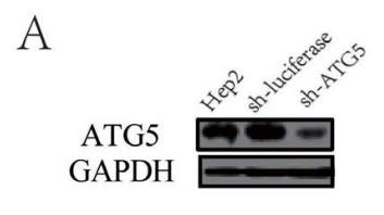
Application: WB    Species: human    Sample: Hep2 cells

Figure 5. |Inhibition of autophagy with specific shRNA targeting ATG5, ATG7 or BECN1 reduces RSV replication. (A-C) Hep2 cells stably expressing specific shRNA against ATG5 (Hep2-sh-ATG5), ATG7 (Hep2-sh-ATG7), BECN1 (Hep2-sh-BECN1) or luciferase (Hep2-sh-luciferase) were established as described in Materials and Methods. The silencing efficiency of ATG5 shRNA (A), ATG7 shRNA (B) and BECN1 shRNA (C) were validated by immunoblotting with anti-ATG5, anti-ATG7, anti-BECN1 and anti-GAPDH antibodies respectively.

4). Dihydroartemisinin Ameliorates Learning and Memory in Alzheimer’s Disease Through Promoting Autophagosome-Lysosome Fusion and Autolysosomal Degradation for Aβ Clearance. Frontiers in Aging Neuroscience (PubMed: 32210783) [IF=4.8]

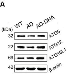
Application: WB    Species: Mouse    Sample: brain tissue

Figure 6 DHA increases the basal level of autophagy. Protein levels of ATG5, ATG12, ATG16L1, Beclin1, ATG14, p-ATG14 (Ser29), Rab7, RILP, Lamp1, CTSB, p-mTOR(Ser2448), mammalian target of rapamycin (mTOR), ULK1, p-GSK3β (Ser9), GSK3β in extracts of whole brain tissue were detected by WB. Representative Western blots are presented in panels (A,C,E,G), and quantification of the results is shown as the mean values ± SD in panels (B,D,F,H). *p < 0.05, **p < 0.01, and ***p < 0.001 between two groups. One-way analysis of variance/Newman–Keuls test was performed for all WB data.

5). Cr (VI)-induced overactive mitophagy contributes to mitochondrial loss and cytotoxicity in L02 hepatocytes. Biochemical Journal (PubMed: 32597464) [IF=4.1]

6). Hypoxia-Induced Autophagy Enhances Cisplatin Resistance in Human Bladder Cancer Cells by Targeting Hypoxia-Inducible Factor-1α. Journal of Immunology Research (PubMed: 33681390) [IF=4.1]

Application: WB    Species: Human    Sample: Bladder Cancer Cells

Figure 5 Expression of related marker proteins and genes under normoxic and hypoxic conditions by Western blotting. (a) Beclin-1 was measured by immunoblotting under normoxia and hypoxia. (b) Caspase-3 was measured by immunoblotting when incubated with HIF-1α inhibitor YC-1. The results indicate that hypoxia might induce cell chemoresistance through the HIF-1α signaling pathway. (c) Beclin-1 was measured by immunoblotting under normoxia and hypoxia. (d) Atg5 was measured by Western blotting and showed a similar trend to caspase-3. (e) HIF-1α was measured with Western blotting. The result revealed that HIF was involved in targeting apoptosis induced by autophagy.

7). Zinc-Deficient Diet Causes Imbalance in Zinc Homeostasis and Impaired Autophagy and Impairs Semen Quality in Mice. BIOLOGICAL TRACE ELEMENT RESEARCH (PubMed: 35713811) [IF=3.9]

8). FAT4 activation inhibits epithelial-mesenchymal transition (EMT) by promoting autophagy in H2228/Cer cells. Medical Oncology (PubMed: 36576661) [IF=3.4]

9). A Feedback Loop between TGF-β1 and ATG5 Mediated by miR-122-5p Regulates Fibrosis and EMT in Human Trabecular Meshwork Cells. Current Issues in Molecular Biology (PubMed: 36975524) [IF=3.1]

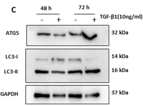
Application: WB    Species: Human    Sample: HTM Cells

Figure 1. TGF-β1 induced ATG5 activation in human trabecular meshwork (HTM cells). TGF-β1 (10 ng/mL) applied to primary HTM cells for 48–72 h, increased relative mRNA expression of (A) ATG-5 and (B) LC3-II. It also increased levels of (C) ATG-5 proteins and lipidation of LC3 II, (D,E) Densitometry analysis of bands. The results represent the averages of three separate tests conducted. The error bars represent the standard deviation

10). HSP90AA1 promotes the inflammation in human gingival fibroblasts induced by Porphyromonas gingivalis lipopolysaccharide via regulating of autophagy. BMC Oral Health (PubMed: 36028869) [IF=2.9]

Application: WB    Species: Human    Sample: HGFs

Fig. 2 Pg-LPS treatment increased HSP90AA1 protein and gene level, and elevated the expressions of autophagy related protein in HGFs. a, b The changes of HSP90AA1 protein and gene level. c Pg-LPS (0.1, 1, 10 μg/mL) treatment increased the expression of autophagy related protein in HGFs. Data were expressed as mean ± SD, n = 3. Compared to the control group, *P 

Load more

Restrictive clause


Affinity Biosciences tests all products strictly. Citations are provided as a resource for additional applications that have not been validated by Affinity Biosciences. Please choose the appropriate format for each application and consult Materials and Methods sections for additional details about the use of any product in these publications.

For Research Use Only.
Not for use in diagnostic or therapeutic procedures. Not for resale. Not for distribution without written consent. Affinity Biosciences will not be held responsible for patent infringement or other violations that may occur with the use of our products. Affinity Biosciences, Affinity Biosciences Logo and all other trademarks are the property of Affinity Biosciences LTD.