
 HepG2 cells were treated with the indicated concentrations of 25-HC for 24 hours before mRNA was extracted and expressions of TLR2, TLR4, TLR5, TLR7 and TLR9 were examined by RT-qPCR. (B) HepG2 cells were treated with the indicated concentrations of 25-HC for 24 hours or treated with 25-HC at 10 μM for the indicated times before protein was collected to detect the TLR2, TLR4, TLR5, TLR7 and TLR9 expressions by Western blotting. HepG2 cells were transfected with siNC or siTLR4 or siTLR9 for 24 hours, (C) then cells were treated with the indicated concentrations of 25-HC for 24 hours before proteins were collected and the expressions of MMP1, MMP2, MMP3 and MMP9 were determined by Western blotting. (D) Or migratory ability of HepG2 cells after treated with 25-HC for 36 hours was determined by Transwell assay. Results were obtained from 3 independent experiments and are expressed as the means ± SEM. Statistical significance was determined by Student’s t-test. *P<0.05, ***P<0.001.")
. Data were expressed as mean ± SD, n = 3. Compared with the control group, ▲P < 0.05 and ▲▲P < 0.01; compared with the COPD group, ★P < 0.05 and ★★P < 0.01.")


 The mRNA expression levels of IL-1α (A), IL-6 (B), COX2 (C), and TGF-α (D) in each group were determined by qPCR. (E,F) Immunofluorescence staining of ICAM1 (E) and TGF-α (F) in lung tissues from each group. Scale bar: 20 μm. (G) TUNEL staining showing the degree of apoptosis. Scale bar: 20 μm. (H) IHC staining of CD86, CD163, and Gr-1 in lung tissues from each group. Scale bar: 20 μm. (I) Protein expression levels of IL-1β, NOS2, TLR2, CD86, CD115, CD206, ARG1, and CD163 in each group, as detected by western blot analysis. Sham: control group; I/RI: mice whose superior mesenteric artery was completely clamped; I/RI + Ger: I/RI group administered with germacrone. Data are shown as the mean ± SD. *P")
 The mRNA expression levels of IL-1α (A), IL-6 (B), COX2 (C) and TGF-α (D) were determined by RT-qPCR in each group, respectively. (E-F) Immunofluorescence staining of ICAM1 (E) and TGF-α (F) in lung tissues of each group(400x), respectively. (G) TUNEL staining was employed to detect apoptosis level (400x). (H) IHC staining of CD86, CD163 and Gr-1 in lung tissues of each group. (I) The protein expression levels of IL-1β, NOS2, TLR2, CD86, CD115, CD206, ARG1, and CD163 were detected by Western blot in each group, respectively. Sham: control group, I/R+Con: chloral hydrate treated group, I/R+Ger: chloral hydrate treated group with Germacone administration. Data were shown as the mean ± SD.")
Control Products

Related Downloads
Protocols
Product Info
*The optimal dilutions should be determined by the end user. For optimal experimental results, antibody reuse is not recommended.
*Tips:
WB: For western blot detection of denatured protein samples. IHC: For immunohistochemical detection of paraffin sections (IHC-p) or frozen sections (IHC-f) of tissue samples. IF/ICC: For immunofluorescence detection of cell samples. ELISA(peptide): For ELISA detection of antigenic peptide.
Cite Format: Affinity Biosciences Cat# DF7002, RRID:AB_2838958.
Fold/Unfold
CD282; CD282 antigen; TIL 4; TIL4; TLR 2; TLR2; TLR2_HUMAN; Toll like receptor 2; Toll like receptor 2 precursor; Toll-like receptor 2; Toll/interleukin 1 receptor like 4; Toll/interleukin 1 receptor like protein 4; Toll/interleukin receptor like protein 4; Toll/interleukin-1 receptor-like protein 4;
Immunogens
A synthesized peptide derived from human TLR2, corresponding to a region within the internal amino acids.
Highly expressed in peripheral blood leukocytes, in particular in monocytes, in bone marrow, lymph node and in spleen. Also detected in lung and in fetal liver. Levels are low in other tissues.
- O60603 TLR2_HUMAN:
- Protein BLAST With
- NCBI/
- ExPASy/
- Uniprot
MPHTLWMVWVLGVIISLSKEESSNQASLSCDRNGICKGSSGSLNSIPSGLTEAVKSLDLSNNRITYISNSDLQRCVNLQALVLTSNGINTIEEDSFSSLGSLEHLDLSYNYLSNLSSSWFKPLSSLTFLNLLGNPYKTLGETSLFSHLTKLQILRVGNMDTFTKIQRKDFAGLTFLEELEIDASDLQSYEPKSLKSIQNVSHLILHMKQHILLLEIFVDVTSSVECLELRDTDLDTFHFSELSTGETNSLIKKFTFRNVKITDESLFQVMKLLNQISGLLELEFDDCTLNGVGNFRASDNDRVIDPGKVETLTIRRLHIPRFYLFYDLSTLYSLTERVKRITVENSKVFLVPCLLSQHLKSLEYLDLSENLMVEEYLKNSACEDAWPSLQTLILRQNHLASLEKTGETLLTLKNLTNIDISKNSFHSMPETCQWPEKMKYLNLSSTRIHSVTGCIPKTLEILDVSNNNLNLFSLNLPQLKELYISRNKLMTLPDASLLPMLLVLKISRNAITTFSKEQLDSFHTLKTLEAGGNNFICSCEFLSFTQEQQALAKVLIDWPANYLCDSPSHVRGQQVQDVRLSVSECHRTALVSGMCCALFLLILLTGVLCHRFHGLWYMKMMWAWLQAKRKPRKAPSRNICYDAFVSYSERDAYWVENLMVQELENFNPPFKLCLHKRDFIPGKWIIDNIIDSIEKSHKTVFVLSENFVKSEWCKYELDFSHFRLFDENNDAAILILLEPIEKKAIPQRFCKLRKIMNTKTYLEWPMDEAQREGFWVNLRAAIKS
Predictions
Score>80(red) has high confidence and is suggested to be used for WB detection. *The prediction model is mainly based on the alignment of immunogen sequences, the results are for reference only, not as the basis of quality assurance.
High(score>80) Medium(80>score>50) Low(score<50) No confidence
Research Backgrounds
Cooperates with LY96 to mediate the innate immune response to bacterial lipoproteins and other microbial cell wall components. Cooperates with TLR1 or TLR6 to mediate the innate immune response to bacterial lipoproteins or lipopeptides. Acts via MYD88 and TRAF6, leading to NF-kappa-B activation, cytokine secretion and the inflammatory response. May also activate immune cells and promote apoptosis in response to the lipid moiety of lipoproteins. Recognizes mycoplasmal macrophage-activating lipopeptide-2kD (MALP-2), soluble tuberculosis factor (STF), phenol-soluble modulin (PSM) and B.burgdorferi outer surface protein A lipoprotein (OspA-L) cooperatively with TLR6. Stimulation of monocytes in vitro with M.tuberculosis PstS1 induces p38 MAPK and ERK1/2 activation primarily via this receptor, but also partially via TLR4. MAPK activation in response to bacterial peptidoglycan also occurs via this receptor. Acts as a receptor for M.tuberculosis lipoproteins LprA, LprG, LpqH and PstS1, some lipoproteins are dependent on other coreceptors (TLR1, CD14 and/or CD36); the lipoproteins act as agonists to modulate antigen presenting cell functions in response to the pathogen. M.tuberculosis HSP70 (dnaK) but not HSP65 (groEL-2) acts via this protein to stimulate NF-kappa-B expression. Recognizes M.tuberculosis major T-antigen EsxA (ESAT-6) which inhibits downstream MYD88-dependent signaling (shown in mouse) (By similarity). Forms activation clusters composed of several receptors depending on the ligand, these clusters trigger signaling from the cell surface and subsequently are targeted to the Golgi in a lipid-raft dependent pathway. Forms the cluster TLR2:TLR6:CD14:CD36 in response to diacylated lipopeptides and TLR2:TLR1:CD14 in response to triacylated lipopeptides. Required for normal uptake of M.tuberculosis, a process that is inhibited by M.tuberculosis LppM (By similarity).
Glycosylation of Asn-442 is critical for secretion of the N-terminal ectodomain of TLR2.
Ubiquitinated at Lys-754 by PPP1R11, leading to its degradation. Deubiquitinated by USP2 (By similarity).
Membrane>Single-pass type I membrane protein. Cytoplasmic vesicle>Phagosome membrane>Single-pass type I membrane protein. Membrane raft.
Note: Does not reside in lipid rafts before stimulation but accumulates increasingly in the raft upon the presence of the microbial ligand. In response to diacylated lipoproteins, TLR2:TLR6 heterodimers are recruited in lipid rafts, this recruitment determines the intracellular targeting to the Golgi apparatus. Triacylated lipoproteins induce the same mechanism for TLR2:TLR1 heterodimers.
Highly expressed in peripheral blood leukocytes, in particular in monocytes, in bone marrow, lymph node and in spleen. Also detected in lung and in fetal liver. Levels are low in other tissues.
Ester-bound lipid substrates are bound through a crevice formed between the LRR 11 and LRR 12.
The ATG16L1-binding motif mediates interaction with ATG16L1.
The TIR domain mediates NAD(+) hydrolase (NADase) activity. Self-association of TIR domains is required for NADase activity.
Belongs to the Toll-like receptor family.
Research Fields
· Cellular Processes > Transport and catabolism > Phagosome. (View pathway)
· Environmental Information Processing > Signal transduction > PI3K-Akt signaling pathway. (View pathway)
· Human Diseases > Infectious diseases: Bacterial > Legionellosis.
· Human Diseases > Infectious diseases: Parasitic > Leishmaniasis.
· Human Diseases > Infectious diseases: Parasitic > Chagas disease (American trypanosomiasis).
· Human Diseases > Infectious diseases: Parasitic > Malaria.
· Human Diseases > Infectious diseases: Parasitic > Toxoplasmosis.
· Human Diseases > Infectious diseases: Parasitic > Amoebiasis.
· Human Diseases > Infectious diseases: Bacterial > Tuberculosis.
· Human Diseases > Infectious diseases: Viral > Hepatitis B.
· Human Diseases > Infectious diseases: Viral > Measles.
· Human Diseases > Infectious diseases: Viral > Herpes simplex infection.
· Human Diseases > Cancers: Overview > Proteoglycans in cancer.
· Human Diseases > Immune diseases > Inflammatory bowel disease (IBD).
· Human Diseases > Immune diseases > Rheumatoid arthritis.
· Organismal Systems > Immune system > Toll-like receptor signaling pathway. (View pathway)
References
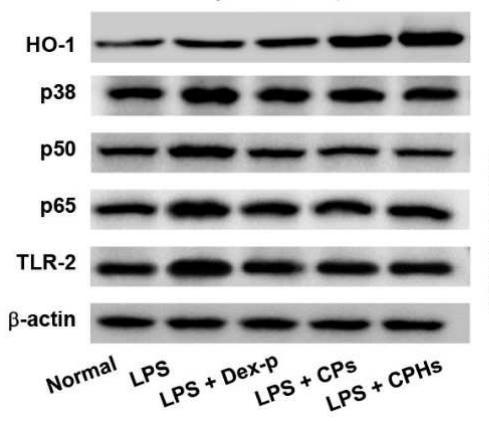
Application: WB Species: mouse Sample: Macrophages

Application: WB Species: Rat Sample:
Application: WB Species: rat Sample:
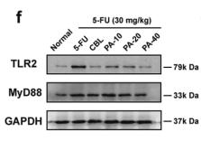
Application: WB Species: mouse Sample: macrophages

Application: WB Species: Mice Sample:
Application: WB Species: Mouse Sample: RAW264.7 cells
Restrictive clause
Affinity Biosciences tests all products strictly. Citations are provided as a resource for additional applications that have not been validated by Affinity Biosciences. Please choose the appropriate format for each application and consult Materials and Methods sections for additional details about the use of any product in these publications.
For Research Use Only.
Not for use in diagnostic or therapeutic procedures. Not for resale. Not for distribution without written consent. Affinity Biosciences will not be held responsible for patent infringement or other violations that may occur with the use of our products. Affinity Biosciences, Affinity Biosciences Logo and all other trademarks are the property of Affinity Biosciences LTD.