, using Phospho-MITF (Ser180) Antibody at 1/1000 dilution.
5ug/NC membrane strip.
Exposure for 2min with Affinity™ ECL Kit(#KF8003).
Bands result from membrane strip incubation.")
, using Phospho-MITF (Ser180) Antibody at 1/1000 dilution.
5ug/NC membrane strip.
Exposure for 1min with Affinity™ ECL Kit(#KF8001).
Bands result from membrane strip incubation.")

| Product: | Phospho-MITF (Ser180) Antibody |
| Catalog: | AF3027 |
| Description: | Rabbit polyclonal antibody to Phospho-MITF (Ser180) |
| Application: | WB IHC IF/ICC |
| Cited expt.: | WB |
| Reactivity: | Human, Mouse, Rat, Monkey |
| Prediction: | Pig, Bovine, Sheep, Rabbit, Dog, Xenopus |
| Mol.Wt.: | 50~80kDa(Observed); 59kD(Calculated). |
| Uniprot: | O75030 |
| RRID: | AB_2834458 |
Control Products

Product Info
*The optimal dilutions should be determined by the end user. For optimal experimental results, antibody reuse is not recommended.
*Tips:
WB: For western blot detection of denatured protein samples. IHC: For immunohistochemical detection of paraffin sections (IHC-p) or frozen sections (IHC-f) of tissue samples. IF/ICC: For immunofluorescence detection of cell samples. ELISA(peptide): For ELISA detection of antigenic peptide.
Cite Format: Affinity Biosciences Cat# AF3027, RRID:AB_2834458.
Fold/Unfold
BHLHE32; bHLHe32; Class E basic helix-loop-helix protein 32; CMM8; Homolog of mouse microphthalmia; Mi; Microphthalmia associated transcription factor; Microphthalmia, mouse, homolog of; Microphthalmia-associated transcription factor; MITF; MITF_HUMAN; mitfa; nacre; WS2; WS2A; z3A.1;
Immunogens
A synthesized peptide derived from human MITF around the phosphorylation site of Ser180.
Isoform M is exclusively expressed in melanocytes and melanoma cells. Isoform A and isoform H are widely expressed in many cell types including melanocytes and retinal pigment epithelium (RPE). Isoform C is expressed in many cell types including RPE but not in melanocyte-lineage cells. Isoform Mdel is widely expressed in melanocytes, melanoma cell lines and tissues, but almost undetectable in non-melanoma cell lines.
- O75030 MITF_HUMAN:
- Protein BLAST With
- NCBI/
- ExPASy/
- Uniprot
MQSESGIVPDFEVGEEFHEEPKTYYELKSQPLKSSSSAEHPGASKPPISSSSMTSRILLRQQLMREQMQEQERREQQQKLQAAQFMQQRVPVSQTPAINVSVPTTLPSATQVPMEVLKVQTHLENPTKYHIQQAQRQQVKQYLSTTLANKHANQVLSLPCPNQPGDHVMPPVPGSSAPNSPMAMLTLNSNCEKEGFYKFEEQNRAESECPGMNTHSRASCMQMDDVIDDIISLESSYNEEILGLMDPALQMANTLPVSGNLIDLYGNQGLPPPGLTISNSCPANLPNIKRELTACIFPTESEARALAKERQKKDNHNLIERRRRFNINDRIKELGTLIPKSNDPDMRWNKGTILKASVDYIRKLQREQQRAKELENRQKKLEHANRHLLLRIQELEMQARAHGLSLIPSTGLCSPDLVNRIIKQEPVLENCSQDLLQHHADLTCTTTLDLTDGTITFNNNLGTGTEANQAYSVPTKMGSKLEDILMDDTLSPVGVTDPLLSSVSPGASKTSSRRSSMSMEETEHTC
Predictions
Score>80(red) has high confidence and is suggested to be used for WB detection. *The prediction model is mainly based on the alignment of immunogen sequences, the results are for reference only, not as the basis of quality assurance.
High(score>80) Medium(80>score>50) Low(score<50) No confidence
Research Backgrounds
Transcription factor that regulates the expression of genes with essential roles in cell differentiation, proliferation and survival. Binds to M-boxes (5'-TCATGTG-3') and symmetrical DNA sequences (E-boxes) (5'-CACGTG-3') found in the promoters of target genes, such as BCL2 and tyrosinase (TYR). Plays an important role in melanocyte development by regulating the expression of tyrosinase (TYR) and tyrosinase-related protein 1 (TYRP1). Plays a critical role in the differentiation of various cell types, such as neural crest-derived melanocytes, mast cells, osteoclasts and optic cup-derived retinal pigment epithelium.
Phosphorylation at Ser-405 significantly enhances the ability to bind the tyrosinase promoter. Phosphorylated at Ser-180 and Ser-516 following KIT signaling, trigerring a short live activation: Phosphorylation at Ser-180 and Ser-516 by MAPK and RPS6KA1, respectively, activate the transcription factor activity but also promote ubiquitination and subsequent degradation by the proteasome.
Ubiquitinated following phosphorylation at Ser-180, leading to subsequent degradation by the proteasome. Deubiquitinated by USP13, preventing its degradation.
Nucleus.
Isoform M is exclusively expressed in melanocytes and melanoma cells. Isoform A and isoform H are widely expressed in many cell types including melanocytes and retinal pigment epithelium (RPE). Isoform C is expressed in many cell types including RPE but not in melanocyte-lineage cells. Isoform Mdel is widely expressed in melanocytes, melanoma cell lines and tissues, but almost undetectable in non-melanoma cell lines.
The leucine zipper region is part of a larger coiled coil.
Belongs to the MiT/TFE family.
Research Fields
· Human Diseases > Cancers: Overview > Pathways in cancer. (View pathway)
· Human Diseases > Cancers: Overview > Transcriptional misregulation in cancer.
· Human Diseases > Cancers: Specific types > Melanoma. (View pathway)
· Organismal Systems > Development > Osteoclast differentiation. (View pathway)
· Organismal Systems > Endocrine system > Melanogenesis.
References

Application: WB Species: snake Sample:
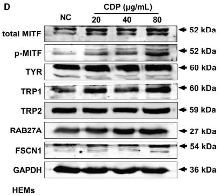
Application: WB Species: human Sample: HEMs

Application: WB Species: clams Sample: haemocytes
Application: WB Species: zebrafish Sample:
Restrictive clause
Affinity Biosciences tests all products strictly. Citations are provided as a resource for additional applications that have not been validated by Affinity Biosciences. Please choose the appropriate format for each application and consult Materials and Methods sections for additional details about the use of any product in these publications.
For Research Use Only.
Not for use in diagnostic or therapeutic procedures. Not for resale. Not for distribution without written consent. Affinity Biosciences will not be held responsible for patent infringement or other violations that may occur with the use of our products. Affinity Biosciences, Affinity Biosciences Logo and all other trademarks are the property of Affinity Biosciences LTD.