Product: MDM2 Antibody
Catalog: AF0208
Description: Rabbit polyclonal antibody to MDM2
Application: WB IHC IF/ICC IP
Reactivity: Human, Mouse, Rat, Monkey
Prediction: Pig, Bovine, Horse, Chicken
Mol.Wt.: 45~55kD,70~95kD; 55kD(Calculated).
Uniprot: Q00987
RRID: AB_2833395

View similar products>>

   Size Price Inventory
 100ul $280 In stock
 200ul $350 In stock

Lead Time: Same day delivery

For pricing and ordering contact:
Local distributors

Product Info

Source:
Rabbit
Application:
WB 1:500-1:3000, IHC 1:50-1:200, IF/ICC: 1:100-1:500
*The optimal dilutions should be determined by the end user.
*Tips:

WB: For western blot detection of denatured protein samples. IHC: For immunohistochemical detection of paraffin sections (IHC-p) or frozen sections (IHC-f) of tissue samples. IF/ICC: For immunofluorescence detection of cell samples. ELISA(peptide): For ELISA detection of antigenic peptide.

Reactivity:
Human,Mouse,Rat,Monkey
Prediction:
Pig(88%), Bovine(88%), Horse(88%), Chicken(86%)
Clonality:
Polyclonal
Specificity:
MDM2 Antibody detects endogenous levels of total MDM2.
RRID:
AB_2833395
Cite Format: Affinity Biosciences Cat# AF0208, RRID:AB_2833395.
Conjugate:
Unconjugated.
Purification:
The antiserum was purified by peptide affinity chromatography using SulfoLink™ Coupling Resin (Thermo Fisher Scientific).
Storage:
Rabbit IgG in phosphate buffered saline , pH 7.4, 150mM NaCl, 0.02% sodium azide and 50% glycerol. Store at -20 °C. Stable for 12 months from date of receipt.
Alias:

Fold/Unfold

ACTFS; Double minute 2 protein; E3 ubiquitin-protein ligase Mdm2; Hdm 2; Hdm2; HDMX; MDM 2; MDM2; MDM2 oncogene E3 ubiquitin protein ligase; Mdm2 p53 E3 ubiquitin protein ligase homolog; Mdm2 transformed 3T3 cell double minute 2 p53 binding protein (mouse) binding protein 104kDa; MDM2_HUMAN; MDM2BP; Mouse Double Minute 2; MTBP; Murine Double Minute Chromosome 2; Oncoprotein Mdm2; p53 Binding Protein Mdm2; p53-binding protein Mdm2; Ubiquitin protein ligase E3 Mdm2; Ubiquitin protein ligase E3 Mdm2;

Immunogens

Immunogen:
Uniprot:
Gene(ID):
Expression:
Q00987 MDM2_HUMAN:

Ubiquitous. Isoform Mdm2-A, isoform Mdm2-B, isoform Mdm2-C, isoform Mdm2-D, isoform Mdm2-E, isoform Mdm2-F and isoform Mdm2-G are observed in a range of cancers but absent in normal tissues.

Description:
MDM2 a ubiquitin ligase for p53, plays a central role in regulation of the stability of p53 via Akt-mediated MDM2 phosphorylation. Phosphorylation of MDM2 increases its interaction with p300, providing a platform to allow the assembly of the protein complex necessary for MDM2-mediated ubiquitination and degradation of p53. Phosphorylation of MDM2 also blocks its binding to p19ARF, increasing the degradation of p53. Facilitates the nuclear export of p53 and targets it for proteasome-mediated proteolysis. Eight alternatively spliced isoforms have been reported.
Sequence:
MCNTNMSVPTDGAVTTSQIPASEQETLVRPKPLLLKLLKSVGAQKDTYTMKEVLFYLGQYIMTKRLYDEKQQHIVYCSNDLLGDLFGVPSFSVKEHRKIYTMIYRNLVVVNQQESSDSGTSVSENRCHLEGGSDQKDLVQELQEEKPSSSHLVSRPSTSSRRRAISETEENSDELSGERQRKRHKSDSISLSFDESLALCVIREICCERSSSSESTGTPSNPDLDAGVSEHSGDWLDQDSVSDQFSVEFEVESLDSEDYSLSEEGQELSDEDDEVYQVTVYQAGESDTDSFEEDPEISLADYWKCTSCNEMNPPLPSHCNRCWALRENWLPEDKGKDKGEISEKAKLENSTQAEEGFDVPDCKKTIVNDSRESCVEENDDKITQASQSQESEDYSQPSTSSSIIYSSQEDVKEFEREETQDKEESVESSLPLNAIEPCVICQGRPKNGCIVHGKTGHLMACFTCAKKLKKRNKPCPVCRQPIQMIVLTYFP

Predictions

Predictions:

Score>80(red) has high confidence and is suggested to be used for WB detection. *The prediction model is mainly based on the alignment of immunogen sequences, the results are for reference only, not as the basis of quality assurance.

Species
Results
Score
Pig
88
Horse
88
Bovine
88
Chicken
86
Dog
75
Sheep
0
Xenopus
0
Zebrafish
0
Rabbit
0
Model Confidence:
High(score>80) Medium(80>score>50) Low(score<50) No confidence

PTMs - Q00987 As Substrate

Site PTM Type Enzyme
Ubiquitination
T4 Phosphorylation
S17 Phosphorylation
K36 Ubiquitination
K39 Ubiquitination
K45 Ubiquitination
Y60 Phosphorylation
K70 Ubiquitination
C77 S-Nitrosylation
K94 Ubiquitination
K98 Ubiquitination
S118 Phosphorylation P48730 (CSNK1D)
S121 Phosphorylation P48730 (CSNK1D)
K146 Ubiquitination
S148 Phosphorylation
S157 Phosphorylation P49137 (MAPKAPK2)
T158 Phosphorylation Q9NQU5 (PAK6)
S160 Phosphorylation
S166 Phosphorylation P48730 (CSNK1D) , P11309 (PIM1) , O00141 (SGK1) , P31749 (AKT1) , P49137 (MAPKAPK2) , P31751 (AKT2) , Q9P1W9 (PIM2) , O43293 (DAPK3)
T168 Phosphorylation
S172 Phosphorylation P48730 (CSNK1D)
S176 Phosphorylation P48730 (CSNK1D)
K182 Acetylation
K185 Acetylation
S186 Phosphorylation Q9NQU5 (PAK6) , Q9P1W9 (PIM2) , P11309 (PIM1) , O43293 (DAPK3) , P31749 (AKT1) , P31751 (AKT2)
S188 Phosphorylation P31749 (AKT1)
S190 Phosphorylation
S192 Phosphorylation
S196 Phosphorylation
T216 Phosphorylation
T218 Phosphorylation
S220 Phosphorylation P48730 (CSNK1D)
S229 Phosphorylation P48730 (CSNK1D)
S240 Phosphorylation P48730 (CSNK1D)
S242 Phosphorylation
S246 Phosphorylation P48730 (CSNK1D)
S253 Phosphorylation P48730 (CSNK1D)
S256 Phosphorylation
S260 Phosphorylation P68400 (CSNK2A1) , P48730 (CSNK1D) , P53350 (PLK1)
S262 Phosphorylation P48730 (CSNK1D)
S269 Phosphorylation P48730 (CSNK1D) , P68400 (CSNK2A1)
Y276 Phosphorylation P00519 (ABL1)
T279 Phosphorylation P48730 (CSNK1D)
Y281 Phosphorylation P12931 (SRC)
S286 Phosphorylation
T288 Phosphorylation
S290 Phosphorylation P48730 (CSNK1D)
Y302 Phosphorylation P12931 (SRC)
T306 Phosphorylation
S307 Phosphorylation
K334 Ubiquitination
K336 Ubiquitination
K338 Ubiquitination
S342 Phosphorylation P48730 (CSNK1D)
K344 Ubiquitination
K346 Ubiquitination
S350 Phosphorylation P48730 (CSNK1D)
T351 Phosphorylation P48730 (CSNK1D)
K363 Ubiquitination
K364 Ubiquitination
T365 Phosphorylation P48730 (CSNK1D)
S373 Phosphorylation P48730 (CSNK1D)
S386 Phosphorylation Q13315 (ATM) , P48730 (CSNK1D)
S388 Phosphorylation
Y394 Phosphorylation A0A173G4P4 (Abl fusion) , P00519 (ABL1)
S395 Phosphorylation Q13315 (ATM)
Y405 Phosphorylation P00519 (ABL1)
S406 Phosphorylation
S407 Phosphorylation Q13535 (ATR)
T419 Phosphorylation P48730 (CSNK1D)
S425 Phosphorylation
S429 Phosphorylation Q13315 (ATM)
K446 Sumoylation
K446 Ubiquitination
K454 Ubiquitination
K466 Ubiquitination
K467 Ubiquitination
K473 Ubiquitination

Research Backgrounds

Function:

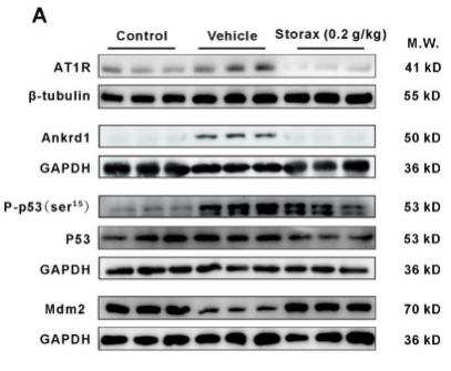
E3 ubiquitin-protein ligase that mediates ubiquitination of p53/TP53, leading to its degradation by the proteasome. Inhibits p53/TP53- and p73/TP73-mediated cell cycle arrest and apoptosis by binding its transcriptional activation domain. Also acts as a ubiquitin ligase E3 toward itself and ARRB1. Permits the nuclear export of p53/TP53. Promotes proteasome-dependent ubiquitin-independent degradation of retinoblastoma RB1 protein. Inhibits DAXX-mediated apoptosis by inducing its ubiquitination and degradation. Component of the TRIM28/KAP1-MDM2-p53/TP53 complex involved in stabilizing p53/TP53. Also component of the TRIM28/KAP1-ERBB4-MDM2 complex which links growth factor and DNA damage response pathways. Mediates ubiquitination and subsequent proteasome degradation of DYRK2 in nucleus. Ubiquitinates IGF1R and SNAI1 and promotes them to proteasomal degradation. Ubiquitinates DCX, leading to DCX degradation and reduction of the dendritic spine density of olfactory bulb granule cells (By similarity). Ubiquitinates DLG4, leading to proteasomal degradation of DLG4 which is required for AMPA receptor endocytosis (By similarity).

PTMs:

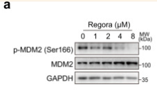
Phosphorylation on Ser-166 by SGK1 activates ubiquitination of p53/TP53. Phosphorylated at multiple sites near the RING domain by ATM upon DNA damage; this prevents oligomerization and E3 ligase processivity and impedes constitutive p53/TP53 degradation.

Autoubiquitination leads to proteasomal degradation; resulting in p53/TP53 activation it may be regulated by SFN. Also ubiquitinated by TRIM13. Deubiquitinated by USP2 leads to its accumulation and increases deubiquitination and degradation of p53/TP53. Deubiquitinated by USP7 leading to its stabilization.

Subcellular Location:

Nucleus>Nucleoplasm. Cytoplasm. Nucleus>Nucleolus.
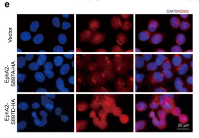
Note: Expressed predominantly in the nucleoplasm. Interaction with ARF(P14) results in the localization of both proteins to the nucleolus. The nucleolar localization signals in both ARF(P14) and MDM2 may be necessary to allow efficient nucleolar localization of both proteins. Colocalizes with RASSF1 isoform A in the nucleus.

Extracellular region or secreted Cytosol Plasma membrane Cytoskeleton Lysosome Endosome Peroxisome ER Golgi apparatus Nucleus Mitochondrion Manual annotation Automatic computational assertionSubcellular location
Tissue Specificity:

Ubiquitous. Isoform Mdm2-A, isoform Mdm2-B, isoform Mdm2-C, isoform Mdm2-D, isoform Mdm2-E, isoform Mdm2-F and isoform Mdm2-G are observed in a range of cancers but absent in normal tissues.

Subunit Structure:

Interacts with p53/TP53, TP73/p73, RBL5 and RP11. Binds specifically to RNA. Can interact with RB1, E1A-associated protein EP300 and the E2F1 transcription factor. Forms a ternary complex with p53/TP53 and WWOX. Interacts with CDKN2AIP, RFWD3, USP7, PYHIN1, and RBBP6. Interacts with ARRB1 and ARRB2. Interacts with PSMA3. Found in a trimeric complex with MDM2, MDM4 and USP2. Interacts with USP2 (via N-terminus and C-terminus). Interacts with MDM4. Part of a complex with MDM2, DAXX, RASSF1 and USP7. Part of a complex with DAXX, MDM2 and USP7. Interacts directly with DAXX and USP7. Interacts (via C-terminus) with RASSF1 isoform A (via N-terminus); the interaction is independent of TP53. Interacts with APEX1; leading to its ubiquitination and degradation. Interacts with RYBP; this inhibits ubiquitination of TP53. Identified in a complex with RYBP and p53/TP53. Also component of the TRIM28/KAP1-MDM2-p53/TP53 complex involved in regulating p53/TP53 stabilization and activity. Binds directly both p53/TP53 and TRIM28. Component of the TRIM28/KAP1-ERBB4-MDM2 complex involved in connecting growth factor responses with DNA damage. Interacts directly with both TRIM28 and ERBB4 in the complex. Interacts with DYRK2. Interacts with IGF1R. Interacts with TRIM13; the interaction ubiquitinates MDM2 leading to its proteasomal degradation. Interacts with SNAI1; this interaction promotes SNAI1 ubiquitination. Interacts with NOTCH1 (via intracellular domain). Interacts with FHIT. Interacts with RFFL and RNF34; the interaction stabilizes MDM2. Interacts with CDK5RAP3 and CDKN2A/ARF; form a ternary complex involved in regulation of p53/TP53. Interacts with MTA1. Interacts with AARB2. Interacts with MTBP. Interacts with PML. Interacts with TBRG1. Interacts with the 5S RNP which is composed of the 5S RNA, RPL5 and RPL11; the interaction is direct, occurs in the nucleoplasm and negatively regulates MDM2-mediated TP53 ubiquitination and degradation. Interacts with ADGRB1; the interaction results in inhibition of MDM2-mediated ubiquitination and degradation of DLG4/PSD95, promoting DLG4 stability and regulating synaptic plasticity (By similarity). Interacts with RPL23A; this interaction may promote p53/TP53 polyubiquitination.

(Microbial infection) Interacts with herpes virus 8 protein v-IRF4.

(Microbial infection) Interacts with and ubiquitinates HIV-1 Tat.

Family&Domains:

Region I is sufficient for binding p53 and inhibiting its G1 arrest and apoptosis functions. It also binds p73 and E2F1. Region II contains most of a central acidic region required for interaction with ribosomal protein L5 and a putative C4-type zinc finger. The RING finger domain which coordinates two molecules of zinc interacts specifically with RNA whether or not zinc is present and mediates the heterooligomerization with MDM4. It is also essential for its ubiquitin ligase E3 activity toward p53 and itself.

Belongs to the MDM2/MDM4 family.

References

1). Regorafenib inhibits EphA2 phosphorylation and leads to liver damage via the ERK/MDM2/p53 axis. Nature Communications (PubMed: 37179400) [IF=16.6]

Application: WB    Species: Human    Sample: HL-7702 cells

Fig. 5 The phosphorylation of EphA2Ser897 regulates p53 by ERK/MDM2 axis. a HL-7702 cells were treated with regorafenib as indicated. The expression levels of p-MDM2 (Ser166) and MDM2 were analyzed by western blot. n = 4 independent experiments. b C57BL/6J mice were treated with or without 400 mg/kg/day regorafenib for 6 weeks. The expression levels of p-MDM2 (Ser166) and p53 in liver tissues were analyzed by western blot (n = 5 per group). c HL-7702 cells treated with 8 μM regorafenib for 24 h (n = 3 independent experiments) or liver tissues (n = 3 per group) were used to detect the expression level of Lamin B1 and MDM2 in whole cell, cytoplasm and nuclei by western blot. d, e HL-7702 cells transfected with vector, EphA2 S897A plasmid or EphA2 S897D plasmid as indicated for 24 h. d The expression levels of HA, p-MDM2 (Ser166) and p53 were analyzed by western blot. e The localization of MDM2 was observed by immunofluorescence. Representative images are shown from three independent experiments. Scale bar: 20 µm. f HL-7702 cells were treated with ALW-II-41-27 or bosutinib as indicated for 24 h. Related proteins were analyzed by western blot. Blots are representative of three independent experiments. g HL-7702 cells were treated with regorafenib as indicated for 24 h. The expression levels of related proteins were analyzed by western blot. The relative level of p-ERK (Tyr204) was analyzed by densitometric analysis. n = 3 independent experiments. h HL-7702 cells were treated with regorafenib as indicated. The expression levels of related proteins were analyzed by western blot. The fold change of pS-EphA2/EphA2 and pY-ERK/ERK were analyzed by densitometric analysis. n = 3 independent experiments. Data were expressed as mean ± SD. Unpaired two-sided Student’s t test for b and c or one way ANOVA followed by Tukey post hoc test for a and g. Source data are provided as a Source Data file. Regora regorafenib, MW molecular weight, WCL whole cell lysate, CE cytoplasmic extraction, NE nuclear extraction.

Application: IF/ICC    Species: Human    Sample: HL-7702 cells

Fig. 5 The phosphorylation of EphA2Ser897 regulates p53 by ERK/MDM2 axis. a HL-7702 cells were treated with regorafenib as indicated. The expression levels of p-MDM2 (Ser166) and MDM2 were analyzed by western blot. n = 4 independent experiments. b C57BL/6J mice were treated with or without 400 mg/kg/day regorafenib for 6 weeks. The expression levels of p-MDM2 (Ser166) and p53 in liver tissues were analyzed by western blot (n = 5 per group). c HL-7702 cells treated with 8 μM regorafenib for 24 h (n = 3 independent experiments) or liver tissues (n = 3 per group) were used to detect the expression level of Lamin B1 and MDM2 in whole cell, cytoplasm and nuclei by western blot. d, e HL-7702 cells transfected with vector, EphA2 S897A plasmid or EphA2 S897D plasmid as indicated for 24 h. d The expression levels of HA, p-MDM2 (Ser166) and p53 were analyzed by western blot. e The localization of MDM2 was observed by immunofluorescence. Representative images are shown from three independent experiments. Scale bar: 20 µm. f HL-7702 cells were treated with ALW-II-41-27 or bosutinib as indicated for 24 h. Related proteins were analyzed by western blot. Blots are representative of three independent experiments. g HL-7702 cells were treated with regorafenib as indicated for 24 h. The expression levels of related proteins were analyzed by western blot. The relative level of p-ERK (Tyr204) was analyzed by densitometric analysis. n = 3 independent experiments. h HL-7702 cells were treated with regorafenib as indicated. The expression levels of related proteins were analyzed by western blot. The fold change of pS-EphA2/EphA2 and pY-ERK/ERK were analyzed by densitometric analysis. n = 3 independent experiments. Data were expressed as mean ± SD. Unpaired two-sided Student’s t test for b and c or one way ANOVA followed by Tukey post hoc test for a and g. Source data are provided as a Source Data file. Regora regorafenib, MW molecular weight, WCL whole cell lysate, CE cytoplasmic extraction, NE nuclear extraction.

2). Xinbao Pill attenuated chronic heart failure by suppressing the ubiquitination of β-adrenergic receptors. Phytomedicine (PubMed: 37149964) [IF=7.9]

3). Pimozide inhibits the growth of breast cancer cells by alleviating the Warburg effect through the P53 signaling pathway. BIOMEDICINE & PHARMACOTHERAPY (PubMed: 35658233) [IF=7.5]

Application: WB    Species: Human    Sample: MCF-7 cells

Fig. 5. Pimozide promotes the expression of p53 through PI3K/Akt/MDM2 pathway. (A-B) Cells were treated with the indicated concentrations of Pimozide for 24 h, the protein expression of p-PI3K, PI3K, p-Akt, Akt, p-MDM2, MDM2 in MCF-7 (A) and MDA-MB-231 (B) cells were determined by Western blot analysis (left panel). Densitometry analysis was performed to assess the protein expression of p-PI3K/PI3K, p-Akt/Akt, p-MDM2/MDM2 (normalized to β-actin expression) (right panel). (C-F) Western blot analysis for p-PI3K, PI3K, p-Akt, Akt, p-MDM2, MDM2, P53, and PKM2 protein levels in MCF-7 (C, E) and MDA-MB-231 (D, F) cells incubated with PI3K inhibitor LY294002 or PI3K agonist SC79 as indicated. (G) After treatment with SC79, colony-forming assay images (left panel) and quantification of colony number percentages (right panel). Data represent mean ± SD from three biological replicates (*p < 0.05, **p < 0.01, ns: not significant).

4). Storax Attenuates Cardiac Fibrosis following Acute Myocardial Infarction in Rats via Suppression of AT1R–Ankrd1–P53 Signaling Pathway. INTERNATIONAL JOURNAL OF MOLECULAR SCIENCES (PubMed: 36361958) [IF=5.6]

Application: WB    Species: Rat    Sample: cardiac tissue

Figure 8 Storax inhibits the AT1R–Ankrd1–P53 pathway in AMI rats against cardiomyocyte apoptosis. (A) WB bands showing the protein expression levels of AT1R, Ankrd1, P-p53 (ser15), P53 and Mdm2 in cardiac tissue. (B–E) Relative protein for AT1R, Ankrd1, P-p53 (ser15) and Mdm2 were quantified by densitometry based on immunoblot images. Results are presented as mean ± SD (n = 3). One-way ANOVA follow by Bonferroni’s post hoc test: # p < 0.05, ## p < 0.01 vs. vehicle group.

5). Casein kinase 1α inhibits p53 downstream of MDM2‑mediated autophagy and apoptosis in acute myeloid leukemia. ONCOLOGY REPORTS (PubMed: 32901886) [IF=4.2]

Application: WB    Species: human    Sample: HEL cells.

Figure 5. |Autophagy inhibitor Spautin-1 aggravates apoptosis induced by D4476, and CK1α inhibits autophagy by targeting the p53/AMPK/mTOR signaling pathway downstream of MDM2 in AML cells. (C) Co‑immunoprecipitation showed that CK1α interacted with MDM2 and p53 in HEL cells. Ten percent whole cell lysate was used as input samples.

6). Linc-ROR promotes arsenite-transformed keratinocyte proliferation by inhibiting P53 activity. Metallomics (PubMed: 32373892) [IF=3.4]

Application: WB    Species: human    Sample: HaCaT cells

Fig. 3 | P53 activity was decreased with arsenite-induced transformation of HaCaT cells. HaCaT cells were continuously maintained in medium containing 1 mM sodium arsenite for 0, 20, 35 and 50 passages. (a) The relative level of P53 mRNA in 0, 20, 35 and 50 passages of cells, with the control group set to 1 (the same hereinafter). (b–d) The western blotting of P53 and MDM2 proteins; GAPDH served as the loading control.

Restrictive clause


Affinity Biosciences tests all products strictly. Citations are provided as a resource for additional applications that have not been validated by Affinity Biosciences. Please choose the appropriate format for each application and consult Materials and Methods sections for additional details about the use of any product in these publications.

For Research Use Only.
Not for use in diagnostic or therapeutic procedures. Not for resale. Not for distribution without written consent. Affinity Biosciences will not be held responsible for patent infringement or other violations that may occur with the use of our products. Affinity Biosciences, Affinity Biosciences Logo and all other trademarks are the property of Affinity Biosciences LTD.