
| Product: | Phospho-4E-BP1 (Thr45) Antibody |
| Catalog: | AF3432 |
| Description: | Rabbit polyclonal antibody to Phospho-4E-BP1 (Thr45) |
| Application: | WB IHC IF/ICC |
| Cited expt.: | WB |
| Reactivity: | Human, Mouse, Rat |
| Prediction: | Pig, Zebrafish, Bovine, Horse, Sheep, Rabbit, Dog, Chicken |
| Mol.Wt.: | 18kDa(Observed); 13kD(Calculated). |
| Uniprot: | Q13541 |
| RRID: | AB_2834874 |
Control Products

Product Info
*The optimal dilutions should be determined by the end user. For optimal experimental results, antibody reuse is not recommended.
*Tips:
WB: For western blot detection of denatured protein samples. IHC: For immunohistochemical detection of paraffin sections (IHC-p) or frozen sections (IHC-f) of tissue samples. IF/ICC: For immunofluorescence detection of cell samples. ELISA(peptide): For ELISA detection of antigenic peptide.
Cite Format: Affinity Biosciences Cat# AF3432, RRID:AB_2834874.
Fold/Unfold
4E-BP1; 4EBP1; 4EBP1_HUMAN; BP 1; eIF4E binding protein 1; eIF4E-binding protein 1; Eif4ebp1; Eukaryotic translation initiation factor 4E-binding protein 1; PHAS-I; PHASI; Phosphorylated heat- and acid-stable protein regulated by insulin 1;
Immunogens
A synthesized peptide derived from human 4E-BP1 around the phosphorylation site of Thr45.
- Q13541 4EBP1_HUMAN:
- Protein BLAST With
- NCBI/
- ExPASy/
- Uniprot
MSGGSSCSQTPSRAIPATRRVVLGDGVQLPPGDYSTTPGGTLFSTTPGGTRIIYDRKFLMECRNSPVTKTPPRDLPTIPGVTSPSSDEPPMEASQSHLRNSPEDKRAGGEESQFEMDI
Predictions
Score>80(red) has high confidence and is suggested to be used for WB detection. *The prediction model is mainly based on the alignment of immunogen sequences, the results are for reference only, not as the basis of quality assurance.
High(score>80) Medium(80>score>50) Low(score<50) No confidence
Research Backgrounds
Repressor of translation initiation that regulates EIF4E activity by preventing its assembly into the eIF4F complex: hypophosphorylated form competes with EIF4G1/EIF4G3 and strongly binds to EIF4E, leading to repress translation. In contrast, hyperphosphorylated form dissociates from EIF4E, allowing interaction between EIF4G1/EIF4G3 and EIF4E, leading to initiation of translation. Mediates the regulation of protein translation by hormones, growth factors and other stimuli that signal through the MAP kinase and mTORC1 pathways.
Phosphorylated on serine and threonine residues in response to insulin, EGF and PDGF. Phosphorylation at Thr-37, Thr-46, Ser-65 and Thr-70, corresponding to the hyperphosphorylated form, is regulated by mTORC1 and abolishes binding to EIF4E.
Ubiquitinated: when eIF4E levels are low, hypophosphorylated form is ubiquitinated by the BCR(KLHL25) complex, leading to its degradation and serving as a homeostatic mechanism to maintain translation and prevent eIF4E inhibition when eIF4E levels are low. Not ubiquitinated when hyperphosphorylated (at Thr-37, Thr-46, Ser-65 and Thr-70) or associated with eIF4E.
The TOS motif mediates interaction with RPTOR, leading to promote phosphorylation by mTORC1 complex.
Belongs to the eIF4E-binding protein family.
Research Fields
· Cellular Processes > Cell growth and death > Cellular senescence. (View pathway)
· Environmental Information Processing > Signal transduction > ErbB signaling pathway. (View pathway)
· Environmental Information Processing > Signal transduction > HIF-1 signaling pathway. (View pathway)
· Environmental Information Processing > Signal transduction > mTOR signaling pathway. (View pathway)
· Environmental Information Processing > Signal transduction > PI3K-Akt signaling pathway. (View pathway)
· Environmental Information Processing > Signal transduction > AMPK signaling pathway. (View pathway)
· Genetic Information Processing > Translation > RNA transport.
· Human Diseases > Drug resistance: Antineoplastic > EGFR tyrosine kinase inhibitor resistance.
· Human Diseases > Infectious diseases: Viral > Human papillomavirus infection.
· Human Diseases > Cancers: Specific types > Acute myeloid leukemia. (View pathway)
· Human Diseases > Cancers: Overview > Choline metabolism in cancer. (View pathway)
· Organismal Systems > Aging > Longevity regulating pathway. (View pathway)
· Organismal Systems > Endocrine system > Insulin signaling pathway. (View pathway)
References
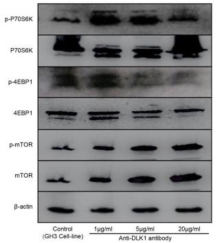
Application: WB Species: Rat Sample: GH3 cell

Application: WB Species: mouse Sample: mouse
Restrictive clause
Affinity Biosciences tests all products strictly. Citations are provided as a resource for additional applications that have not been validated by Affinity Biosciences. Please choose the appropriate format for each application and consult Materials and Methods sections for additional details about the use of any product in these publications.
For Research Use Only.
Not for use in diagnostic or therapeutic procedures. Not for resale. Not for distribution without written consent. Affinity Biosciences will not be held responsible for patent infringement or other violations that may occur with the use of our products. Affinity Biosciences, Affinity Biosciences Logo and all other trademarks are the property of Affinity Biosciences LTD.