

| Product: | SDF 1 Antibody |
| Catalog: | AF5166 |
| Description: | Rabbit polyclonal antibody to SDF 1 |
| Application: | WB IHC |
| Cited expt.: | WB, IHC |
| Reactivity: | Human, Mouse, Rat |
| Prediction: | Pig, Bovine, Horse, Sheep, Dog, Chicken, Xenopus |
| Mol.Wt.: | 10 kDa(Observed); 11kD(Calculated). |
| Uniprot: | P48061 |
| RRID: | AB_2837652 |
Control Products

Related Downloads
Protocols
Product Info
*The optimal dilutions should be determined by the end user. For optimal experimental results, antibody reuse is not recommended.
*Tips:
WB: For western blot detection of denatured protein samples. IHC: For immunohistochemical detection of paraffin sections (IHC-p) or frozen sections (IHC-f) of tissue samples. IF/ICC: For immunofluorescence detection of cell samples. ELISA(peptide): For ELISA detection of antigenic peptide.
Cite Format: Affinity Biosciences Cat# AF5166, RRID:AB_2837652.
Fold/Unfold
12-O-tetradecanoylphorbol 13-acetate repressed protein 1; AI174028; C-X-C motif chemokine 12; Chemokine (C-X-C motif) ligand 12 (stromal cell-derived factor 1); Chemokine (C-X-C motif) ligand 12; Chemokine CXC motif ligand 12; cxcl12; hIRH; hSDF-1; Intercrine reduced in hepatomas; IRH; OTTHUMP00000019491; PBSF; Pre-B cell growth-stimulating factor; SCYB12; SDF 1; SDF-1; SDF-1-alpha(3-67); SDF-1a; SDF-1b; SDF1_HUMAN; SDF1A; SDF1B; Stromal cell-derived factor 1; Stromal cell-derived factor 1 delta; Stromal cell-derived factor 1 gamma; Stromal cell-derived factor 1a; Stromal cell-derived factor-1 alpha; Thymic lymphoma cell-stimulating factor; Tlsf; TLSF-a; TLSF-b; Tlsfa; Tlsfb; TPAR1;
Immunogens
A synthesized peptide derived from human SDF 1, corresponding to a region within C-terminal amino acids.
Isoform Alpha and isoform Beta are ubiquitously expressed, with highest levels detected in liver, pancreas and spleen. Isoform Gamma is mainly expressed in heart, with weak expression detected in several other tissues. Isoform Delta, isoform Epsilon and isoform Theta have highest expression levels in pancreas, with lower levels detected in heart, kidney, liver and spleen.
- P48061 SDF1_HUMAN:
- Protein BLAST With
- NCBI/
- ExPASy/
- Uniprot
MNAKVVVVLVLVLTALCLSDGKPVSLSYRCPCRFFESHVARANVKHLKILNTPNCALQIVARLKNNNRQVCIDPKLKWIQEYLEKALNKRFKM
Predictions
Score>80(red) has high confidence and is suggested to be used for WB detection. *The prediction model is mainly based on the alignment of immunogen sequences, the results are for reference only, not as the basis of quality assurance.
High(score>80) Medium(80>score>50) Low(score<50) No confidence
Research Backgrounds
Chemoattractant active on T-lymphocytes and monocytes but not neutrophils. Activates the C-X-C chemokine receptor CXCR4 to induce a rapid and transient rise in the level of intracellular calcium ions and chemotaxis. SDF-1-beta(3-72) and SDF-1-alpha(3-67) show a reduced chemotactic activity. Binding to cell surface proteoglycans seems to inhibit formation of SDF-1-alpha(3-67) and thus to preserve activity on local sites. Also binds to atypical chemokine receptor ACKR3, which activates the beta-arrestin pathway and acts as a scavenger receptor for SDF-1. Binds to the allosteric site (site 2) of integrins and activates integrins ITGAV:ITGB3, ITGA4:ITGB1 and ITGA5:ITGB1 in a CXCR4-independent manner. Acts as a positive regulator of monocyte migration and a negative regulator of monocyte adhesion via the LYN kinase. Stimulates migration of monocytes and T-lymphocytes through its receptors, CXCR4 and ACKR3, and decreases monocyte adherence to surfaces coated with ICAM-1, a ligand for beta-2 integrins. SDF1A/CXCR4 signaling axis inhibits beta-2 integrin LFA-1 mediated adhesion of monocytes to ICAM-1 through LYN kinase. Inhibits CXCR4-mediated infection by T-cell line-adapted HIV-1. Plays a protective role after myocardial infarction. Induces down-regulation and internalization of ACKR3 expressed in various cells. Has several critical functions during embryonic development; required for B-cell lymphopoiesis, myelopoiesis in bone marrow and heart ventricular septum formation. Stimulates the proliferation of bone marrow-derived B-cell progenitors in the presence of IL7 as well as growth of stromal cell-dependent pre-B-cells (By similarity).
Processed forms SDF-1-beta(3-72) and SDF-1-alpha(3-67) are produced after secretion by proteolytic cleavage of isoforms Beta and Alpha, respectively. The N-terminal processing is probably achieved by DPP4. Isoform Alpha is first cleaved at the C-terminus to yield a SDF-1-alpha(1-67) intermediate before being processed at the N-terminus. The C-terminal processing of isoform Alpha is reduced by binding to heparin and, probably, cell surface proteoglycans.
Secreted.
Isoform Alpha and isoform Beta are ubiquitously expressed, with highest levels detected in liver, pancreas and spleen. Isoform Gamma is mainly expressed in heart, with weak expression detected in several other tissues. Isoform Delta, isoform Epsilon and isoform Theta have highest expression levels in pancreas, with lower levels detected in heart, kidney, liver and spleen.
Belongs to the intercrine alpha (chemokine CxC) family.
Research Fields
· Environmental Information Processing > Signaling molecules and interaction > Cytokine-cytokine receptor interaction. (View pathway)
· Environmental Information Processing > Signal transduction > NF-kappa B signaling pathway. (View pathway)
· Human Diseases > Cancers: Overview > Pathways in cancer. (View pathway)
· Human Diseases > Immune diseases > Rheumatoid arthritis.
· Organismal Systems > Immune system > Chemokine signaling pathway. (View pathway)
· Organismal Systems > Development > Axon guidance. (View pathway)
· Organismal Systems > Immune system > Leukocyte transendothelial migration. (View pathway)
· Organismal Systems > Immune system > Intestinal immune network for IgA production. (View pathway)
References
Application: WB Species: rat Sample: EPCs
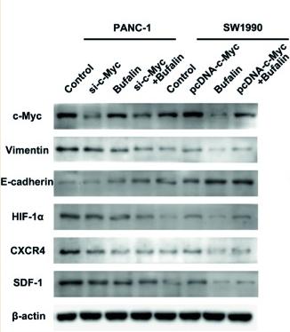
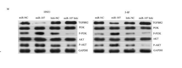
Application: WB Species: Human Sample: pancreatic cancer cells

Application: WB Species: Rat Sample: CIHH rats
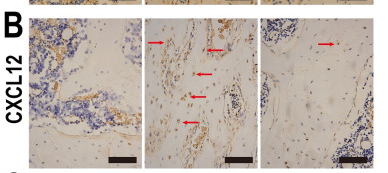
Application: IHC Species: Rat Sample:
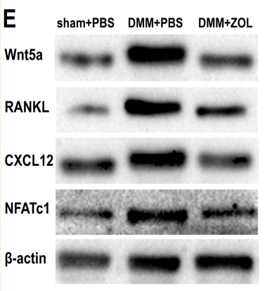
Application: WB Species: Rat Sample:
Restrictive clause
Affinity Biosciences tests all products strictly. Citations are provided as a resource for additional applications that have not been validated by Affinity Biosciences. Please choose the appropriate format for each application and consult Materials and Methods sections for additional details about the use of any product in these publications.
For Research Use Only.
Not for use in diagnostic or therapeutic procedures. Not for resale. Not for distribution without written consent. Affinity Biosciences will not be held responsible for patent infringement or other violations that may occur with the use of our products. Affinity Biosciences, Affinity Biosciences Logo and all other trademarks are the property of Affinity Biosciences LTD.