, using Cleaved-Caspase 9 (Asp353) Antibody at 1/1000 dilution.
5ug/NC membrane strip.
Exposure for 10s with Affinity™ ECL Kit(#KF8001).
Bands result from membrane strip incubation.")

 After incubating GRh2-loaded cells with or without 1 μM of Leu. Caspase activity was determined as indicated in section 2. Enzymatic activity is expressed as fold induction with respect to non-treated cells and represents the mean values ± SD. of three different experiments. *p < 0.001 significantly different from the control; #p < 0.001 significantly different from the cells treated with GRh2 in the absence of Leu. (B) Western blot assays of active fragments of caspase-3 and -9. The results from one representative experiment are shown. (C) The histogram represents quantification of caspase-3 and caspase-9 protein expression levels in GRh2 stimulated HepG2 cell samples using ImageJ64 software (levels of control cells/β-actin defined as 1). Results are presented as mean ± S.D. with triplicate measurement. *P < 0.01 vs. the control group.")
, caspase 9, cleaved caspase 9 (phosphorylated form), Bcl-2, Bax, and β-actin was determined using Western blotting.")
 and (B): The relative luciferase activities were analyzed in 293T cells upon co-transfection of miR-153-5p mimic and luciferase reporter psiCHECK2/circPAN3-mut153, or co-transfection of miR-183-5p mimic and psiCHECK2/circPAN3-mut183.")
 MIN6-PC and MIN6-MKP5 cellswere exposed to GP for 24 h, after which the activation of caspase-3, -9, -8, and the expression of PARP-1 were assessed by western blotting (A). Relative Bcl-2/Bax expression was determined by real-time PCR (B).")
 THP-1/ADM and K562/ADM cells were pre-incubated with 5 mM 3-MA for 24 h before exposure to 2 μg/mL of ADM for additional 24 h. Apoptosis-related proteins were detected and quantified by Western blot. All data are presented as means ± SD of three independent experiments. *P < 0.05, **P < 0.01, and ***P < 0.001, compared to parental cell line (THP-1 or K562), negative control, or newly diagnosed patients. ADM: doxorubicin; GFP, green fluorescent protein; mRFP, monomeric red fluorescent protein; OD: optical density.")
 Western blot assay showed that DDTC downregulated the expression of caspase 3 and caspase 9 protein levels and upregulated the expression of cleaved caspase 3 and cleaved caspase 9 protein levels in SKOV3 cells andA2780 cells.")

 by western blotting analysis (A–F).
The date is expressed as the means ± S.E.M. (n = 5 in every groups). *P < 0.05, **P < 0.01 and ***P < 0.001 vs model group.")
 the cell viability was examined by CCK-8 assay and (f, g) the proteins were examined by western
blotting. Data are expressed as mean ± SD (n = 3). * p < 0.05, ** p <
0.01 compared with control group. ++ p < 0.01 represent the significant difference between HepG2 and HUVEC cells at uniform
concentration. ## p < 0.01 represent the significant difference between LS and LS + z-VAD-fmk. LS, lappaconitine sulfate.")
 FACS analysis of apoptotic cells by PI and Annexin V double staining. (b) Quantitative measurement of the proportion of early and late stage apoptotic cells. Data are presented as means ± SD from three independent experiments. Significance: *P< 0.05, **P< 0.01 vs. negative control. (c) Western blot of apoptosis-related proteins.")
 was used to stain the nuclei, PI (red) was used to stained dead neurons. Scale bar = 40 μm.b. The expression of cleaved-caspase-3 and cleaved-caspase-9 in brain tissues of rats from each group was measured by Western blot. β-actin was used as a loading control.Data are expressed as mean± SD. ##p < 0.01 compared to Control group; **p < 0.01 compared to Ethanol+Lv-control group.")

was detected and the statistic results was analysis. The date is expressed as the means ± SD,
n = 3 for all groups. For all results: *P < 0.05
verses OGD-0 h, *P < 0.05 represents a significant difference. CM mean: conditioned
medium prepared from BV-2 cells supernatant.")
 BxPC3 cells were treated with indicated concentrations of SSD for 48 hours and expression levels of cleaved caspase 9, cleaved caspase 3, p53, and FoxO3a determined using Western blotting. β-actin served as the internal control. (B) Semiquantitative immunoblot-staining analysis of the indicated apoptosis-regulatory proteins on SSD treatment with an image analyzer. Grayscale-scan analysis of Western blot images was from three independent experiments, and fold changes included normalization to β-actin. *P<0.05, **P<0.01 denote significant differences compared with the control group for each protein.")
 Protein expression levels of Bax, Bcl‑2, cleaved caspase‑9, cleaved caspase‑3 and β‑actin in H9C2 cells treated with LPS, TA or TA + LPS were determined by western blot analysis.")

 Whole cell lysates were prepared following stimulation with LPS, NAC and TA. The protein expression levels of apoptosis-associated proteins (p-JNK, JNK, Bax, cleaved caspase-9 and cleaved caspase-3) and an anti-apoptotic protein (Bcl-2) were detected in H9C2 cells treated with LPS, NAC + LPS, TA + LPS and TA + NAC + LPS by western blot analysis. Semi-quantification of the protein expression levels of (B) p-JNK/JNK and cleaved caspase-3, and (C) Bax/Bcl-2 and cleaved caspase-9 in H9C2 cells. The mRNA expression levels of (D) Bax, (E) caspase-3, (F) caspase-12 and (G) Cyt c in H9C2 cells normalized to Gapdh. Data are presented as the mean ± standard error of the mean (n=3). *P<0.05, **P<0.01 and ***P<0.001 vs. control; #P<0.05 and ##P<0.01 vs. LPS group; §P<0.05 and §§P<0.01 vs. TA + LPS. TA, tannic acid; LPS, lipopolysaccharide; NAC, N-acetylcysteine; p, phosphorylated; JNK, c-Jun N-terminal kinase; Cyt c, cytochrome c.")
, cleaved-caspase-9 (C-Caspas 9) and cytochrome c (Cyto c) in the Jurkat Clone E6-1 cells were measured by Western blotting. (A) Total cellular extracts and cytosolic fractions were analyzed by Western blot analysis using antibodies against Bax, Bcl-2, cleaved-caspase-3 (C-Caspase 3), cleaved-caspase-9 (C-Caspase 9), and cytochrome c (Cyto c), (B) bax protein content of Jurkat Clone E6-1 cells in different doses of hirsutine, (C) bcl-2 protein content of Jurkat Clone E6-1 cells in different doses of hirsutine, (D) histogram of bax/bcl-2 ratio, (E) cleaved-caspase-9 (C-Caspase 9) protein content of Jurkat Clone E6-1 cells in different doses of hirsutine, (F) cleaved-caspase-3 (C-Caspase 3) protein content of Jurkat Clone E6-1 cells in different doses of hirsutine, (G) cytochrome c (Cyto c) protein content of Jurkat Clone E6-1 cells in different doses of hirsutine, *p < 0.05, **p < 0.01 (n = 3).")
. #P<0.05, compared with the control group; *P<0.05, compared with OGD/R group. CT, costunolide; OGD/R, oxygen-glucose deprivation/reperfusion.")

Cells were stimulated with 2 mM NaF for the
indicated times (0, 4, 8, 12, 16, and 24 h).
Representative Western blot images showing
the changes of protein level of activated
caspases. (B) BEAS-2B cells were transfected
with the scramble or caspase-8 siRNA for 48
h, and then treated with NaF (2 mM) for 24
h. After treatment, BEAS-2B cells were harvested, and the proteins were isolated. Then,
Western blot was performed to analysis the
proteins expression related to cleaved
caspase-8 and -3. (C) Summarized data
showing the changes of caspase-3 activity in
NaF-treated cells with or without caspase-8
siRNA. Representative immunofluorescence
images (D) and summarized data (E)
showing the effect of caspase-8 siRNA on
NaF-induced apoptosis in TUNEL assay.
Scale bar represents 100 μm. Each data
shows mean ± S.D of at least three independent experiments. **p < 0.01.")
. Migration of LA795 cells in the presence of 0, 3, 10, 30, 100, and 300 μM silibinin assessed by wound healing assay (n = 4). (B). Statistical results of the percentage of wound area in (A) (n = 4) (C). Statistical results of LA795 cells viability incubated with different concentrations of silibinin (n = 6). (D). Expression of β-catenin, E-cadherin, N-cadherin, and vimentin of LA795 cells after treated with 200 μM silibinin for 24 h (n = 3). (E). Representative real-time images of LA795 cells incubated with different concentrations of silibinin (n = 6). (F). Representative images of apoptotic cells after 200 μM silibinin treatment for 24 h detected by annexin V apoptosis assay (n = 3). (G). Expression of cleaved-caspase 3 and cleaved-caspase 9 of LA795 cells after 200 μM silibinin treatment for 24 h (n = 3).")
 Flow cytometry was used to determine cell apoptotic rate using Annexin V-FITC/PI staining. Apoptotic cells included Annexin V+/PI- and Annexin V+/PI+ cells. (B) Quantitative analysis of the apoptotic rate of cells treated with LPS, TA and TA + LPS. (C) Protein expression levels of Bax, Bcl-2, cleaved caspase-9, cleaved caspase-3 and β-actin in H9C2 cells treated with LPS, TA or TA + LPS were determined by western blot analysis. (D) Semi-quantification of the protein expression levels of Bax/Bcl-2, cleaved caspase-9 and cleaved caspase-3 in the LPS, TA and TA + LPS groups. Data are presented as the mean ± standard error of the mean (n=3). *P<0.05, **P<0.01 and ***P<0.001 vs. Cont group; #P<0.05 and ##P<0.01 vs. LPS group. TA, tannic acid; LPS, lipopolysaccharide; Cont, control; FITC, fluorescein isothiocyanate; PI, propidium iodide.")
 Western blot was used to determine Bax, BCL-2, active caspase 3 and active caspase 9 expressions in LX-2 cells. **P < 0.01 vs. control group; ##P < 0.01 vs. AA group; n = 3.")
 Cell viability of K562-R cells was determined by MTT assay after the exposure of CPT and dasatinib for 48 h. (B) The effects of CPT and nilotinib on the cell viability of K562-R cells were measured by MTT assay. Data was represented as the mean ± SD obtained from three independent experiments. (C) Protein expression of cleaved caspase-3, caspase-9 and PARP after the treatment of CPT and three TKIs were analysed by western blot. (D) The semi-quantitative immunoblotting staining analysis of the indicated apoptosis related proteins by Image J. (E) The phosphorylation and total protein expression levels of Bcr-Abl, Src, STAT3 and eIF4E were determined after the combination treatment of CPT and imatinib, dasatinib and nilotinib in K562-R cells. (F) The indicated protein expression levels were quantified by Image J. Data were expressed as the mean ± SD, n = 3. *p < 0.05, and **p < 0.01 denote significant differences compared with the single TKI treatment group.")
 DAPI staining (blue) was used to observe the changes in the nucleus of HT-29 and SW620 cells under OMT or DOX treatment. (B) Cell apoptosis was detected by Annexin V-FITC/PI binding assay after OMT and/or DOX treatment in HT-29 and SW620 cells. (C) Western blotting was used to analyze the expression of Bcl-2, Bax, cleaved caspase-3, and cleaved caspase-9. (D) Statistical analysis of total apoptosis rates including early and late apoptosis (Q2 + Q4, respectively). (E–J) The densitometric analysis of protein bands was performed and normalized with the corresponding GAPDH content. Data are presented as the mean ± SD of three independent experiments. *p < 0.05, **p < 0.01, ***p < 0.001 vs. the control group; # p < 0.05, ## p < 0.01, ### p < 0.001 vs. the OMT (5 mM) group or DOX (0.3 μM) group.")
 Representative TUNEL images in each group after treatment with various concentrations of MAF. Scale bars, 250 µm. (B) Western blot analysis and quantification of Bcl-2, Bax, cleaved-caspase-3 and cleaved-caspase-9 in MAF treatment. ***P<0.001 vs. Control; ##P<0.01 and ###P<0.001 vs. H/R. MAF, mangiferin; H/R, hypoxia-reoxygenation.")

 Original bands of Bax, Bcl-2, Mcl-1, Bcl-xL, cytochrome c, and cleaved
caspase-3 and -9, together with p-JNK (p means phosphorylation) and JNK. β-Actin was used as an internal control. (B) Relative expression of
these proteins. The data are represented as mean ± SD for three independent experiments (**p < 0.01 and ***p < 0.001 compared with control
group).")

 SLC25A21 promoted the efflux of α-KG from the mitochondria to the cytosol. (B) The levels of α-KG in the mitochondria affected succinate production. (C) The upregulation of SLC25A21 promoted ROS accumulation in BCa cells. (D) The upregulation of SLC25A21 decreased Δψm in both BCa cell lines. (E) Western blot assays showed that SLC25A21 induced cyto C transfer from the mitochondria to the cytosol and increased the activation of caspase-9 and caspase-3 in BCa cells. (F) Immunohistochemistry showed that SLC25A21 increased the activation of caspase-9 and caspase-3 in BCa xenograft tissues. (G) Schematic diagram showing the mechanism of action of SLC25A21 on cell apoptosis in BCa. The results were reproducible in three independent experiments. *P < 0.05, **P < 0.01, ***P < 0.001 and ****P < 0.0001.")
 Cell apoptosis results of LA795 cells incubated by 50 μM HHT for 24 h detected with Annexin-V assay (n = 3). (B) Cell apoptosis results of LA795 cells with TMEM16A shRNA transfection and added 50 μM HHT (n = 3). (C) Cell apoptosis results of 2BS cells with TMEM16A transfection and added 50 μM HHT (n = 3). (D–F) Expression of cleaved-caspase 3 and cleaved-caspase 9 with LA795 cells incubated by 50 μM HHT (D), or LA795 cells with TMEM16A shRNA transfection and added 50 μM HHT (E), or 2BS cells with TMEM16A transfection and added 50 μM HHT (F) (n = 3).")
.")
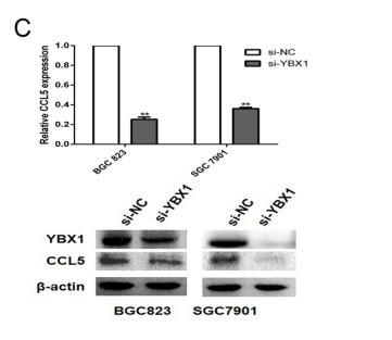
 Cell proliferation activity in lung cancer (OD value). (b) Detection of lung cancer cell apoptosis by flow cytometry. (c) Detection of lung cancer cell migration by transwell chamber (200×). (d) Detection of Nrf2, HO-1, MMP-9, MMP-2, Cleaved Caspase-3, Cleaved Caspase-9, and protein expression in lung cancer cells by western blot; compared with the control group and si-NC group, ∗P < 0.05.")


 Representative western blot of Nrf2, p-PI3K, PI3K, p-AKT, AKT, and β-Actin in RS4:11 cells. The cells were pretreated with or without VCR (0.5,1,2,4 and 8 μM, 24 h). (B) Representative western blot of Nrf2, p-PI3K, PI3K, p-AKT, AKT, and β-Actin in Nalm-6 cells. The cells were pretreated with or without VCR (0.5,1,2,4 and 8 μM, 24 h). (C, D) The relative grey values were shown in the histogram. (E) After treatment with or without 10 μM MK2206 for 24 h in RS4:11 cells, protein expression levels of Nrf2, p-AKT, AKT, BAD, Bcl-2, Caspase-9, Cleaved-Caspase9 and CREB was evaluated by western blot analysis in the Nrf2-overexpression and EV groups. (F) After treatment with or without 10 μM MK2206 for 24 h in Nalm-6 cells, protein expression levels of Nrf2, p-AKT, AKT, BAD, Bcl-2, Caspase-9, Cleaved-Caspase9 and CREB was evaluated by western blot analysis in the Nrf2-overexpression and EV groups. (G, H) The relative gray values were shown in histogram. Data are presented as the mean ± SD of three independent experiments. *P < 0.05, **P < 0.01, ***P < 0.001, n.s, no significance. (I) Schematic representation of the molecular mechanisms proposed in the positive effect of regulate via Nrf2 of PI3K/Akt/Nrf2 signaling pathway in ALL cells.")
 Confocal microscopy analysis of α-tubulin polymerization and F-actin cytoskeletal remodeling in CRC cells with CX43 overexpression and knock-down. (c) The young’s modulus of CX43 overexpressed DLD1 cells was measured by atomic mechanics microscope. Mechanical curve (left) and quantitative analysis (right) are displayed. (d) Western blot analyses expression of stem cell characteristic related proteins in CRC cells with CX43 overexpression. (e) The tumor cell spheroidizing ability of CX43 overexpression cells was detected by tumor sphere formation assays. (f) Western blot was used to survey the relative expression of apoptosis pathway related proteins in CX43 overexpression cells. The downregulation of CX43 could rescue the above phenotypes (d, e and f).")
-6, and IL-1β levels in astrocyte supernatants were determined by enzyme-linked immunosorbent assay. Intracellular (B) and mitochondrial (C) reactive oxygen species levels were assessed using the DCFDA probe and MitoSOX probe. D Mitochondrial membrane potential was assessed using the JC-1 probe. E Isolation and identification of primary neurons by light microscopy and fluorescent staining for neuron-specific enolase. F Cell viability of neurons was determined by cell counting kit-8 assay. G Apoptosis of neurons was determined by flow cytometry. H Apoptosis-related protein levels (cleaved caspase-3 and cleaved caspase-9) in neurons were determined by western blotting. Data are shown as the mean ± standard deviation of three independent experiments. **p")
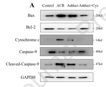
, apoptosis percentages (B), the expression of BAX, Cyto-C, BCL-2 (C), as well as cleaved PARP, cleaved caspase 3 and 9 (D) in acrolein-induced LO2 cells. The data in C, D were expressed as relative intensity to GAPDH; Error bars represent standard deviation (n = 3); Different letters indicate significant differences (p < 0.05) between treatments; Acr, acrolein.")
 Bar graphs of the mRNA levels of Bax and Bcl-2 in NCI-H1299 cells. (C) Bar graphs of the Bax/Bcl-2 ratio in NCI-H1299 cells. (D) The relative protein levels of cleaved caspase-3/9 in NCI-H1299 cells treated with HTE were measured by western blotting. (E and F) The ratios of protein levels were normalized to the value of the control group. All data are expressed as the mean ± SD of three independent experiments. **P")
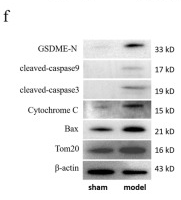
 inhibited myocardial cells apoptosis after acute myocardial infarction (AMI). Rats were pretreated with HQR followed by AMI. Sham rats were used as control. Sham rats were used as control. Representative apoptosis of myocardial cells, TUNEL‐positive cells, the scale bars represents a length of 300 μm on histology (A), the activity of caspase‐9/3 (B, C) and the mRNA levels of caspase‐9/3, and protein levels of cleaved caspase‐9/3 (D) were evaluated under different groups. Data are shown as mean ± SD. *p < 0.05 versus sham group, # p < 0.05 versus AMI (obese) group, Δ p < 0.05 versus HQR (4.5 g/kg.d) group, ▲ p < 0.05 versus HQR (9.0 g/kg.d) group. (n = 3)")
 inhibited renal cells apoptosis after renal ischemia-reperfusion (IR). Rats were pre-treated with TIIA followed by removing the right kidney and clamping of the left renal artery for 30 min and reperfusion for 24 h. Sham rats were used as control. Sham rats were used as control. Representative apoptosis of renal cells, TUNEL positive cells, the scale bars represent a length of 200 μm on histology (A), the activity of renal caspase-9/3 (B, C), and the protein expression of cleaved caspase-9/3 (D) were evaluated under different groups. Data are shown as mean ± SD. *p < 0.05 versus sham group, #p < 0.05 versus IR (obese) group, ∆p < 0.05 versus TIIA (5 mg/kg.d) group, ▲p < 0.05 versus TIIA (10 mg/kg.d) group. (n=3).")
 Flow cytometry was used to determine cell apoptotic rate using Annexin V-FITC/PI staining. Apoptotic cells included Annexin V+/PI- and Annexin V+/PI+ cells. (B) Quantitative analysis of the apoptotic rate of cells treated with LPS, TA and TA + LPS. (C) Protein expression levels of Bax, Bcl-2, cleaved caspase-9, cleaved caspase-3 and β-actin in H9C2 cells treated with LPS, TA or TA + LPS were determined by western blot analysis. (D) Semi-quantification of the protein expression levels of Bax/Bcl-2, cleaved caspase-9 and cleaved caspase-3 in the LPS, TA and TA + LPS groups. Data are presented as the mean ± standard error of the mean (n=3). *P")
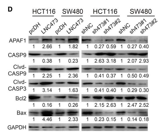
 and Transwell assays (scale = 50 μm), respectively. 2 F: Changes in the levels of proteins related to the proliferation, apoptosis, and migration of HCT116 and SW480 cells after lncRNA RP11-197K6.1 knockdown, as analyzed by western blotting assay (**P")
 Apoptotic cells were detected by TUNEL staining. (B–F) Representative images of Caspase-3 activity, Cleaved-caspase 3, Cleaved-caspase 9, and Bax and Bcl-2 expression were examined by western blot and the fold activation data analysis. Data are expressed as mean ± SD (at least n = 6/group), #p < 0.05(vs. Sham group), *p < 0.05 (vs. CLP group).")
 HT22 cells were exposed to 20 mM glutamate with varying concentrations of lutein (1.25, 2.5, 5, and 10 μM) and subjected to the FITC test (one-step TUNEL apoptosis test kit) to assess the number of apoptotic and dead cells, respectively. The bar graph indicates the percentage of apoptotic cells. Data are presented as the mean value ± S.E.M of three independent experiments. (B) Representative images of JC-1 staining were detected using fluorescence microscopy. The bar graph indicates the percentage of cells exhibiting mitochondrial depolarization. Data are presented as the mean value ± S.E.M of three independent experiments. (C) Western blot analysis was conducted. β-Actin serving as a loading control. The bars represent the fold-increase in the levels of cleaved caspase 3 and cleaved caspase 9 compared to control cells. Data are presented as the mean value ± S.E.M of three independent experiments. Statistical significance is denoted as follows: ###p")
 or 100 μM, 150 μM, or 200 μM genipin (experimental groups) for 24 h. (a) Cell death was detected using Annexin V/PI double staining and flow cytometry. The sum of the percentage of cells in quadrants Q2-2 and Q2-4 was calculated and is represented in a histogram. (b) The expression levels of apoptosis-related proteins were detected in SK-N-SH cells using Western blotting. The blots were cropped and the original uncropped ones are shown in Supplementary Fig. S3. (c) Caspase-3 activity was measured, and the percentage in each experimental group relative to the control group was calculated. The results were then represented in a histogram. The density values of the protein bands in the images were measured using ImageJ software, and the results are represented in a histogram. The data are presented as the means ± SDs. *p")
 Bar graph of the relative cell viability in each group (n = 6). (B) Light microscopic images of morphological changes in H9c2 cells of each group ( × 400). (C) The ΔΨm change, determined using the JC-1 assay, is expressed as the ratio of JC-1 aggregates to monomers (red to green) fluorescence intensity ( × 400). (D) Bar graph of relative florescence intensity of JC-1. (E) ROS production levels measured by flow cytometry in each group ( × 400). (F) Summary data showing the ROS cells in each group. (G) Western blot showing the Bax, Bcl-2, Bid, and cleaved-caspase 9 protein expression. (H) Fold change of Bax and Bid expression relative to GAPDH expression measured in western blots (n = 3). (I) Fold change of Bcl-2 and cleaved-caspase 9 expression relative to GAPDH expression measured in western blots (n = 3). *indicates a significant difference compared to the control group; # indicates a significant difference compared to the H/R model group: *p < 0.05, **p < 0.01, #p < 0.05, ##p < 0.01. DHC: dehydrocorydaline, H/R: hypoxia/reoxygenation, ROS: reactive oxygen species, ΔΨm: mitochondrial membrane potential. (For interpretation of the references to color in this figure legend, the reader is referred to the Web version of this article.)")
 The Mlg cells were incubated with TGF-β1 and/or PZ (2 or 4 μM) for 24 hours. The expression levels of caspase3, cleaved-caspase3, caspase7, cleaved-caspase7, caspase9 and cleaved-caspase9, were detected via using Western blot. The loading control was β-tubulin. (B,C) Mlg cells were exposed to Baf a1/CQ and/or PZ (2 or 4 μM) for 24 hours. The p62 expression level was evaluated by Western blot. GAPDH was used as a loading control. (D,E) The plasmid of GFP-LC3B and GFP-Cherry-LC3B were transfected to NIH-3T3 with PEI, and fluorescence microscopy was used to examine the number of autophagosomes (green puncta) and autolysosome (red puncta) after exposing to TGF-β1 and/or PZ for 12 hours. Scale bars: 50 µm. Data in (A-C) are mean ± standard deviation. ###, P")


. The values displayed represent the mean ± standard error of the mean derived from data collected in three independent experiments. ##P")
 Western blot showing expression levels of EMT-related proteins E-cadherin, N-cadherin, Vimentin, Snail, Slug, and Twist1 after ZDHHC9 knockdown in 143B and U2OS cells. (B) Western blot analysis of apoptosis-related proteins Cleaved Caspase-3, Cleaved Caspase-9, Bcl-2, Bax, and cell cycle-related proteins CDK4 and Cyclin D1 after ZDHHC9 knockdown in 143B and U2OS cells. (C) Flow cytometry analysis showing apoptosis rates of 143B and U2OS cells after ZDHHC9 knockdown. (D) Flow cytometry analysis of cell cycle distribution in 143B and U2OS cells after ZDHHC9 knockdown. (*P")
 and MLg (B) cells were exposed to oleuropein and/or TGF-β1 (5 ng/mL) for 24 h, after which samples were collected for Western blot analysis to evaluate the protein levels of cleaved Caspase 9, Caspase 9, cleaved Caspase 3, and Caspase 3 (original images can be found in Figure S1). The results are presented as Mean ± SD, n = 3, where # indicates differences between the control group and the TGF-β1 group, with ## p < 0.01, ### p < 0.001 and #### p < 0.0001, and * indicates differences between the TGF-β1 group and the oleuropein treatment group, with *** p < 0.001, and **** p < 0.0001. (C) MLg cells were exposed to oleuropein and/or TGF-β1 (5 ng/mL) for 24 h, after which cells were digested and collected for flow cytometry analysis using the Annexin V-FITC/PI apoptosis detection method.")
 Hippo pathway related proteins expression was determined by Western blot and qRT-PCR. n = 3; (D) Cell viability assay; (E) Optical microscope images of MCMECs morphological changes; (F) Cell damage assay; (G) TUNEL assay to assess cell apoptosis rate in vitro. n = 3; (H) Transmission electron microscope images of MCMECs; (I,K) Caspase-9, Bcl-2, VEGF, FGF2 protein expression was determined by Western blot. n = 3; (J) Caspase-9, Bcl-2, VEGF, FGF2 mRNA level was assessed by qRT-PCR. n = 3.")
| Product: | Cleaved-Caspase 9 (Asp353) Antibody |
| Catalog: | AF5240 |
| Description: | Rabbit polyclonal antibody to Cleaved-Caspase 9 (Asp353) |
| Application: | WB IHC IF/ICC |
| Cited expt.: | WB, IF/ICC |
| Reactivity: | Human, Mouse, Rat |
| Mol.Wt.: | 17/38 kDa(mature), 47kDa(precursor)(Observed); 46kD(Calculated). |
| Uniprot: | P55211 |
| RRID: | AB_2837726 |
Control Products

Related Downloads
Protocols
Product Info
*The optimal dilutions should be determined by the end user. For optimal experimental results, antibody reuse is not recommended.
*Tips:
WB: For western blot detection of denatured protein samples. IHC: For immunohistochemical detection of paraffin sections (IHC-p) or frozen sections (IHC-f) of tissue samples. IF/ICC: For immunofluorescence detection of cell samples. ELISA(peptide): For ELISA detection of antigenic peptide.
Cite Format: Affinity Biosciences Cat# AF5240, RRID:AB_2837726.
Fold/Unfold
APAF-3; APAF3; Apoptosis related cysteine peptidase; Apoptotic protease Mch-6; Apoptotic protease-activating factor 3; CASP-9; CASP9; CASP9_HUMAN; Caspase 9 apoptosis related cysteine peptidase; Caspase 9 Dominant Negative; Caspase 9c; Caspase-9; Caspase-9 subunit p10; ICE LAP6; ICE like apoptotic protease 6; ICE-LAP6; ICE-like apoptotic protease 6; MCH6; PPP1R56; protein phosphatase 1, regulatory subunit 56; RNCASP9;
Immunogens
A synthesized peptide derived from human Caspase 9 (Cleaved-Asp353).
Ubiquitous, with highest expression in the heart, moderate expression in liver, skeletal muscle, and pancreas. Low levels in all other tissues. Within the heart, specifically expressed in myocytes.
- P55211 CASP9_HUMAN:
- Protein BLAST With
- NCBI/
- ExPASy/
- Uniprot
MDEADRRLLRRCRLRLVEELQVDQLWDALLSRELFRPHMIEDIQRAGSGSRRDQARQLIIDLETRGSQALPLFISCLEDTGQDMLASFLRTNRQAAKLSKPTLENLTPVVLRPEIRKPEVLRPETPRPVDIGSGGFGDVGALESLRGNADLAYILSMEPCGHCLIINNVNFCRESGLRTRTGSNIDCEKLRRRFSSLHFMVEVKGDLTAKKMVLALLELAQQDHGALDCCVVVILSHGCQASHLQFPGAVYGTDGCPVSVEKIVNIFNGTSCPSLGGKPKLFFIQACGGEQKDHGFEVASTSPEDESPGSNPEPDATPFQEGLRTFDQLDAISSLPTPSDIFVSYSTFPGFVSWRDPKSGSWYVETLDDIFEQWAHSEDLQSLLLRVANAVSVKGIYKQMPGCFNFLRKKLFFKTS
Research Backgrounds
Involved in the activation cascade of caspases responsible for apoptosis execution. Binding of caspase-9 to Apaf-1 leads to activation of the protease which then cleaves and activates caspase-3. Promotes DNA damage-induced apoptosis in a ABL1/c-Abl-dependent manner. Proteolytically cleaves poly(ADP-ribose) polymerase (PARP).
Isoform 2 lacks activity is an dominant-negative inhibitor of caspase-9.
Cleavages at Asp-315 by granzyme B and at Asp-330 by caspase-3 generate the two active subunits. Caspase-8 and -10 can also be involved in these processing events.
Phosphorylated at Thr-125 by MAPK1/ERK2. Phosphorylation at Thr-125 is sufficient to block caspase-9 processing and subsequent caspase-3 activation. Phosphorylation on Tyr-153 by ABL1/c-Abl; occurs in the response of cells to DNA damage.
Ubiquitous, with highest expression in the heart, moderate expression in liver, skeletal muscle, and pancreas. Low levels in all other tissues. Within the heart, specifically expressed in myocytes.
Belongs to the peptidase C14A family.
Research Fields
· Cellular Processes > Cell growth and death > p53 signaling pathway. (View pathway)
· Cellular Processes > Cell growth and death > Apoptosis. (View pathway)
· Cellular Processes > Cell growth and death > Apoptosis - multiple species. (View pathway)
· Environmental Information Processing > Signal transduction > PI3K-Akt signaling pathway. (View pathway)
· Human Diseases > Drug resistance: Antineoplastic > Platinum drug resistance.
· Human Diseases > Neurodegenerative diseases > Alzheimer's disease.
· Human Diseases > Neurodegenerative diseases > Parkinson's disease.
· Human Diseases > Neurodegenerative diseases > Amyotrophic lateral sclerosis (ALS).
· Human Diseases > Neurodegenerative diseases > Huntington's disease.
· Human Diseases > Infectious diseases: Bacterial > Legionellosis.
· Human Diseases > Infectious diseases: Parasitic > Toxoplasmosis.
· Human Diseases > Infectious diseases: Bacterial > Tuberculosis.
· Human Diseases > Infectious diseases: Viral > Hepatitis B.
· Human Diseases > Infectious diseases: Viral > Influenza A.
· Human Diseases > Cancers: Overview > Pathways in cancer. (View pathway)
· Human Diseases > Cancers: Specific types > Colorectal cancer. (View pathway)
· Human Diseases > Cancers: Specific types > Pancreatic cancer. (View pathway)
· Human Diseases > Cancers: Specific types > Endometrial cancer. (View pathway)
· Human Diseases > Cancers: Specific types > Prostate cancer. (View pathway)
· Human Diseases > Cancers: Specific types > Small cell lung cancer. (View pathway)
· Human Diseases > Cancers: Specific types > Non-small cell lung cancer. (View pathway)
· Human Diseases > Cardiovascular diseases > Viral myocarditis.
· Organismal Systems > Endocrine system > Thyroid hormone signaling pathway. (View pathway)
References
Application: WB Species: human Sample: HepG2
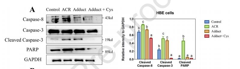
Application: WB Species: Human Sample: HBE (A) and Caco-2 (B) cells
Application: WB Species: Human Sample: HBE and Caco-2 cells
Application: WB Species: human Sample: HT-29 cells
Application: WB Species: mouse Sample: HSC apoptosis
Application: WB Species: Mouse Sample: B cells
Restrictive clause
Affinity Biosciences tests all products strictly. Citations are provided as a resource for additional applications that have not been validated by Affinity Biosciences. Please choose the appropriate format for each application and consult Materials and Methods sections for additional details about the use of any product in these publications.
For Research Use Only.
Not for use in diagnostic or therapeutic procedures. Not for resale. Not for distribution without written consent. Affinity Biosciences will not be held responsible for patent infringement or other violations that may occur with the use of our products. Affinity Biosciences, Affinity Biosciences Logo and all other trademarks are the property of Affinity Biosciences LTD.