


 and mouse anti-beta tubulin Ab(T0023 1:200) for 1 hour at 37°C. An AlexaFluor594 conjugated goat anti-rabbit IgG(H+L) Ab(Red) and an AlexaFluor488 conjugated goat anti-mouse IgG(H+L) Ab(Green) were used as the secondary antibody.
The nuclear counter stain is DAPI(blue).")
| Product: | DBT Antibody |
| Catalog: | DF13569 |
| Description: | Rabbit polyclonal antibody to DBT |
| Application: | WB IHC IF/ICC |
| Cited expt.: | WB |
| Reactivity: | Human, Mouse, Rat |
| Prediction: | Zebrafish, Bovine, Horse, Sheep, Rabbit, Dog, Xenopus |
| Mol.Wt.: | 53kDa(Observed); 53kD(Calculated). |
| Uniprot: | P11182 |
| RRID: | AB_2846588 |
Control Products

Related Downloads
Protocols
Product Info
*The optimal dilutions should be determined by the end user. For optimal experimental results, antibody reuse is not recommended.
*Tips:
WB: For western blot detection of denatured protein samples. IHC: For immunohistochemical detection of paraffin sections (IHC-p) or frozen sections (IHC-f) of tissue samples. IF/ICC: For immunofluorescence detection of cell samples. ELISA(peptide): For ELISA detection of antigenic peptide.
Cite Format: Affinity Biosciences Cat# DF13569, RRID:AB_2846588.
Fold/Unfold
BCATE2; BCKAD E2 subunit; Branched chain acyltransferase, E2 component; Dihydrolipoamide branched chain transacylase (E2 component of branched chain keto acid dehydrogenase complex; maple syrup urine disease); Dihydrolipoamide branched chain transacylase; Dihydrolipoamide branched chain transacylase E2; Dihydrolipoyl transacylase; Dihydrolipoyllysine residue (2 methylpropanoyl)transferase; E2; E2 component of branched chain alpha keto acid dehydrogenase complex; E2B; Lipoamide acyltransferase component of mitochondrial branched chain alpha keto acid dehydrogenase complex; Mitochondrial branched chain alpha keto acid dehydrogenase transacylase subunit (E2b);
Immunogens
A synthesized peptide derived from human DBT, corresponding to a region within C-terminal amino acids.
- P11182 ODB2_HUMAN:
- Protein BLAST With
- NCBI/
- ExPASy/
- Uniprot
MAAVRMLRTWSRNAGKLICVRYFQTCGNVHVLKPNYVCFFGYPSFKYSHPHHFLKTTAALRGQVVQFKLSDIGEGIREVTVKEWYVKEGDTVSQFDSICEVQSDKASVTITSRYDGVIKKLYYNLDDIAYVGKPLVDIETEALKDSEEDVVETPAVSHDEHTHQEIKGRKTLATPAVRRLAMENNIKLSEVVGSGKDGRILKEDILNYLEKQTGAILPPSPKVEIMPPPPKPKDMTVPILVSKPPVFTGKDKTEPIKGFQKAMVKTMSAALKIPHFGYCDEIDLTELVKLREELKPIAFARGIKLSFMPFFLKAASLGLLQFPILNASVDENCQNITYKASHNIGIAMDTEQGLIVPNVKNVQICSIFDIATELNRLQKLGSVGQLSTTDLTGGTFTLSNIGSIGGTFAKPVIMPPEVAIGALGSIKAIPRFNQKGEVYKAQIMNVSWSADHRVIDGATMSRFSNLWKSYLENPAFMLLDLK
Predictions
Score>80(red) has high confidence and is suggested to be used for WB detection. *The prediction model is mainly based on the alignment of immunogen sequences, the results are for reference only, not as the basis of quality assurance.
High(score>80) Medium(80>score>50) Low(score<50) No confidence
Research Backgrounds
The branched-chain alpha-keto dehydrogenase complex catalyzes the overall conversion of alpha-keto acids to acyl-CoA and CO(2). It contains multiple copies of three enzymatic components: branched-chain alpha-keto acid decarboxylase (E1), lipoamide acyltransferase (E2) and lipoamide dehydrogenase (E3). Within this complex, the catalytic function of this enzyme is to accept, and to transfer to coenzyme A, acyl groups that are generated by the branched-chain alpha-keto acid decarboxylase component.
Mitochondrion matrix.
Belongs to the 2-oxoacid dehydrogenase family.
Research Fields
· Metabolism > Amino acid metabolism > Valine, leucine and isoleucine degradation.
· Metabolism > Carbohydrate metabolism > Propanoate metabolism.
· Metabolism > Global and overview maps > Metabolic pathways.
References
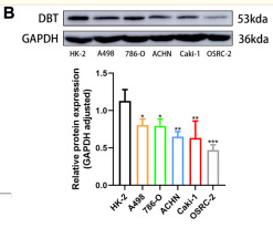
Application: WB Species: Human Sample: HK-2 cells
Restrictive clause
Affinity Biosciences tests all products strictly. Citations are provided as a resource for additional applications that have not been validated by Affinity Biosciences. Please choose the appropriate format for each application and consult Materials and Methods sections for additional details about the use of any product in these publications.
For Research Use Only.
Not for use in diagnostic or therapeutic procedures. Not for resale. Not for distribution without written consent. Affinity Biosciences will not be held responsible for patent infringement or other violations that may occur with the use of our products. Affinity Biosciences, Affinity Biosciences Logo and all other trademarks are the property of Affinity Biosciences LTD.