Cleaved-Caspase 3 (Asp175), p17 Antibody - #AF7022
- WB
- IHC
- IF/ICC
- Cites
;
Lane 3: MCF7 (etoposide treated, 25uM 5h);
Lane 4: Mouse heart;
Lane 5: Mouse spleen.")

, p17 Antibody.
Lane 1: Rat liver, blocked with antigen-specific peptides.
Lane 2: Rat liver.
Lane 3: Rat spleen.")

 and mouse anti-beta tubulin Ab(T0023 1:200) for 1 hour at 37°C. An AlexaFluor594 conjugated goat anti-rabbit IgG(H+L) Ab(Red) and an AlexaFluor488 conjugated goat anti-mouse IgG(H+L) Ab(Green) were used as the secondary antibody.
The nuclear counter stain is DAPI(blue).")
 The results of western blots showing the levels of caspase-3 and cleaved caspase-3 protein increased after TCF3 shRNA treatments in both cells. (B, D) Histograms showing quantization of western blot and the data was normalized against β-actin. Results of at least three independent experiments. *p<0.05, **p")
 After incubating GRh2-loaded cells with or without 1 μM of Leu. Caspase activity was determined as indicated in section 2. Enzymatic activity is expressed as fold induction with respect to non-treated cells and represents the mean values ± SD. of three different experiments. *p < 0.001 significantly different from the control; #p < 0.001 significantly different from the cells treated with GRh2 in the absence of Leu. (B) Western blot assays of active fragments of caspase-3 and -9. The results from one representative experiment are shown. (C) The histogram represents quantification of caspase-3 and caspase-9 protein expression levels in GRh2 stimulated HepG2 cell samples using ImageJ64 software (levels of control cells/β-actin defined as 1). Results are presented as mean ± S.D. with triplicate measurement. *P < 0.01 vs. the control group.")


 Western-blot of Bcl2 protein in A549 and SPC-A-1 cells after transfection.")
 Apoptosis detection of HT-29 cells in different groups by flow cytometry, SFM without drug as control; (B) Apoptosis rates of HT-29 cells in different groups. (mean value ± SD, n=3, **p < 0.01, ***p < 0.001, compared with the control group); (C) Western blotting of the Bcl-2, p53, cleaved caspase-9, cleaved caspase-8,cleaved caspase-3; (D) Relative amount of these apoptosis-related proteins in different groups(mean value ± SD, n=3, *p < 0.05, **p < 0.01, ***p < 0.001, compared with the control group).")
 Immunofluorescent images and (B) Quantitative expression analysis of apoptosis-related proteins including Bcl-2, cleaved caspase-3 and p53 in control and SPORE-CUR-FA groups (mean value ± SD, n=5 * p < 0.05, ** p < 0.01, *** p < 0.001, compared with control group). (C) Mean curcumin plasma concentration-time profiles in rats after oral administration of CUR and SPORE-CUR-FA at a dose of 80 mg/kg (mean value ± SD, n= 3). (D)Western blotting of Bcl-2, cleaved caspase-3 and p53 in different groups, physiological saline as control; (E) Relative expression amount of these apoptosis-related proteins in different groups(mean value ± SD, n=3, **p < 0.01, ***p < 0.001, compared with control group).")
. e qRT-PCR analysis of the average relative mRNA expression levels of apoptosis-related genes in the no-chemotherapy group and chemosensitive group (*p<0.05)")
. e qRT-PCR analysis of the average relative mRNA expression levels of apoptosis-related genes in the no-chemotherapy group and chemosensitive group (*p<0.05)")
. e qRT-PCR analysis of the average relative mRNA expression levels of apoptosis-related genes in the no-chemotherapy group and chemosensitive group (*p<0.05)")
 measured by PicoGreen staining in LX2 cells; scale bar,10 μm. b Representative western blots of TOM20 with GAPDH serving as the internal reference. Bar graph represents the mean ± SEM. *P < 0.05 vs.the indicated groups. c TUNEL staining of LX2 cells from the indicated groups; scale bar, 25 μm. Bar graph represents the mean ± SEM. *P < 0.05 vs.the indicated groups. D Annexin V-FITC/PI double-staining and flow cytometry analysis of LX2 cells. Bar graph represents the mean ± SEM. **P < 0.01 vs. the indicated groups. e Representative western blots of cleaved caspase3, cleaved caspase9, collagen I and α-SMA. Bar graph represents the mean± SEM of three different experiments. *P < 0.05 and **P < 0.01 vs. the indicated groups")
, caspase 9, cleaved caspase 9 (phosphorylated form), Bcl-2, Bax, and β-actin was determined using Western blotting.")
 Representative images of TUNEL assays (200x magnification). (b)Apoptosis index (n = 6). (c)Representative western blots of Bcl-2 and Bax. (d–f) Quantitative analysis of the immunoreactive band displayed by a bar graph (n = 3). (g) Representative western blots of cleaved caspase 3 and caspase 3. (H) Quantitative analysis of the immunoreactive band displayed by a bar graph (n = 3). (a) Sham group, (b) I/R group, (c) AD-Scramble group, (d) AD-miR-145 group,the (e) IPC group.")


 Diagram of groups. (b) Apoptosis was analyzed by using the annexin V FITC/PI assay. (c) Bar graphs show the relative mean of Caco-2 cells apoptosis rate in different view fields. (d) The releasing levels of LDH were detected. (e) Representative images of TUNEL stainings of Caco-2 cells (green, TUNEL positive; blue, DAPI). Scale bar = 50 um. (f) Immunoblot analysis of protein extracts from Caco-2 cells with the indicated antibodies..")
 Representative images of TUNEL stainings of colon sections on day 3 after colitis induction (red, TUNEL positive; blue, DAPI). Scale bar = 50 um (up) or 20 um (down). (b) Representative images of immunohistochemical stainings of FADD, RIPK1 and cCASP3 in the colon sections on day 3 after colitis induction. Scale bar = 50 um or 20 um (downmost).")
 p53 and p-p53. (B) Bax,
Bcl-2, and Bad. (C) Total Caspase 3 and Cleaved-Caspase 3. (D) Full PARP and
Cleaved-PARP. Comparison between all groups was evaluated through One-way
ANOVA. Mean±SD. n=4. **p<0.01, *p<0.05, compared with control group. ##p<0.01,
#p<0.05, compared with Cd group. ns, not significant.")

 PBS, (b) Dextran, (c)
Phycocyanin, (d) Cordycepin, (e) the Phycocyanin/Cordycepin mixture, or (f) Phy-Dex-Cord micelles for 24
h. (A) Trypan blue staining was used to observe cell morphology (Bar: 100 µm). (B) Apoptotic cells were
detected with flow cytometry. Subcellular localization (C1) cleaved caspase-3 and (C2) cleaved PARP in C6
cells, as determined by Confocal laser scanning microscopy. (D1) Apoptosis-related protein, and (D2, D3)
quantitative analysis of Bax, Bcl-2, p53, cleaved caspase-3, cleaved PARP, and PARP levels in C6 cells.
Significant increase or decrease at labels (*) (p < 0.05), labels (**) (0.001< p < 0.01) and labels (***) p <
0.001, are identified in comparison with the Control group. (E) Cell counting determination of apoptotic rate
with Trypan blue and statistical analysis of FCM results.")
. b CircGCNL1 with/
without miR-330-3p mimics was transfected into co-cultured synoviocytes. After 2 days of co-culture, proteins were extracted from chondrocytes in
the lower chamber, and WB was performed to detect the levels of MMP13, MMP3, COL2A1, ADAMTS4, TNF-α, p65, Bcl-2, Bax, caspase-3, and cleaved
caspase-3. N = 4 (four independent experiments). Data are presented as mean ± S.D. One-way ANOVA with Bonferroni test was performed. WB
western blotting, NC normal control, MMP matrix metalloproteinase, TNF tumor necrosis factor, Bcl-2 B-cell lymphoma-2, Bax BCL2-associated X,
COL2A1 collagen type II alpha 1.")
 Representative western blot and
quantitation of the level of cleaved caspase‐3. *p < .05 versus PBS (24 hr). Data are expressed as the mean ± standard deviation (n = 3). Scale
bar = 100 μm (b). GAPDH, glyceraldehyde 3‐phosphate dehydrogenase; PBS, phosphate‐buffered saline; RPL41, ribosomal protein L41; TUNEL,
TdT‐mediated dUTP nick‐end labeling")
 Western blot analysis of autophagy flux markers (ATG5, Beclin1, VPS34, C-CTSD,
SQSTM1/p62, UB and LC3) and ER stress-induced apoptosis markers (CHOP, CASP12, C-CASP12, C-CASP3) in spinal cord lesions from ATG5-/+ mice and ATG5+/+ mice with and without SCI, at Day3 after SCI. (B, C) The levels of the autophagy flux makers from (A) normalized to loading control GAPDH. (D) The expressions of ER stress-induced
apoptosis markers from (A) normalized to loading control GAPDH. (E) Image (30×) of spinal cord sections from the indicated groups at Day 3 stained with antibodies against
LC3II/NeuN, p62/NeuN, CHOP/NeuN,and CASP3/NeuN, respectively. Scale bar: 25 µm. (F) Quantification of immunofluorescence data from (E) showing the mean number of
LC3II in motor neurons at the spinal cord. (G-I) Quantification of immunofluorescence data from (E) showing the mean optical density of p62, CHOP, and CASP3, respectively,
in motor neurons of spinal cord. n=6, ns stands for not significant, *P<0.05, **P<0.01.")
 Western blot analysis of the expression of Bad and cleaved caspase-3 by 4T1 cells after treatment with
CODox, morusin and COMBO. Quantitative analysis of the fold changes of (B) Bad and (C) cleaved caspase-3 accumulated in 4T1 cells after treatment with CODox,
morusin and COMBO (n = 2). (D) Western blot analysis of the expression of Alix, IRE1α, CHOP and p-eIF2α by 4T1 cells after treatment with CODox, morusin and
COMBO. Quantitative analysis of the fold changes of (E) Alix, (F) IRE1α, (G) CHOP and (H) p-eIF2α accumulated in 4T1 cells after treatment with CODox, morusin
and COMBO (n = 2). The cells were incubated with CODox, morusin or COMBO at the equivalent concentrations of DOX (0.6 mg/L), morusin (30 mg/L) and Cu2+
(1.2 mg/L). *P < 0.05, **P < 0.01 and ***P < 0.005 were tested via a Student’s t-test.")
 were used as negative control. (C) Immunoblot analysis to assess the expression of cleaved-PARP, cleaved-caspase3 and Bax. Semiquantitative analyses of protein expression in following histogram (mean ± S.E.M., three independent replicates per groups). * p < 0.05; ** p < 0.01.")
 Protein levels of Bcl‑2, Bax were detected with western blotting when MDA‑MB‑231 and MCF‑7 cells were treated with 0, 20 mM MET, 100 µM FLA and the combination of 20 mM MET:100 µM FLA for 24 h (*P<0.05, 20 mM MET vs. control, 20 mM MET:100 µM FLA vs. 20 mM MET), caspase3 were detected no significance and cleaved caspase3 were detected when MDA‑MB‑231 (*P<0.05, 20 mM MET vs. control; **P<0.01, 20 mM MET:100 µM FLA vs. 20 mM MET) and MCF‑7 cells (*P<0.05, 20 mM MET vs. control, 20 mM MET:100 µM FLA vs. 20 mM MET) were the same treatment, (B) and their densitometry results over β‑actin in at least three separate experiments. FLA, flavone; MET, metformin.")
 RT-qPCR and Western blot were used to detect the level of TIPE2 mRNA and protein,
respectively. RT-qPCR (B) and Western blot (C) were used to detect the expression of TLR4, MyD88, NF-κB p65, p-NF-κB p65Ser276, cleaved Caspase-3 in TIPE2-OE
cells. RT-qPCR (E) and Western blot (F) were used to assay the expression of TLR4, MyD88, NF-κB p65, p-NF-κB p65Ser276, cleaved Caspase-3 in TIPE2-KD cells. n = 3,
Data are presented as mean ± SD from three independent experiments. *P < 0.05, **P < 0.01, ***P < 0.001; TIPE2-NC: TIPE2-negative control, TIPE2-OE: TIPE2-
overexpression, TIPE2-KD: TIPE2-knockdown.")
. C: the levels of apoptotic molecule in renal tissue of rats. The data are presented as the means ± S.D. (n
þ¼
CP-25 groups compared to MTX group.
6). ##P < 0.01 compared with normal
group;**P < 0.01 compared with the model group.")
 The protein expression of cleaved caspase-3 (CC3), claudin-3 (Clau-3) and E-cadherin (E-cad)assessed by Western blotting and the intensity of CC3 (F), Clau-3 (G), and E-cad (H) in Ctrl, NEC, and FTY720 groups. (n ¼ 4e6/group). Data are expressed as mean ± SD. *p < 0.05, **p < 0.01, ***p < 0.001 versus control or NEC groups.")
 Flow cytometry determined the rate of apoptosis in rat NP cells transfected with agomir-145-CTR or agomir-145 in the presence of H2O2 for 48 h, with untransfected cells serving as the controls. (C, D) Western blotting (C) and subsequent densitometric analysis (D) showed the protein expression of Bcl-2, Bax,caspase-3 and activated caspase-3 (A-caspase 3); GAPDH was used as a loading control. The data are presented as the mean ± SD. *P < 0.05 vs. the control group.")
 inhibited cell proliferation and induced apoptosis in tumor tissues. (A) Ki-67 staining of tumor sections obtained from mice treated with (a) vehicle control or (b) QX. Quantification of Ki-67-positive cells in the tumor sections (c). (B)TUNEL staining of tumor sections obtained from mice treated with (a) vehicle control or (b) QX. Quantification of TUNEL-positive cells in the tumor sections (c). (C) Western blotting of proapoptotic proteins Bim, FasL, and cleaved caspase-3 expression in tumor tissues of QX-treated mice or vehicle control-treated mice. Data are presented as mean ± SD. *P < .05, **P < .01 versus vehicle control.")
 HUVECs were pretreated for 2h with 10 mM 3-MA, 20μM Z-VAD-FMK, or 10
mM NAC, and then treated with 5μg/ml HMME and light dose of 2 J/cm2
. Cell viability
was detected by CCK8 assay 24h after HMME-PDT.
** p<0.01. (B) Autophagosome
formation was observed in HUVECs 6h after HMME-PDT. Scale bar= 25μm. (C) The
number of early autophagosomes (yellow puncta) per cell was calculated, and more
than 10 cells were quantified for each condition.
** p<0.01. (D) TUNEL staining of
HUVECs was performed 24h after HMME-PDT. Scale bar=100μm. (E) Apoptosis ratio
Journal Pre-proof
was calculated according to the TUNEL staining of HUVECs.
** p<0.01. (F) Western
blotting analysis was used to measure the expression levels of Cytochrome C, Cleaved
Caspase 3, PARP, Cleaved PARP, Beclin 1, LC3, and β-actin in HUVECs 24h after
HMME-PDT")
 THP-1/ADM and K562/ADM cells were pre-incubated with 5 mM 3-MA for 24 h before exposure to 2 μg/mL of ADM for additional 24 h. Apoptosis-related proteins were detected and quantified by Western blot. All data are presented as means ± SD of three independent experiments. *P < 0.05, **P < 0.01, and ***P < 0.001, compared to parental cell line (THP-1 or K562), negative control, or newly diagnosed patients. ADM: doxorubicin; GFP, green fluorescent protein; mRFP, monomeric red fluorescent protein; OD: optical density.")
 and erythropoietin (EPO) inhibit apoptosis in spinal cord injury (SCI).(C) Cleaved caspase 3 protein expression for the sham, SCI, HBO, EPO, and HBO+EPO groups at 21 days.")
 Representative Western blot results for Caspase3 and Cleaved-Caspase3 in the testes of rats from all four groups.")
. Data are presented as mean ± S.D. One-way ANOVA with Bonferroni test was performed. WB western blotting, NC normal control, MMP matrix metalloproteinase, TNF tumor necrosis factor, Bcl-2 B-cell lymphoma-2, Bax BCL2-associated X,COL2A1 collagen type II alpha 1.")
 The expression of death receptor 4 (DR4), death receptor 5 (DR5), cysteinyl aspartate specific proteinase 8(caspase 8) and cysteinyl aspartate specific proteinase 3 (caspase 3) induced by Amuc_1434* in LS174T cells was dependent on tumor necrosis factor-related apoptosis-inducing ligand (TRAIL). (a) LS174T cells were treated with 8 and 64 µg/mL Amuc_1434* for 24 h, respectively. The cell lysates were analyzed by Western blot.")

 reduced the expressions of Cleaved caspase 3 and BAX/BCL‐2, and increased the expressions of Nrf2 and its downstream antioxidative genes, compared with H2O2 treatment. The relative gene expressions were normalized to GAPDH. Data are presented as mean ± SEM of three independent experiments. Different letters indicate significant differences (P < .05)")

 or NS intraperitoneally every 3 days (n = 6 per group).F. IHC of MARK2, P-gp, and cleaved caspase 3 protein levels in xenograft tumor sections collected from mice treated with cisplatin. Magnification: 400 ×, scale bars: 100 μm. NS,normal saline; IHC, immunohistochemistry.")
. c, d Expressions of cleaved caspase-3 and cleaved PARP in SiHa and HeLa cells by western blotting. **p<0.01 versus cells transfected with NC shRNA")
 in each group was normalized to that in Control group. Data were expressed as mean ± SD (n = 3).")
 Western blot assay showed that DDTC downregulated the expression of caspase 3 and caspase 9 protein levels and upregulated the expression of cleaved caspase 3 and cleaved caspase 9 protein levels in SKOV3 cells andA2780 cells.")
.")
 could overcome the resistance of CAL-62 cells.E Western blotting of active caspase-3 and caspase-3 after treatment with a combination of 80 nM PP and 150 μM ART for 48 h. Gels/blots are cropped for a clear presentation of results. Protein samples derived from the same experiment and gels/blots were processed in parallel. Black line delineates the boundary between gels. Ratio, the ratio of integrated density(treatment and untreated cells); N, groups that were not treated with ART; ART, groups treated with 150 μM ART; PP + ART, groups treated with a combination of 80 nM PP and 150 μM ART. Blue,apoptosis; red, G1/G0; yellow, G2/M; slash, S")
 the cell viability was examined by CCK-8 assay and (f, g) the proteins were examined by western
blotting. Data are expressed as mean ± SD (n = 3). * p < 0.05, ** p <
0.01 compared with control group. ++ p < 0.01 represent the significant difference between HepG2 and HUVEC cells at uniform
concentration. ## p < 0.01 represent the significant difference between LS and LS + z-VAD-fmk. LS, lappaconitine sulfate.")

: Immunofluorescent staining of
beclin1 and cleaved-caspase3 in lung tissue. (B): Bar graph of levels of beclin1 and cleaved-caspase3 expression in lung tissue. (C):
Western blot bands of beclin1 and cleaved-caspase3 protein in lung tissue. (D): Bar graph of Western blot bands in Figure 5 (C).
Data are representative of at least three separate experiments. (*p < 0.05, **p < 0.01 vs. control group; #
p < 0.05, ##p < 0.01 vs. model
group).")
 attenuates myocardial apoptosis in isoproterenol (ISO)-induced cardiomyopathy rats. (A) Western blotting of Cyto Cyt C in cytoplasmic lysates, Mito CytC in mitochondrial lysates, and Cleaved Caspase-3 and Pro-Caspase-3 in total cell lysates (n = 3). (B) Flow cytometry was employed to analyze the mitochondrial membrane potential (MMP) of myocardial cells based on JC-1 staining (n = 3). (C) The MMP was quantified as the red-to-green fluorescence ratio. (D) Representative DNA fragmentation assays measured by DAPI and TUNEL staining. (E) The apoptosis rate was computed as the positive cell percentage. Statistics were calculated from 5 views from every group (n = 5). Data are represented as means ± SD, ##P < 0.01 compared with the ISO group, **P < 0.01 compared with the control group.")


 The apoptotic profiles were evaluated by flow cytometric analysis after Annexin V-FITC/PI staining. (B) Comparison of the percentages of apoptotic cells with different treatments showed that bioengineered nCAR/miR-34a-5p largely induced apoptosis (P < 0.0001, two-way ANOVA). (C–D) Immunofluorescence assessment of apoptotic biomarker cleaved-casepase-3 in ES A673 cells further confirmed the effect of bioengineered nCAR/miR-34a-5p on apoptosis, which was quantitated as over 100% increase in the number of cleaved-caspase-3-positive cells treated with nCAR/miR-34a-5p, as compared to either control RNA or vehicle treatment (P < 0.01, one-way ANOVA). Values are mean ± SD of triplicate treatments. *P < 0.05; **P < 0.01; ***P < 0.001; ****P < 0.0001.")
 Cell viability was measured by CCK-8 assay when exposed to allicin at doses
of 0, 5, 10, 20, and 50 μg/ml. (B) General morphological observations of
HUVECs under an inverted phase-contrast microscope. Scale bar: 200 μm. (C)
The EdU positive index was expressed as the percentage of cell counts. (D) Laser
confocal microscopy of Edu staining. Nuclei were stained blue with DAPI. The
red indicated the cells undergoing proliferation. Scale bar: 200 μm.")
 Representative images of the flow cytometry detection of negative control cells. (B) Representative images of the flow cytometry detection of FASN knockdown
cells. (C) Quantitative analysis results of the flow cytometry of cell apoptosis in HepG2 cells. (D) Quantitative analysis results and representative images of the western blot
results for cleaved caspase-3, Bax, and Bcl-2 in HepG2 cells.")
 HT-22 cells were exposed to 0, 0.25, 0.5, 1.0 and 2.0 mM of METH for 24 h or 0, 1 h,
3 h, 6 h, 12 h and 24 h of 1mM METH. Western blotting analysis of IRE1α and p-IRE1α
expression levels (n = 3). (B) HT-22 cells were treated with STF-083010 (50 M) for 2 h followed
by METH (1 mM) treatment for 24 h. Western blotting analysis for XBP1-s, CHOP, cleavedcaspase-3 and cleaved-GSDME-NT expression levels (n = 3). Levels of LDH release (n = 4) and
SYTOX green acid positive cells (n = 6) were determined by cell death. (C) HT-22 cells were
transfected with siTRAF2 or siScr for 8h followed by METH (1 mM) treatment for 24 h. Western
blotting analysis of TRAF2, CHOP, cleaved-caspase-3 and cleaved-GSDME-NT expression levels
(n = 3). Levels of LDH release (n = 4) and SYTOX green acid positive cells (n = 6) were
determined by cell death assays. Data were expressed as the mean ± SEM. * p < 0.05, ** p < 0.01,***p < 0.001.")
 in monolayer culture. (A) Schematic of the monolayer experiments in b–f. (B) Gross observation (left; scale bar: 5 mm) and microscope images (right; scale bar: 500 μm) of OA chondrocytes after treatment with 2.5μM ABT263 with SA-β-gal staining. (C) Representative flow cytometric plots measuring cell apoptosis from two independent experiments. (D) Immunofluorescence of HMGB1 in cultured OA chondrocytes at day 3 and day 10. Scale bar: 100 μm. (E) Statistical analysis of the percentage of apoptotic cells and fluorescence intensity of HMGB1 from three independent experiments are shown. (F) Western blot analysis of proteins level for c-caspase3 and p21 at Day 3 and Day 10. (G) qPCR analysis of mRNA levels for CDKN1A and CDKN2A (N = 3 for each group).")
 was used to stain the nuclei, PI (red) was used to stained dead neurons. Scale bar = 40 μm.b. The expression of cleaved-caspase-3 and cleaved-caspase-9 in brain tissues of rats from each group was measured by Western blot. β-actin was used as a loading control.Data are expressed as mean± SD. ##p < 0.01 compared to Control group; **p < 0.01 compared to Ethanol+Lv-control group.")
 Band intensity of total protein levels of pro-caspase-3, cleaved caspase-3, Bax and Bcl-2. β-actin was used as a loading control. (B) Relative intensity of pro-caspase-3, (C) cleaved caspase-3, (D) Bax and (E) Bcl-2 were quantified by scanning densitometry and normalised to control. Data are shown as mean ± SEM. * p ≤ 0.05, ** p ≤ 0.01, and *** p ≤ 0.001 compared to control. Control: untreated SCC15; Crude EtOAc: crude ethyl acetate; Fr.6: fraction no. 6; Fr.6.17.2: sub-fraction no. 6.17.2; BPC6: base peak chromatogram no. 6; 3-HBI: 3-hydroxy-β-ionone.")
, ART 2 µM for 24 h, ART 8 µM for 24 h, ART 2 µM for 48 h, and ART 8 µM for 48 h; the expression of PRL and apoptosis-related proteins was detected by Western blot. (A) PRL protein expression decreased after treatment with 2 µM and 8 µM ART for 24 h, and continued to decrease significantly in the 48 h group. (B) The PRL mRNA expressions of MMQ exposed to ART detected by qPCR. ART treatment decreased the expression of PRL mRNA. (*, P<0.05, vs. control group; mean ± SD, n=3). (C) ART treatment increased the protein expression of BAX, cleaved caspase 3, and cleaved PARP, and decreased the expression of BCL-2 compared with the control group. ART, artesunate; PRL, prolactin.")
. The
expression of apoptotic proteins in the liver was confirmed by immunofluorescence (IF)
staining. (A) Representative images indicating the expression of caspase3 (green) are
shown. (B) Representative images indicating the expression of cleaved caspase3 (red)
are shown. Magnification, 400×; scale bar represents 10 µm;4’,6-diamidino-2-phenylindole (DAPI): blue.")
 Western blotting and quantification of SOD1, Cytc and HO-1 expression in each group at 3 days after SCI in mice, n = 5. (E–H) Western blotting and quantification of Bcl-2, BAX and cleaved caspase-3 expression in each group at 3 days after SCI in mice, n = 5. (I) Representative immunofluorescence co-stained with cleaved caspase-3 (green) and NeuN (red) at 3 days after SCI in mice. Scale bar = 50 μm, scale bar (enlarged) = 10 μm. (J) The quantitative assessment of the fluorescence intensity of cleaved caspase-3 in each group, n = 5. (K) Representative immunofluorescence staining for TUNEL (green) from the spinal cord section in each group at 7 days after injury. Scale bar = 100 μm, scale bar (enlarged) = 20 μm. (L) The quantification of TUNEL-positive cells from cephalic and caudal area around lesion site, n = 10. GAPDH was the loading control. *P < 0.05, **P < 0.01, ***P < 0.001. Data were expressed as means ± SD.")
 Western blotting and quantification of SOD1, Cytc and HO-1 expression in each group at 3 days after SCI in mice, n = 5. (E–H) Western blotting and quantification of Bcl-2, BAX and cleaved caspase-3 expression in each group at 3 days after SCI in mice, n = 5. (I) Representative immunofluorescence co-stained with cleaved caspase-3 (green) and NeuN (red) at 3 days after SCI in mice. Scale bar = 50 μm, scale bar (enlarged) = 10 μm. (J) The quantitative assessment of the fluorescence intensity of cleaved caspase-3 in each group, n = 5. (K) Representative immunofluorescence staining for TUNEL (green) from the spinal cord section in each group at 7 days after injury. Scale bar = 100 μm, scale bar (enlarged) = 20 μm. (L) The quantification of TUNEL-positive cells from cephalic and caudal area around lesion site, n = 10. GAPDH was the loading control. *P < 0.05, **P < 0.01, ***P < 0.001. Data were expressed as means ± SD.")
 polymerase")
 Western blot analysis showing the protein levels of Bcl‑2, Bax, cleaved caspase‑3, caspase‑3, p‑c‑Met and c‑Met in different groups.")
 Western blot analysis showed that NUDCD1 knockdown decreased the expression of Bcl-2 and increased the expression of Bax and Cleaved Caspase-3 in PANC-1 and Patu8988 cells. **p<0.01, ***p<0.001,****p<0.0001.")
 expression levels in rat heart. (A) Protein expression levels of caspase-3 and cleaved caspase-3 were measured by western blot analysis and (B) intensity was normalized to β-actin. (C) Protein expression levels of Bcl-2 were measured by western blot analysis and (D) intensity was normalized to β-actin. (E) Protein expression levels of NF-κB (p65) were measured by western blot analysis and (F) intensity of was normalized to β-actin. Data are presented as the mean ± SEM (n=3). *P<0.05 and **P<0.01 vs. Control; #P<0.05 and ##P<0.01 vs. ATO. TA, tannic acid; ATO, arsenic trioxide; L, low dose; H, high dose.")

 Representative and quantitative analysis by Western blotting of HO-1, SOD1 and NQO1 in chondrocytes treated as above for 6 hours. (E–H) Western blotting and quantification of apoptotic markers in each group as above. (I and J) IF staining and quantitative intensity of cleaved caspase 3 in each chondrocyte group as above. Scale bar = 100 μm; scale bar (enlarged) = 50 μm; n = 5; GAPDH was the loading control; significance: *P<0.05, **P<0.01, ***P<0.001.")
 Representative and quantitative analysis by Western blotting of HO-1, SOD1 and NQO1 in chondrocytes treated as above for 6 hours. (E–H) Western blotting and quantification of apoptotic markers in each group as above. (I and J) IF staining and quantitative intensity of cleaved caspase 3 in each chondrocyte group as above. Scale bar = 100 μm; scale bar (enlarged) = 50 μm; n = 5; GAPDH was the loading control; significance: *P<0.05, **P<0.01, ***P<0.001.")
 attenuates myocardial apoptosis in isoproterenol (ISO)-induced cardiomyopathy rats. (A) Western blotting of Cyto Cyt C in cytoplasmic lysates, Mito CytC in mitochondrial lysates, and Cleaved Caspase-3 and Pro-Caspase-3 in total cell lysates (n = 3). (B) Flow cytometry was employed to analyze the mitochondrial membrane potential (MMP) of myocardial cells based on JC-1 staining (n = 3). (C) The MMP was quantified as the red-to-green fluorescence ratio. (D) Representative DNA fragmentation assays measured by DAPI and TUNEL staining. (E) The apoptosis rate was computed as the positive cell percentage. Statistics were calculated from 5 views from every group (n = 5). Data are represented as means ± SD, ##P < 0.01 compared with the ISO group, **P < 0.01 compared with the control group.")
 TUNEL staining for detecting the apoptotic cells in renal tissue of the rats. (B)
Western blot analysis for Bcl-2 and Bax in renal tissue of the rats. Relative protein expression of (C) Bcl-2 and (D) Bax. (C) Western blot analysis for caspase 3 and
cleaved caspase 3 in renal tissue of the rats. Relative protein expression of (F) caspase 3 and (G) cleaved caspase 3. +++ p < 0.001, compared with Control group. **
p < 0.01 and *** p < 0.001, compared with KSD + LV-control group.")
 The protein levels of Mcl-1, Bcl-2, Bax and cleaved caspase-3 were
measured by western blot after HI injury. (B–E) Quantitative analysis of the protein levels of Bcl-2, Mcl-1, Bax and cleaved caspase-3 based on the
western blot results. (F) Representative images of terminal deoxynucleotidyl transferase dUTP nick end labeling (TUNEL) staining (green) and nucleus
(blue) were labeled with DAPI after HI brain injury. Scale bar = 50 mm. (G) Quantification data of TUNEL staining in vivo. **P < 0.01 and ***P < 0.001
vs. the sham group; #P < 0.05 and ##P < 0.01 vs. the HI group. All data are presented as mean ± SEM, n = 3.")
 After treatment with various concentrations of crizotinib or sunitinib for 48 h, the loss of MMP in cells was measured by JC-1 staining (n = 3). (B) The induction of apoptosis in L02 cells was determined by Annexin V/PI flow cytometry following treatment with increasing concentrations of crizotinib for 48 h, which was also employed to quantify the percentages of apoptotic and necrotic cells (n = 4). (C–E) The changes of PARP, c-caspase3, Bcl2, Bax and cytc protein expression were observed (n = 3). *p < 0.05, **p < 0.01 and ***p < 0.001 vs. control group.")
 After cells were transfected with miR-136-5p inhibitor or inhibitor-NC for 24 h, as well as with cisplatin (2.6 µM) and/or Y27632 (25 µM, ROCK inhibitor) for 24 h, cell viability was detected using MTT assays, n=6. (B) Flow cytometric detection of apoptosis using annexin V-FITC/PI staining and (C) quantification of apoptosis. (D) The protein expression levels of ROCK1, p-GSK-3β (Ser9)/GSK-3β, cleaved caspase-3, Bax and Bcl-2 were measured using western blot analysis and normalized to the levels of β-actin (D). Data are presented as the mean ± SD, n=3. *P<0.05. GSK-3β, glycogen synthase kinase-3β; miR, microRNA; NC, negative control; p-, phosphorylated; ROCK, Rho-associated coiled-coilcontaining protein kinase.")
 The effect of DBD on metastatic tumor was analyzed by Western blot. Figures (B and C, E and F) showed the expression of Bax, Bcl2, Cas3 and C-cas3 in metastatic tumor among R, NR, DBD I and DBD II groups, with β-actin as internal control. (D) Western blot analysis showed the ratio of Bax/Bcl2 in Metastatic tumor among groups R, NR, DBD I and DBD II. All data was presented as mean ± standard deviation. (*p<0.05, **p<0.01, ***p<0.001, ****p<0.0001).")
 Cell viability was examined using a Cell Counting Kit-8 assay. Absorbance was measured at 450 nm. (B) Capacity of cell proliferation was assessed using an EdU assay. (C) Total cells were stained by Hoechst 33342 (blue). EdU-positive cells were labeled in red fluorescence. The percentage of EdU-positive cells was calculated in the merged image. Scale bar, 50 µm. (D and E) Cell apoptosis was examined by flow cytometry via Annexin V-FITC/PI staining. (F) Western blotting was used to determine the relative expression levels of (G) cleaved caspase-3, (H) cleaved PARP and (I) Bcl-2. GAPDH was used as the internal control. Data are presented as the mean ± SD. *P<0.05. #P<0.05. EdU, 5-ethynyl-2′-deoxyuridine; Hyp, hyperoside; PARP, poly(ADP)-ribose polymerase; NC, negative control; OD, optical density.")
 Cell apoptosis was determined using Annexin V-FITC/PI double staining. Green, Annexin V;
Red, PI; scale bar: 100 μm. (B) The percent of apoptotic cells was quantified by counting the green-positive cells. (C) The mRNA levels of Bax and Bcl-2 were
determined using RT-qPCR. (D) The mRNA level of HSP70 was determined using RT-qPCR. (E–H) The protein level of Bax, Bcl-2 and cleaved-caspase 3 were
examined using western blotting. Data are presented as the mean ± SEM (n = 3), *P < 0.05; **P < 0.01; ***P < 0.001. (For interpretation of the references to colour
in this figure legend, the reader is referred to the web version of this article.)")
 between samples of different treatments are marked with different letters on each column.")

 RT-qPCR detecting the expression of miR-202-5p in H/R H9C2 cells treated with Tet and/or miR-202-5p inhibitor. (b) The transfection of miR-202-5p inhibitor was evaluated in H9C2 cells by RT-qPCR. (c, d) Western blot showing the protein expression of TRPV2 in H9C2 cells transfected with miR-202-5p inhibitor. (e) RT-qPCR detecting the mRNA expression of TRPV2 in H/R H9C2 cells treated with Tet and/or miR-202-5p inhibitor. (f–k) Western blot showing the protein expression of TRPV2, pro-caspase3, cleaved caspase3, Bax, and Bcl-2 in H/R H9C2 cells treated with Tet and/or miR-202-5p inhibitor. ns: not significant; ∗∗p < 0.01; ∗∗∗p < 0.001; ∗∗∗∗p < 0.0001.")
 A498 cell apoptosis and (B) 769-P cell apoptosis. (C) Effect of SEC61G knockdown on BCL-2, Bax, Cleaved caspase-3 protein expression levels in A498 cells. (D) Effect of SEC61G knockdown on BCL-2, Bax, Cleaved caspase-3 protein expression levels in 769-P cells. Data are presented as the mean ± SD of three independent experiments. ***P<0.001 vs. NC. SEC61G, SEC61 translocon subunit-γ; si, small interfering RNA; NC, negative control.")
 Cells were stimulated by 1 μm insulin for 48 h, and then treated with 10 or 50 μm fasudil for 48 h. The
morphological changes were detected by Hoechst staining. (b) The protein levels of bax, bcl-2 and cleaved-caspase-3 were evaluated utilising immunoblotting.
Results were expressed as mean ± SD (n = 3). ###P < 0.001 compared with Control group; *P < 0.05, **P < 0.01 and ***P < 0.001 compared with Insulin group.
L-fasudil, 10 μm fasudil; H-fasudil, 50 μm fasudil.")
 Photograph of the xenograft tumours from different groups. (B) Tumour volumes at the indicated
time points and (C) tumour weight are presented. (D) Apoptosis in tumour tissues was evaluated by TUNEL assay. (E) Expression of Ki-67 in tumour tissues was
determined by immunohistochemical staining. (F) The protein levels of Bax, Bcl-2, cleaved Caspase 3, p21, and cyclin D1 in tumour tissues was assessed by western
blotting. All data are expressed as mean ± standard deviation. *p<0.05, **p<0.01, ***p<0.001 versus the indicated group.")

 Representative fluorescent microscopic images
of OHC loss from cochlear surface preparations stained with phalloidin processed 24 h after noise exposure (n = 3 per group); scale bar = 50 mm.
(B) Representative transmission electron microscopy (TEM) images of cochlear mitochondria (n = 3 per group); scale bar = 20 mm. (C) Western
blot analysis of cleaved caspase-3, Bax, and Bcl-2 expression in the cochlea after noise exposure (n = 3 per group). b-actin was used as the
loading control. The data represent the mean ± SEM; *P < 0.05 vs. Control group, #
P < 0.05 vs. NE group.")
. Migration of LA795 cells in the presence of 0, 3, 10, 30, 100, and 300 μM silibinin assessed by wound healing assay (n = 4). (B). Statistical results of the percentage of wound area in (A) (n = 4) (C). Statistical results of LA795 cells viability incubated with different concentrations of silibinin (n = 6). (D). Expression of β-catenin, E-cadherin, N-cadherin, and vimentin of LA795 cells after treated with 200 μM silibinin for 24 h (n = 3). (E). Representative real-time images of LA795 cells incubated with different concentrations of silibinin (n = 6). (F). Representative images of apoptotic cells after 200 μM silibinin treatment for 24 h detected by annexin V apoptosis assay (n = 3). (G). Expression of cleaved-caspase 3 and cleaved-caspase 9 of LA795 cells after 200 μM silibinin treatment for 24 h (n = 3).")
 and Cx3cr1CreERT2-EYFP/+; Sykfl/fl (cKO) mice. The mRNA level of Aif1 (the coding gene of Iba 1) (A), microglia number (Iba 1+) (B, C) and macroglia number (GFAP+) (F and G) in retina were determined by RT-PCR and immunofluorescent staining, respectively. (Green, Iba 1 or GFAP; Blue, DAPI). Scale bar = 50 µM. And the average process number (D) and length (E) of microglia were quantified. The protein level of cleaved Caspase 3 was determined by western blot (H and I). There were five mice for the control groups and six mice for the diabetic groups. n.s., not significant. *P < 0.05, **P < 0.01, ***P < 0.001, ****P < 0.0001.")
 miR‐140‐3p expression in CRC cell lines determined by real‐time PCR analysis. (B) miR‐140‐3p expression in LoVo cells following miR‐140‐3p mimic or negative control transfection. (C) Cell viability was tested at designated times. (D) Cell migration assay in LoVo cells following miR‐140‐3p overexpression. Scale bar, 100 μm. (E) Cell invasion assay in LoVo cells following miR‐140‐3p overexpression. Scale bar, 100 μm. (F) The expression of Vimentin detected by immunofluorescence staining. Scale bar, 50 μm. (G) Western blot analysis of EMT‐associated markers in LoVo cells following miR‐140‐3p overexpression. (H) Apoptosis was determined by FACS. (I) Hoechst staining was employed to determine apoptosis. Scale bar, 50 μm. (J) Western blot analysis was performed to examine the expression of apoptosis‐related markers. Data in B, D, E, G, H, I, and J were represented as mean ± SD (n = 3, two‐tailed unpaired t test), with repeated measures ANOVA for C")
 The cell viability of chondrocytes at 24 hours after different concentrations of TubA treatment. (B) The cell viability of chondrocytes treated with TubA at a concentration of 100 μM in a time-dependent manner. (C) The cell viability of chondrocytes at 24 hours after co-treatment of TBHP and TubA for 6 hours. (D–F) Western blotting and quantification of HDAC6 and acetyl-α-tubulin in each group. Chondrocytes were treated TBHP or/and TubA for 6 hours. (G–J) Western blotting and quantification of apoptotic markers in each chondrocyte group as above. (K, L) IF staining and quantification of cleaved caspase 3 in each chondrocyte group as above, scale bar = 100 μm, scale bar (enlarged) = 20 μm. N = 5, GAPDH was the loading control, significance: *P<0.05, **P<0.01, ***P<0.001.")
 The cell viability of chondrocytes at 24 hours after different concentrations of TubA treatment. (B) The cell viability of chondrocytes treated with TubA at a concentration of 100 μM in a time-dependent manner. (C) The cell viability of chondrocytes at 24 hours after co-treatment of TBHP and TubA for 6 hours. (D–F) Western blotting and quantification of HDAC6 and acetyl-α-tubulin in each group. Chondrocytes were treated TBHP or/and TubA for 6 hours. (G–J) Western blotting and quantification of apoptotic markers in each chondrocyte group as above. (K, L) IF staining and quantification of cleaved caspase 3 in each chondrocyte group as above, scale bar = 100 μm, scale bar (enlarged) = 20 μm. N = 5, GAPDH was the loading control, significance: *P<0.05, **P<0.01, ***P<0.001.")
 Representative western blot bands. (B–G) The relative protein expressions of ALDH-2 (F3,16 = 9.839, P = 0.001), 4HNE (F3,16 = 6.776, P = 0.004), Caspase-3 (F3,16 = 0.199, P = 0.895), cleaved caspase-3 (F3,16 = 5.436, P = 0.009), Bax (F3,16 = 6.609, P = 0.004), and Bcl-2 (F3,16 = 6.705, P = 0.004) are expressed as the optical density ratio of a target protein to β-actin. *P < 0.05, **P < 0.01.")
 polymerase")
 or 50
ng/mL WNT10A for 24 h. A: Representative flow cytometry dot plots of cells for Annexin V-PI counterstaining (upper) and immunohistochemistry staining for
caspase3 (middle) and the protein levels of cleaved caspase3, caspase3, cleaved caspase9, and caspase9 (lower, n = 5); scale bars = 100 μm; B: SA-β-Gal and caspase3
immunofluorescence colocalization staining (n = 5). DAPI was used for nuclear staining. The SA-β-Gal staining images were treated with fluorescence and then
merged; scale bars = 100 μm; *P<0.05.")
 Western blot was used to determine Bax, BCL-2, active caspase 3 and active caspase 9 expressions in LX-2 cells. **P < 0.01 vs. control group; ##P < 0.01 vs. AA group; n = 3.")
, Cleaved-Caspase 3, EdU and TUNEL expression(400X, Bar: 25um) in the kidney tumors of CLDN10 overexpression and the control groups (D) and quantification of these picture (E).")

 The relative expression of apoptosis-related proteins Bcl-2, Bax, and Cleaved Caspase-3 in NSC34 neuronal cells under the CM (c) or Transwell (d) coculture system analyzed by Western blot.")
 and different concentrations of mangiferin (100 μM/ml and 500 μM/ml). (a) The mitochondrial morphology in HNPCs was observed using TEM. Scale bars: 5 μm and 1 μm. (b–e) The levels of OPA1, TFAM, and Drp1 were assayed using Western blotting. (f, g) JC-1 was used to detect the mitochondrial membrane potential in HNPCs. Scale bar: 100 μm. (h, i) ROS levels were detected in HNPCs from each treatment group. Scale bar: 100 μm. (j, k) MitoTracker was used to detect the mitochondrial membrane potential of HNPCs. Scale bar: 25 μm. The expression of (l) Bax, (m) Bcl-2, and (n) c-caspase-3 in each indicated group was assayed using real-time PCR. (o–r) The levels of Bax, Bcl-2, and c-caspase-3 were assayed using Western blotting. (s, u, v) Hoechst 33342 and (t, w) TUNEL staining of the HNPCs in each indicated group. Scale bar: 100 μm. All the experiments were repeated at least three times. Significant differences are indicated as follows: ns P > 0.05, ∗P < 0.05, ∗∗P < 0.01, ∗∗∗P < 0.001, and ∗∗∗∗P < 0.0001.")
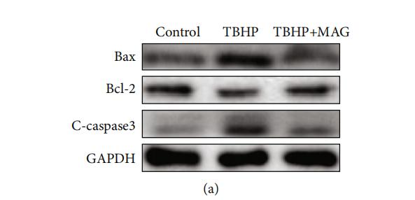
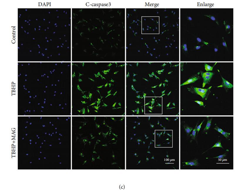
 The expression of Nrf2 from different concentrations of t-BHQ-treated NPCs. (c) Representative images of Nrf2 in NPCs with t-BHQ and TBHP treatment for 24 h. (d, e) Successful knockdown of Nrf2. (f, g) The expression of cleaved caspase-3 in NPCs. (h) TUNEL and ROS assays were performed in NPCs. (i) Intracellular MDA level was assessed by using the MDA assay kit. (j) Intracellular SOD2 level was assessed by using the SOD2 assay kit. All experiments were performed three times in duplicate, and data are reported as the mean ± standard deviation. ∗p < 0.05, ∗∗p < 0.01.")
 Representative images of western blotting. Quantitative analysis of the (B) Bax, and (C) Bcl levels and the (D) cleaved caspase: caspase 3 and (E) p-Akt: Akt ratio and the (F) beclin-1 and (G) LC3BII: LC3BI ratio (n=3). NRG, naringin; I/R, ischemia reperfusion; LY, PI3K/Akt inhibitor LY294002; NRG100, 100 mg/kg naringin; p-Akt, phosphorylated Akt; LC3B, microtubule-associated protein 1B-light chain 3. aP<0.05 vs. sham group; bP<0.05 vs. I/R group; cP<0.05 vs. NRG100 + I/R group.")
 Flow cytometry assays detected the effect of altered HOXA13 expression and 5-FU treatment on GC cells apoptosis. (C, D) The protein levels of cleaved caspase-9 and cleaved caspase-3 in GC cells were determined by Western blot. **P < 0.01, ***P < 0.001.")
 Cell apoptosis in different transfection groups was assessed using a TUNEL assay. Magnification, x100. (B) Western blot analysis showing the protein levels of Bcl-2, Bax, cleaved caspase-3, caspase-3, p-c-Met and c-Met in different groups. ***P<0.001 vs. shRNA-NC; #P<0.05 and ###P<0.001 vs. shRNA-B3GALT5-AS1-1 + ov-NC. B3GALT5-AS1, β-1,3-galactosyltransferase 5-AS1; CSNK2A1, casein kinase 2 a1; p-, phosphorylated; Ov, expression; shRNA, short hairpin RNA; NC, negative control.")
 promotes apoptosis induction of lenalidomide (Len) in MM cells. (A and B) Annexin V-FITC/PI double staining analysis of ARP1 and RPMI-8226 cells treated with 1 μM chidamide and/ or 4 μM lenalidomide for 24 hours. Percentages of MM cell apoptosis based on three independent experiments. (C and D) MM cells derived from 3 MM patients were treated with 1 μM chidamide and/ or 4 μM lenalidomide in vitro for 24 hours, using flow cytometry for analysis. (E and F) expression of apoptosis biomarkers cleaved caspase-3, cleaved PARP were determined using Western blotting, β-actin as reference control. *, p < 0.05; **, p < 0.01 vs combination.")
 The images of tumor tissues. (b) The changes of tumor volume over
time. (c) Tumor weight was recorded. (d) NPR3 expression was measured by immunohistochemistry (400×, magnification, Scale bar = 50 μm). (e and f) Relative
protein levels of NPR3, cyclin D, cleaved caspase 3, PI3K, p-AKT, and AKT were detected in tumor tissues. **P < 0.01 and ***P < 0.001 vs vector group. n = 6.")
 The mRNA and protein level of SGLT1 in the left ventricular of mice. (C) HE staining of longitudinal (upper panel, scale bar: 25 μm) and transverse (lower
panel, scale bar: 25 μm) left ventricle section. (D) Representative photomicrographs of TUNEL staining on left ventricle slices. The white arrows indicated TUNELpositive cells. Scale bar: 50 μm. (E) Western blot analysis of Bcl-2, Bax, and cleaved-caspase 3 in left ventricle, and quantitative analysis of these proteins. Data in
panel A, B, and E were shown as the mean ± SD (n = 6), with one-way ANOVA followed by post hoc Tukey’s test.")
 Histological results of the testis in rats treated
with Cdcl2, melotonin (Mel, SIRT3 activator), 3-MA (autophagy inhibitor targeting PI3K), 3-TYP (SIRT3 inhibitor). Scale bar, 100 mm in the upper line, and 25 mm in the lower line. (B)
Activities of testicular marker enzymes LDH, ACP, AKP, g-GT. (C) A representative immunoblot of SIRT3, autophagy markers (LC3B, BECN1), apoptosis markers (Bcl-2, Caspase3,
Cleaved-caspase3). (D) Quantification analysis of SIRT3. (E) Quantification analysis of LC3B-I, LC3B-II, LC3B-II/LC3B-I, BECN1. (F) Quantification analysis of Bcl-2, Caspase3, Cleavedcaspase3, Cleaved-Caspase3/Caspase3). *p < 0.05; ns shows p > 0.05, indicating no significant difference.")
 (A and B). Immunohistochemistry of cleaved caspase-3 and P-JNK identified the effect of H2 on the degradation of cartilage matrix in OA mice (n = 15) (C). Quantification of cleaved caspase-3 and P-JNK-positive cells in the cartilage samples (D and E). All data are presented as mean ±SEM. ###P < 0.001 vs the sham group; ***P < 0.001 vs the DMM group; n = 15.")
 and
adjacent nontumorous renal tissues from patients who underwent radical renal resection (CON group, n = 12) was detected using immunoblot (A), and (B)
immunohistochemistry, Scale bar: 200 μm. (C) TUNEL staining was performed to examine apoptosis in renal tissues from KS patients and normal controls, scale bar:
100 μm. (D) Protein expression of Cleaved-caspase3 (p17), Bax, and Bcl-2 in renal tissues from KS patients and normal controls was examined using Western blot
analysis. Quantification of their blots normalized to β-actin was shown. Data were expressed as means ± SEM (n = 6), *P < 0.05, **P < 0.01.")
 Cell viability was measured by CCK-8 assay at 24 h post-PQ treatment. (b, c) Cell apoptosis was determined by Annexin V-FITC-PI staining at 24 h post-PQ treatment. (d) The protein expression of TOLLIP, cleaved caspase-3, and cleaved PARP at 24 h post-PQ treatment. ∗p < 0.05, ∗∗p < 0.01, and ∗∗∗p < 0.001 (n = 3).")
 The mRNA and protein expression of TOLLIP in the kidney tissues at 24 h post-PQ treatment. (c) The level of BUN and creatinine in the serum at 24 h post-PQ treatment. (d) Representative images of H&E staining of the kidney tissues at 24 h post-PQ treatment. (e) Representative images of cleaved caspase-3 immunofluorescence staining of the kidney tissues at 24 h post-PQ treatment. ∗∗p < 0.01 and ∗∗∗p < 0.001 (n = 8).")
 DAPI staining (blue) was used to observe the changes in the nucleus of HT-29 and SW620 cells under OMT or DOX treatment. (B) Cell apoptosis was detected by Annexin V-FITC/PI binding assay after OMT and/or DOX treatment in HT-29 and SW620 cells. (C) Western blotting was used to analyze the expression of Bcl-2, Bax, cleaved caspase-3, and cleaved caspase-9. (D) Statistical analysis of total apoptosis rates including early and late apoptosis (Q2 + Q4, respectively). (E–J) The densitometric analysis of protein bands was performed and normalized with the corresponding GAPDH content. Data are presented as the mean ± SD of three independent experiments. *p < 0.05, **p < 0.01, ***p < 0.001 vs. the control group; # p < 0.05, ## p < 0.01, ### p < 0.001 vs. the OMT (5 mM) group or DOX (0.3 μM) group.")
 The MTT results of PC12 cells stimulated by H2O2 (n = 6). (e) Photos of PC12 cells treated with different concentrations of H2O2 for different time. (f) Using MTT to detect the cell viability of PC12 cells after zinc treatment (n = 6). (g–i) The expression levels of cleaved caspase 3, Bax, and Bcl-2 proteins were detected by WB. β-Actin was used as internal controls for WB (n = 6). (j, k) Apoptosis was detected using the MMP and Apoptosis Detection Kit (n = 6, scale bar = 50 μm). (l, m) Flow cytometry results of apoptosis (n = 6). Data are represented as the means ± SD. ∗p < 0.05, ∗∗p < 0.01, and ∗∗∗p < 0.001.")
 were fed an MCD diet in the presence and absence of VTE (1 g 100 g−1 diet) for 6 weeks (A) TUNEL staining in the livers from the MCS diet-, MCD diet-, and MCD + VTE-fed mice. Scale bars, 100 μm (B) TUNEL positive cells (‰). Data are statistically analysed as means ± SEM (n = 6 per group) (C, D) Protein levels of cle-CASP3 and CHOP in mice were analysed by immunoblotting. The relative protein level was shown (E) The relative mRNA expression of CHOP in mouse liver. β-ACTIN was used as an internal control for normalizing the mRNA levels and protein levels. n = 5 for all groups. # p < 0.05, MCD compared with the MCS group. *p < 0.05. MCD compared with the MCD + VTE group.")
 Flow cytometric analysis revealed that NUDCD1 knockdown increased the rate of apoptosis in Patu8988 and PANC-1 cells. (E–N) Western blot analysis showed that NUDCD1 knockdown decreased the expression of Bcl-2 and increased the expression of Bax and Cleaved Caspase-3 in PANC-1 and Patu8988 cells. **p<0.01, ***p<0.001, ****p<0.0001.")
 or directly cocultured with 200 μg/mL AGEs and ROS inhibitor 10 μM NAC for 36 h. (a, b) The intracellular ROS levels were detected using the fluorescent probe DCFH-DA and measured by flow cytometry. (c, d) After labeled with DCFH-DA fluorescent probe, representative fluorescent images were acquired under a fluorescence microscope, scale bar: 100 μm. (g–j) Representative western blot bands of p16, p53, and cleaved caspase 3 and relative band density were quantified. (k, l) Representative images of immunofluorescence staining for p16 and cleaved caspase-3 in each group, with the relative fluorescence intensity quantified, scale bar: 50 μm. Data are represented as mean ± SD. ∗∗P < 0.01, ∗P < 0.05.")
.")
 and different concentrations of mangiferin (100 μM/ml and 500 μM/ml). (a) The mitochondrial morphology in HNPCs was observed using TEM. Scale bars: 5 μm and 1 μm. (b–e) The levels of OPA1, TFAM, and Drp1 were assayed using Western blotting. (f, g) JC-1 was used to detect the mitochondrial membrane potential in HNPCs. Scale bar: 100 μm. (h, i) ROS levels were detected in HNPCs from each treatment group. Scale bar: 100 μm. (j, k) MitoTracker was used to detect the mitochondrial membrane potential of HNPCs. Scale bar: 25 μm. The expression of (l) Bax, (m) Bcl-2, and (n) c-caspase-3 in each indicated group was assayed using real-time PCR. (o–r) The levels of Bax, Bcl-2, and c-caspase-3 were assayed using Western blotting. (s, u, v) Hoechst 33342 and (t, w) TUNEL staining of the HNPCs in each indicated group. Scale bar: 100 μm. All the experiments were repeated at least three times. Significant differences are indicated as follows: ns P > 0.05, ∗P < 0.05, ∗∗P < 0.01, ∗∗∗P < 0.001, and ∗∗∗∗P < 0.0001.")
 ARP-1 cells were pretreated with or without NAC (15.0 mmol/L) for 2 hours at 37°C and then incubated with CHI (1.0 µmol/L) and/or BTZ (5.0 nmol/L) for 24 hours, then ROS generation was detected. (B) ARP-1 cells were pretreated with or without 15 mmol/L NAC and then treated with Chidamide or Bortezomib alone or in combination, and cell viabilities were evaluated using CCK-8 assays. (C) The expression of γ-H2AX in ARP-1 cells treated with CHI (1.0 µmol/L) and/or BTZ (5.0 nmol/L) were determined by Western blot. (D) Representative images of γ-H2AX (Red) and nuclei (Blue) in ARP-1 cells treated with single agent or combination for 24 hours by immunofluorescence assay. Scale bars represent 20 µm. (E, F) Western blot analysis of the expressions of cleaved caspase3, cleaved caspase8, cleaved PARP-1 and HDAC1 in XG1 (E) and ARP-1 (F) cells after 48 hours treatment with single agent or in combination. Error bars indicate mean ± SD. **p < 0.01.")
 Efficacy of single-agent and combination treatment of CHI with BTZ in a human myeloma model in B-NDG mice. Tumor volumes, tumor weight and mice weight in ARP-1 cells xenografted mice model following treatment with single agent CHI and combinations with BTZ were shown (n=3). (C) Immunohistochemical analyses with anti-cleaved caspase-3, anti-cleaved PARP and anti-Ki-67 induced by the treatment with single agent and in combinations in vivo. (D) Western blot analysis of the expression of HDAC1 in the xenografts treated with single agent and in combinations. Error bars indicate mean ± SD. **p < 0.01; NS, not significant.")
 by shRNA or dichloroacetate (DCA) sensitizes diffuse large B‐cell lymphoma (DLBCL) cells to rituximab (RTX). A, B, Annexin V‐phycoerythrin (PE)/7‐AAD double staining analysis of DLBCL cells treated with RTX (50 μg/mL). Interference with PDK4 shRNA increased RTX‐induced cell apoptosis and caspase‐3 activation in U2932 and OCI‐Ly8 cell lines. C, PDK4 inhibitor dichloroacetate (DCA) enhanced the RTX‐induced apoptosis in DLBCL cell lines U2932, OCI‐ly7, and OCI‐Ly8. cop‐GFP, copepod super green fluorescent protein. *P < .05, **P < .01")
 apoptosis- and (B) autophagy-associated proteins. *P<0.05 and **P<0.01. miR, microRNA; H/R, hypoxia/reoxygenation; LC, light chain; NC, negative control.")
 Western blot analysis of MOF, MDR1 and HIF-1α protein expression and Caspase 3 cleavage after treatment with DMSO vehicle, 10 μM sorafenib or 10 μM 5-FU treatment for 72 h. (B,C) Cytotoxicity assays for sorafenib (B) or 5-FU (C). The experiments in legends (B,C) were performed on Huh-7 cells, which were treated with different concentration of the indicated drugs for 72 h. Cells with LW6 treatment were treated with 15 μM LW6. ∗P < 0.05, ∗∗∗P < 0.001 and ns, not significant as compared to shVector cells; n.s., not significant as compared to shVector + LW6 cells. V, shVector; #1, shMOF #1; #2, shMOF #2.")
 The expression of Bxl2-2, Bax and cleaved caspase 3 were measured by western blot after the treatment of MHO7.")
 The effect of ketamine (KET) on autophagy- and apoptosis-related marker expressions in rats was evaluated by quantitative real-time polymerase chain reaction (qRT-PCR) and western blot. β-actin was used as the internal control. (e–g) The effect of KET on ERS-related marker expressions in rats was evaluated by western blot. We used β-actin as the internal control. All experiments have been performed in triplicate and data were expressed as mean ± SD. *P < 0.05, ***P < 0.001 vs. Sham KET-L: 5 mg/kg KET; KET-H: 50 mg/kg KET")
 Representative TUNEL images in each group after treatment with various concentrations of MAF. Scale bars, 250 µm. (B) Western blot analysis and quantification of Bcl-2, Bax, cleaved-caspase-3 and cleaved-caspase-9 in MAF treatment. ***P<0.001 vs. Control; ##P<0.01 and ###P<0.001 vs. H/R. MAF, mangiferin; H/R, hypoxia-reoxygenation.")

 SLC25A21 promoted the efflux of α-KG from the mitochondria to the cytosol. (B) The levels of α-KG in the mitochondria affected succinate production. (C) The upregulation of SLC25A21 promoted ROS accumulation in BCa cells. (D) The upregulation of SLC25A21 decreased Δψm in both BCa cell lines. (E) Western blot assays showed that SLC25A21 induced cyto C transfer from the mitochondria to the cytosol and increased the activation of caspase-9 and caspase-3 in BCa cells. (F) Immunohistochemistry showed that SLC25A21 increased the activation of caspase-9 and caspase-3 in BCa xenograft tissues. (G) Schematic diagram showing the mechanism of action of SLC25A21 on cell apoptosis in BCa. The results were reproducible in three independent experiments. *P < 0.05, **P < 0.01, ***P < 0.001 and ****P < 0.0001.")
 along with the quantification of positive cells (B), scale bar: 50 mm.
(C) The protein levels of Bcl-2, Bax and cleaved caspase-3 were detected by western-blot. All data were presented as
mean + SD of six separate experiments. Versus control group: #
P < 0.05, ##P < 0.01, ###P < 0.001, ####P < 0.0001; Versus
CS group, *P < 0.05, **P < 0.01, ***P < 0.001, ****P < 0.0001.")
 were generated as described in “Materials and methods”. After confirming the overexpression or knockdown efficiencies of GPR17, cells were cultured and harvested for the subsequent experiments. A CCK-8 assay was performed to examine viable cell numbers. B Immunofluorescent staining against BrdU was performed to examine the numbers of proliferating cells, scale bar, 400 μm. The proportions of the BrdU+ cell in the total cell number were displayed in the lower panel. C Western blot was performed to examine intracellular cleaved-caspase3 levels. Densitometric quantification of cleaved-caspase3/caspase3 ratio from at least three independent assays was indicated on top of each band, respectively. D Expression of GPR17 in IDHwt (n = 233) and IDHmut (n = 428) glioma patients from the TCGA cohort. E Flow cytometry analysis was performed to assess mitochondrial ROS level. F–H Control and U87/U251-GPR17 cells were treated with vehicle or N-acetyl-l-cysteine (5 mM) for 48 h, CCK-8 assays (F), immunofluorescent staining against BrdU (G), and western blotting against cleaved-caspase3 (H) were performed as mentioned above. Scale bar, 400 μm. Densitometric quantification of cleaved-caspase3/caspase3 ratio from at least three independent assays was indicated on top of each band, respectively. For all panels, data represent the means ± SEM from three independent experiments. *p < 0.05, **p < 0.01, ***p < 0.001, Student’s t-test.")
 Normal group; (II) Model group; (III) Scu 3 μM; (IV) Scu 10 μM; (V) Scu 30 μM. (A) Cell morphology were observed by Hoechst 33258. One
representative image of three individual experiments is shown. (B–C) The number of apoptotic cells in HUVECs were analyzed via using the Annexin V FITC-PI
apoptosis detection kit and detected by flow cytometry. The expression of Bcl-2 (D), Bax (E), Cyt.C (F) and Cleaved caspase-3 (G) was measured by western blotting. ##P < 0.01 vs. untreated group; *P < 0.05, **P < 0.01 vs. HG only treated group. One-way ANOVA followed by Tukey’s test.")
 Cell apoptosis results of LA795 cells incubated by 50 μM HHT for 24 h detected with Annexin-V assay (n = 3). (B) Cell apoptosis results of LA795 cells with TMEM16A shRNA transfection and added 50 μM HHT (n = 3). (C) Cell apoptosis results of 2BS cells with TMEM16A transfection and added 50 μM HHT (n = 3). (D–F) Expression of cleaved-caspase 3 and cleaved-caspase 9 with LA795 cells incubated by 50 μM HHT (D), or LA795 cells with TMEM16A shRNA transfection and added 50 μM HHT (E), or 2BS cells with TMEM16A transfection and added 50 μM HHT (F) (n = 3).")
 for 24 or 48 h could significantly induce AKI and inflammatory response in mouse model. A and B, Creatinine and urea
nitrogen in serum indicate a significant decrease in renal function in cisplatin-treated mice. C. Renal tissues were collected for HE stains, TUNEL assay and
Immunohistochemistry analysis of the KIM-1 and NGAL protein expression levels to record tubular injury and renal function. D. Expression levels of apoptosis-related
proteins (cleaved caspase-3, Bax and Bcl-2) in the control and cisplatin-treated groups. E. Semi-quantification of tubular damage, apoptosis and expression levels of
KIM-1 and NGAL. F. Semi-quantification of cleaved caspase-3 and Bax/Bcl-2. G. Concentration of cytokines TNF-α, IL-1β and IL-6, respectively, in renal tissues as
detected by ELISA. The data in A, B, E, F and G are expressed as the mean ± SD. *P < 0.05 vs. the control group.")
, Bax (C), Bcl-2 (D), Bax/Bcl-2 (E), Bad (F), cytosol cytochrome C (G), mitochondrial cytochrome C (H), caspase-9 (cleaved) (I), caspase-3 (cleaved) (J), PARP (K), and PARP (cleaved) (L) in H22 ascites tumor-bearing mice. Data are expressed as the
mean ± SEM (n = 3), * p < 0.05, ** p < 0.01 vs the model group.")
 The flow cytometry was applied to detect apoptosis induced by compound
26, and calculate the percentage of apoptotic cells. (b) (c) Perform immunoblotting on the whole cell lysate and probe with Bcl-2, Bax, caspase-3/9, cleaved-caspase3/9 antibodies, and β-actin as loading control, 15 μg proteins were loaded in each lane to quantify the results. (* p < 0.05, ** p < 0.01, *** p < 0.001, **** p <
0.0001 vs control group).")
 The protein expression of cleaved caspase 3 in ICR mice treated with vehicle, crizotinib, or sunitinib treatment (n = 6). *P < 0.05 or **P < 0.01 (the crizotinib or sunitinib alone vs. control).")
. B Quantitative analysis of intracellular lipid droplets. C The expressions of TNFα, IL-1, and Cleaved-caspase3 in control, PA, and C14 group (n = 6), as detected using western blotting. D The results of TNFα, IL-1, and Cleaved-caspase3 expression. E The free fatty acids of H9c2 cells in each group was measured (n = 6) and compared with the treated and control group. F The data revealed positive expression of TNFα and cleaved-caspase 3 in H9c2. (G-H) Immunofluorescence staining results showing the levels of TNF-α and Cleaved-caspase3 in H9c2 (n = 6, Scale bar = 100 μm). Data are represented as means ± SD, where two-way ANOVA followed by Tukey’s post hoc test employed. *P < 0.05, **P < 0.01, and ***P < 0.001")
. B Quantitative analysis of intracellular lipid droplets. C The expressions of TNFα, IL-1, and Cleaved-caspase3 in control, PA, and C14 group (n = 6), as detected using western blotting. D The results of TNFα, IL-1, and Cleaved-caspase3 expression. E The free fatty acids of H9c2 cells in each group was measured (n = 6) and compared with the treated and control group. F The data revealed positive expression of TNFα and cleaved-caspase 3 in H9c2. (G-H) Immunofluorescence staining results showing the levels of TNF-α and Cleaved-caspase3 in H9c2 (n = 6, Scale bar = 100 μm). Data are represented as means ± SD, where two-way ANOVA followed by Tukey’s post hoc test employed. *P < 0.05, **P < 0.01, and ***P < 0.001")

 and the PI3K/AKT/mTOR/NF-κB (b) signaling pathway of protein levels in tumor tissues. Data was expressed as mean ± SD. Compared with the model group, ★P < 0.05, ★★P < 0.01.")
. for 3–4 independent experiments.")
 The representative images of H&E, Nissl and FJ-B staining of ipsilateral cerebral cortex at day 3 from Sham, TBI and TBI+Mino group. Scale bar= 500 µm (4×), Scale bar=100μm (20×). (B) Quantification of Nissl staining for neuronal loss analysis at 3 day after TBI. (C) Quantification of FJ-B immunofluorescence staining for neuronal apoptosis analysis at 3 day after TBI. (D)Representative images of TUNEL (green) immunofluorescence staining in perilesional cortex 3 day post-TBI. Scale bar= 100 µm. (E) Quantification of TUNEL staining at 3 day after TBI. (F) Representative western blot analysis of cleaved-caspase 3, Bax and Bcl-2 in ipsilateral cerebral cortex 3 day post-TBI. (G, H) Quantification of cleaved-caspase 3 and the ratio of Bcl-2/Bax from western blot analysis. All data represent the mean ± SEM, n = 3, *P < 0.05, **P < 0.01, ***P < 0.001 vs. the indicated group.")
 Representative Western blot brands. (B) Quantitative analyses of phospho‐PI3K/PI3K, phospho‐AKT/AKT, Bcl‐2/Bax, cleaved‐Caspase 3. Mean ± SD from three individual experiments. *p < 0.05, **p < 0.01, versus the sh‐NT‐HeLa group")
 The ICR mice hepatocyte apoptosis in liver tissue (TUNEL, 20×). The blue fluorescence indicates nuclei, and the green fluorescence indicates apoptotic cells. (B) TUNEL-positive cells were quantified. (C, D) The protein expression of cleaved caspase 3 in ICR mice treated with vehicle, crizotinib, or sunitinib treatment (n = 6). *P < 0.05 or **P < 0.01 (the crizotinib or sunitinib alone vs. control).")
.")
.")
 Cell viability of ETH (200 mm) and ETH + ROF groups after treatment for 24 h. The apoptotic rate (C), Bax expression (D), and Bcl-2 expression (E) in LO2 cells. The protein levels of caspase 3 and cleaved caspase 3 (F). Mean ± SEM (n = 3–6). *p < 0.05, **p < 0.01 and ***p < 0.001 vs. the ETH group. ###p < 0.001 vs. the CON group.")
 Apoptosis of the NPCs detected by flow cytometry analysis. (c, d) Typical fluorescence photomicrograph and quantitation of TUNEL staining of NPCs (scale bar: 100 μm). (e, f) Representative western blot bands and quantitation of the expression of cleaved-caspase 9, cleaved-caspase 3, Bax, and Bcl-2 in the NPCs. (g, h) Representative western blot bands and quantitation of the expression of anabolic mediators (aggrecan and collagen II) and catabolic mediators (MMP3 and MMP9). The data are represented as the mean ± SD from at least 3 independent experiments. ∗P < 0.05 and ∗∗P < 0.01 vs. the TBHP group.")
 TUNEL staining for the detection of cell apoptosis in OC tumor tissues (magnification: ×200). (b) Protein expression levels of Bax, Bcl-2, cleaved caspase 3, and caspase 3 in tumor tissues were detected by Western blot assay, and GAPDH was used as the internal control. (c) Serum levels of IL-6, TNF-α, and CA125 were detected by ELISA. (d) Flow cytometry was used to detect the CD3+CD4+CD3+/CD8+ T lymphocyte percentages. The data are presented as mean ± SD. ▲▲p < 0.01, compared to the NC group; ∗p < 0.05, ∗∗p < 0.01, compared to the vehicle group. OC: ovarian cancer.")
 Representative immunofluorescent images of JC-1 is visible either as JC-1 monomers (green), JC-1 aggregates (red), and both channels merged (200x magnification), whereas more JC-1 aggregates (red) were seen in quercetin treatment while more JC-1 monomers (green) in ethanol group. (b) Relative fluorescence intensity calculated as red to green ratio. (c) Representative immunofluorescence images co-stained with γH2AX (red), Mito-Tracker (green) and both channels merged (400x magnification). The graphs (down panel) show the fluorescence intensity profiles in two fluorescence channels along the arrow, whereas a weaken fluorescence intensity of γH2AX is seen in quercetin treatment and enhanced by ethanol. (d) The mRNA expression that encodes the enzyme (TWNK, MTCO1, and MFND) responsible for mtDNA transcription levels. (e) Western blot analysis of Bax, Bcl2, caspase3, and cleaved-caspase3 protein abundance. Data are expressed as mean ± SD (n = 6). Different subscript letters indicate significant differences among the groups (p < 0.05).")

 The apoptosis was measured by TUNEL. (b) The protein expression of Bax, Cleaved-caspase 3, Bcl-2, and Cyclin D1 was detected by western blot. ▲P < 0.05 and ▲▲P < 0.01vs. the model. Results were presented as mean ± SD. n = 3. Note: RAC: Radix Actinidia chinensis; RCC: renal cell carcinoma; TUNEL: terminal deoxynucleotidyl transferase dUTP nick end labeling.")
 Cardiac apoptosis was assessed by TUNEL staining (magnification, ×200). (b) Positive apoptosis cells in each group. (c) Western blot analysis and statistical analysis of (d) Bax, (e) cleaved caspase-3, and (f) Bcl-2 proteins in each group. ∗P < 0.05 and ∗∗P < 0.01 vs. sham group. ▲P < 0.05 and ▲▲P < 0.01 vs. IR group.")
 Western blot study of NTN1, DAPK1, P-DAPK1(Ser308), P-P53(ser20), and caspase 3(p17) protein in U251 cells infected with sh-Luci and sh-PTBP1 for 3 and 7 days. (n = 3). As an internal reference protein, GAPDH was used. (F) Activity of PP2A during reprogramming. (n = 3). (G-I) TUJ1 and KI67 positive rates of six distinct groups constituted of lentivirus (sh-Luci or sh-PTBP1) and TC-DAPK6 (vehicle, 100 nM, 250 nM, and 500 nM; nine random fields from triplicate samples were recorded for quantification; TUJ1+ (%) = TUJ1+ M-cherry+/M-cherry+; KI67+ (%) = KI67+ M-cherry+/M-cherry+; M-cherry+ cells = 156-553 for each condition). The data are presented as mean ± SD. *P < 0.05, *** P < 0.001 vs. sh-Luci-3d or sh-PTBP1 + vehicle group. Dpi (d): days post infection; ND: not detected; NS: no significance. Scale: 100 µm.")

 WB analysis of the antiapoptotic and caspase proteins in HepG2 cells treated with C20/C22 at 2 μM for 0, 6, 12, and 24 h. The reported values correspond to the mean ± SD for three independent experiments. (D) Immunofluorescence assays employed to determine the expression of caspase3/7 in cells with indicated treatments. Scale bars, 10 µm. (E,F) Apoptotic cells detected by flow cytometry after indicated treatments (n = 3). The p-value was analyzed by one-way ANOVA followed by Tukey’s test using GraphPad Prism version 8.00. ** p < 0.01, *** p < 0.001, and **** p < 0.0001 vs. 0 h/control group.")
 Effects of miR-192 downregulation on the mRNA expression of Acvr2a. (B) Effects of miR-192 downregulation on the protein expression of ACVR2A. (C) Effects of miR-192 downregulation on the mRNA expression of caspase-3. (D) Effects of miR-192 downregulation on the protein levels of cleaved caspase-3. Data are shown as mean ± SE. (n = 3). * p < 0.05, ** p < 0.01 vs. the H2O2-free group (control); # p < 0.05 vs. the H2O2 plus inhibitor control.")
. Bcl-2 (B), Bax (C), Bax/Bcl-2 (D), caspase-3 (E), and cleaved-caspase-3 (F) levels were calculated by grayscale analysis. Values are displayed as mean ± SEM (n=3). Significant differences are shown as *P < 0.05, **P < 0.01 in comparison to the Con group. #P < 0.05, ##P < 0.01 in comparison to the Iso group.")
 Immunofluorescence staining of iNOS and F4/80 in the lung tissues of each group. (C,D) TUNEL staining presented apoptotic cells in the lung tissues of WT and Nrf2−/− mice before or after CLP treatment. (E,F) Western blot analyses of apoptosis-associated proteins such as Bcl-2, Bax, and Cleaved—caspase3 in the lung tissues of each group. ** p < 0.01, *** p < 0.001.")
. B Cell viability of podocytes was analyzed using an MTT assay following different treatments (n = 3). C Western blot assay showed the expression of ANGPTL3 at different time points in human podocytes after PAN treatment. *p < 0.05, **p < 0.01 compared with the Ctrl group. D Relative expression of ANGPTL3, BAX, Bcl2, caspase 8, cleaved caspase 9 and cleaved caspase3 in different groups (n = 3). *p < 0.05, **p < 0.01 compared with the PAN group.")
 Western blot analysis of caspase-3 and PARP expression in 100 μM glabridin- or 1 μg/ml doxorubicin-treated cells. (B) Representative images of nuclear staining. MDA-MB-231 and MCF7 cells were treated with 100 μM glabridin for 24 h, and the nuclei were stained with DAPI (C) DNA fragmentation assay. Genomic DNA isolated from glabridin- or doxorubicin-treated cells subjected to agarose gel electrophoresis. (D) Cells were treated with 60 μM glabridin in the absence or presence of Z-VAD-FMK (20 μM), and the percentage of cell death was determined using PI staining (E) Cells were treated with 60 μM glabridin in the absence or presence of Z-VAD-FMK (10 μM or 20 μM), and the cell viability was determined using CCK-8 assay. The results are presented as the means ± SD of three independent experiments (vs. control: ∗P < 0.05; NS, not significant). The uncropped images of (A) and (C) were referred to in Supplementary Figure S8 (A) and (B). NT, no treatment; GLA, glabridin; Doxo, doxorubicin; Z, Z-VAD-FMK; Z10, 10μM Z-VAD-FMK; Z20, 20μM Z-VAD-FMK.")

 treatment reduces HI-induced neuronal apoptosis. a Western blot evaluation of the protein levels of Bcl2, Bax, caspase-8, caspase-8 p18, cleaved-caspase-3 and caspase-3 24h after HI injury. b Quantification of western blot data of Bcl2/Bax. c Quantification of western blot data of caspase-8 p18/caspase-8. d Quantification of western blot data of cleaved-caspase-3/caspase-3. ∗∗P < 0.01, ∗∗∗P < 0.001 vs. the sham group. #P < 0.05, ###P < 0.001 vs. the HI group. &P < 0.05, &&P < 0.01 vs. the CA treatment group. n = 3")
 The expression of Ki67 was detected by immunohistochemistry at days 3 (n = 6) and 7 (n = 6). (b) TUNEL assay of testicular tissues after torsion-detorsion injury at days 3 (n = 6) and 7 (n = 6). (c) The expression of Cleaved Caspase-3 was detected by immunofluorescence at days 3 (n = 6) and 7 (n = 6). (d) Quantitative analysis of Ki67, TUNEL, and Cleaved Caspase-3 expression. Bars, 50 μm. Data are represented as mean ± SD. ∗∗∗P < 0.001, ∗∗∗∗P < 0.001.")
 Representative images of terminal deoxynucleotidyl-transferase-mediated dUTP nick end labeling (TUNEL) staining. TUNEL-positive apoptotic cells were shown in green and DAPI nuclear staining was shown in blue (scale bar = 50 μm). (B) Quantitative analysis of TUNEL-positive cells (n = 4). (C) The protein expression levels of Bax, Bcl-2 and cleaved Caspase-3 in rat cardiac tissue of each group were shown by Western blotting (WB) bands. (D–F) Relative protein for Bax, Bcl-2 and cleaved Caspase-3 were quantified by densitometry based on immunoblot images. Results are presented as mean ± SD (n = 3). One-way ANOVA followed by Bonferroni’s post hoc test: # p < 0.05, ## p < 0.01 vs. vehicle group.")
 Heatmap of miRNA microarray results in RAW264.7 cells treated with LPS and genistein. (B) qRT-PCR of miR-155, miR-21 and miR-125a expression in RAW264.7 cells with the treatments of LPS and genistein. (C) qRT-PCR of miR-155, miR-21 and miR-125a expression in RAW264.7 cells. (D) qRT-PCR of miR-21 in stably expressing miRNA RAW264.7 cells. (E and F) Cell viability and Western blot of pro-apoptotic proteins CHOP, BAX, cleaved Caspase 3, cleaved Caspase 8 and anti-apoptotic proteins BCL-2 in RAW264.7 cells with stable miR-21 overexpression or repression treated by LPS and genistein (10 μM, 2 h). (G and H) qRT-PCR and Western blot of iNOS and COX-2 in RAW264.7 cells with the indicated treatments after miR-21 overexpression or knockdown. Data are shown as mean ± SD of three independent experiments. LPS, lipopolysaccharides; GEN, genistein; *P < 0.05, **P < 0.01, ***P < 0.001.")
 The viability of hepatocytes was analyzed by MTT after L02 cells were incubated with 5, 10, 15 μM Sunitinib for 48 h in the presence or absence of 20 μM CQ pretreatment for 2 h (n = 6). (B) Levels of ALT, AST, and LDH released into the supernatant after L02 cells were incubated with 10 μM Sunitinib for 48 h in the presence or absence of 20 μM CQ pretreatment for 2 h (n = 3). (C) Protein expression of PARP, c-Caspase3, P62, and LC3B (n = 3). (D) Protein expression of LC3B after L02 cells were incubated with 10 μM Sunitinib for 48 h in the presence or absence of 25 μM Z-VAD-FMK pretreatment for 2 h (n = 3). ** p < 0.01 and *** p < 0.001 vs. Control group. # p < 0.05, ## p < 0.01, and ### p < 0.001 vs. Sunitinib group.")
 and MDA-MB-231 cells (D–F). G The expression of BAX, Bcl-2, and Cleaved-Caspase3. H–I Syringin increased the expression of BAX and Cleaved-Caspase3 and decreased the expression of Bcl-2. *P < 0.05 and **P < 0.01")
 and those cultured in regular media for the same course (Ctrl) were analyzed. (A) CCK-8 assay was applied to assess cell viability for 6 days. (B) The migration of cells was studied and quantified by transwell assay. (C) Cytotoxicity assay was used to examine apoptosis after 3 days of growth factor deprivation stress (Q3). (D) Western blot analysis showed altered expression of apoptotic markers in cells subjected to 5 days of growth factor deprivation (Q5). (E) Flow cytometry analysis was performed to study cell death in response to 48 h treatment of 5-FU (6 μM). (F) Protein levels of E-cadherin, N-cadherin, and vimentin were determined by Western blot analysis. Data were representatives of at least three independent experiments, shown as mean ± SD. Significance threshold determined by one-way ANOVA or Student's t-test:")
 of cleaved caspase3 (A, B) were analyzed. The proteins expression of P62 (C, D), LC3-II/I (C, E), Beclin-1 (C, F), and cleaved caspase3 (C, G) in DEX-treated MC3T3-E1 cells were determined by western blot. (H, I) The apoptosis was determined by flow cytometer. **P˂0.01. NC, negative control group; DEX, model rat group treated with DEX (5 mg/kg).")
 of cleaved caspase3 (A, B) were analyzed. The proteins expression of P62 (C, D), LC3-II/I (C, E), Beclin-1 (C, F), and cleaved caspase3 (C, G) in DEX-treated MC3T3-E1 cells were determined by western blot. (H, I) The apoptosis was determined by flow cytometer. **P˂0.01. NC, negative control group; DEX, model rat group treated with DEX (5 mg/kg).")
. There was no difference in M1 population (F4/80+/CD80+/CD206- cells) between WT and LDLR−/− mice without or with NAC treatment (B). M2 population (F4/80+/CD80-/CD206+) in the aorta was significantly decreased in aging LDLR−/− mice (12 months of age) on ND compared with age-matched WT mice, and this was effectively reversed with 3 months of NAC treatment (C). Similar findings on M2 population in the aorta of aging LDLR−/− mice were observed using immunostaining of F4/80-AF594+/CD206-AF647+ (D–F). Immuno-histological analysis of the aortic root cross sections of aging LDLR−/− mice also showed that CD80 level remained the same with or without NAC treatment (H and I). However, NAC treatment significantly decreased the overall level of caspase 3 in the aorta of aging LDLR−/− mice on ND (H and J). No significant difference in caspase-3 level was observed in CD80-positive cells (H and K). WT: wild-type mice; NAC: N-acetylcysteine; LDLR−/−: LDLR−/− mice; LDLR−/− + NAC: LDLR−/− mice treated with NAC. Data are presented as mean ± SE, n = 6–10.")
 treatment through activating heat shock factor 1 (HSF1). (a) Identification of novel HSF1 activator in PA-inducted HepG-2 cells based on high-content screening system by using HSF1 antibody. Representative images were captured and presented. Scale bar, 100 μm. (b) Cellular triglyceride (TG) levels and survival cell determination. (c) Expression of proteins related to oxidative stress, apoptosis, inflammation and HSF1; protein levels were quantified and normalized to actin. (d) HSF1 translocation and distribution assay. Actin was selected as whole cell lysate or cytosol loading control, and Lamin B was selected for nucleus. (e–h) HSF1 knockout diminished the hepatoprotective effects of SYSU-3d in PA-treated cells. The HSF1−/−-HepG-2 cells were exposed to PA (0.75 mM) induction for 24 h in the presence or absence of SYSU-3d (1 μM); cells were harvested and indicated parameters were analysed. (e) Expression of HSF1 and TG levels was determined by using HSF1 antibody and Nile red dye, respectively. (f) TG level. (g) Cell survival. (h) Expression of HSF1, apoptosis and inflammation markers; protein levels were quantified and normalized to Actin. (i–l) Overexpression of HSF1 restored the hepatoprotective effect of SYSU-3d. The HSF1−/−-HepG-2 cells were transfected with AdHSF1 for 24 h and then exposed to palmitic acid (PA) induction with or without SYSU-3d (1 μM) treatment for another 24 h. TG levels (i), cell survival (j), caspase 3 activity (k) and expression of HSF1 and related proteins (l) were determined; protein levels were quantified and normalized to GAPDH. N = 5 independent experiments. *P")
 FJC staining of the ischemic penumbra area of brain tissues in MCAO/R rats. (B) Quantification of FJC-positive cells (green). (C) Immunofluorescence staining of neurons (NeuN, FITC, green) and TUNEL (red) staining in the ischemic penumbra area in MCAO/R rats. MCAO/R rats showed greater number of TUNEL-positive cells than sham rats, but inhibition of Vav1 decreased this effect. Nuclei were stained with DAPI (blue). Scale bars: 50 μm. (D) Quantification of NeuN and TUNEL double positive cells. (E, F) Bcl-2, Bax, and cleaved caspase-3 protein levels (normalized to the sham group) were analyzed by western blot. Data are expressed as the mean ± SD (n = 6), and were analyzed by one-way analysis of variance followed by Tukey’s multiple comparisons test. The experiments were repeated three times. DAPI: 4′,6-Diamidino-2-phenylindole; FITC: fluorescein isothiocyanate; FJC staining: Fluoro-Jade C staining; MCAO/R: middle cerebral artery occlusion/reperfusion; NeuN: neuronal nuclear; TUNEL: terminal-deoxynucleotidyl transferase mediated nick end labeling; Vav1: Vav guanine nucleotide exchange factor 1.")
 Relative mRNA expression levels, (B) representative western blotting images of FGF23 and (C) FGF23/GAPDH level. (D) Semi-quantification of apoptotic rates and (E) representative flow cytometry plots of Annexin V/PI staining. (F) Representative western blotting images and (G) relative protein expression levels of C-caspase3 and Bcl-2. (H) Representative western blotting images and (I) relative expression ratios of p-PI3K/PI3K and p-AKT/AKT. GAPDH was used as a loading control. *P")
 NADPH production in CRC cells transfected with the NC construct, shELFN1-AS1 or shELFN1-AS1+G6PD. (B) Flow cytometry was used to assess ROS accumulation in CRC cells transfected with the NC construct, shELFN1-AS1 or shELFN1-AS1+G6PD. The histogram shows the fluorescence changes in CRC cells. (C) Flow cytometry detection of apoptosis in different groups. The histogram shows the apoptotic rate of CRC cells. (D) Knockdown of ELFN1-AS1 in CRC cells inhibited the expression of Bcl-2 and promoted the expressions of cleaved caspase-3 and Cyt-C; however, overexpression of G6PD blocked these effects. Data are shown as the mean±SD.")
 The apoptosis of BGCs which were treated with 0, 0.6, and 1.2 mM NEFA was determined by flow cytometry and analyzed using FlowJo. (C–F) The apoptosis−related mRNA levels of Bax, Bcl−2, and Caspase−3 were determined using qRT−PCR. (G,H) The apoptosis−related proteins Bax, Bcl−2, and Caspase−3 were analyzed by Western blot. The quantification of the relative protein levels (performed using Image J) is shown under the band (protein/β−Tubulin). This experiment was repeated 3 times, and data are expressed as mean ± standard errors of the mean (SEM)")
 The Annexin V-FITC/PI double labelling technique was used to determine the MC3T3-E1 cells' apoptosis rate in each group. (b) Bax and Bcl2 protein expressions were found using a Western blot analysis. (c) Caspase-3, cleaved PARP, PARP, cleaved caspase-3, and β-actin marker expressions were found using a Western blotting in MC3T3-E1 cells between groups. (d) We compared the levels of Bcl-2 and Bax mRNA expressions in MC3T3-E1 cells across treatments using RT-PCR. (e) Each group's MC3T3-E1 cells had their mitochondrial membrane potential measured using flow cytometry. Bcl-2 stands for B-cell lymphoma-2, PARP stands for poly ADP-ribose polymerase, and cleaved PARP stands for cleaved poly ADP-ribose polymerase;")
 The apoptosis of DB and SU-DHL-4 cells was measured using flow cytometry with Annexin V and PI staining. Annexin V+PI− cells were considered as early apoptotic cells, and Annexin V+PI+ cells as late apoptotic cells. (C) The levels of several apoptosis markers, cleaved caspase-3, cleaved caspase-9 and cleaved PARP-1, were examined in CNN3-silenced cells. **P")
 Flow cytometry analysis of neutrophil apoptosis after BHB treatment. Neutrophils were treated with 0, 0.6, 2, and 3 mM BHB for 10 h, respectively. Annexin V-fluorescein isothiocyanate (FITC)/propidium iodide staining was used to identify apoptosis cells. Annexin V+ cells were regarded as total apoptotic neutrophils. The numbers in the top-right and bottom-right quadrants indicate the percentages of late and early apoptotic cells, respectively. (B) Quantification of the total apoptotic rate of neutrophils in A. One-way ANOVA with subsequent Bonferroni correction was used to analyze the data. Linear increase (P < 0.0001) and quadratic effect (P < 0.0001) for the apoptosis rate were found, due to the increased dose of BHB. Data expressed as LSM ± SEM. (C) Flow cytometry analysis of the effects of different duration times of BHB on neutrophil apoptosis. Neutrophils were treated with 2 mM BHB multiple times (0, 2, 4, 6, 8, and 10 h). Stimulation with 2 mM BHB for 8 h was selected as ideal conditions for subsequent experiments. (D) Quantification of the total apoptotic rate of neutrophils in C. One-way ANOVA with subsequent Bonferroni correction was used to analyze the data. Linear increase (P < 0.0001) and quadratic effect (P < 0.0001) were found for the apoptosis rate with increased incubation time. Data expressed as LSM ± SEM. (E) Protein abundance of cleaved caspase 3 and caspase 3. The 3 wells of each group represent 3 samples from different replicates, respectively. (F) Quantification of protein abundance in E. Data were analyzed with independent-samples t-tests. Data expressed as mean ± SEM. (G) mRNA abundance of CASP3. Data were analyzed with independent-samples t-tests. Data expressed as mean ± SEM. For western blotting experiments, β-actin was used to normalize protein abundance; for quantitative reverse-transcription PCR experiments, ACTB and YWHAZ were used to normalize mRNA abundance. Statistical significance was declared at P < 0.05, and P < 0.01 was declared to be highly significant.")
 Confocal microscopy analysis of α-tubulin polymerization and F-actin cytoskeletal remodeling in CRC cells with CX43 overexpression and knock-down. (c) The young’s modulus of CX43 overexpressed DLD1 cells was measured by atomic mechanics microscope. Mechanical curve (left) and quantitative analysis (right) are displayed. (d) Western blot analyses expression of stem cell characteristic related proteins in CRC cells with CX43 overexpression. (e) The tumor cell spheroidizing ability of CX43 overexpression cells was detected by tumor sphere formation assays. (f) Western blot was used to survey the relative expression of apoptosis pathway related proteins in CX43 overexpression cells. The downregulation of CX43 could rescue the above phenotypes (d, e and f).")
 The effect of Ecn on the apoptosis of OS cells (Hoechst 33258 staining, 200 ×, 400 ×). (B) The effect of Ecn on the apoptosis of OS cells (Flow cytometry). (C) The effect of Ecn on the protein level of apoptosis-related molecules in OS cells (Western blot). Data are presented as mean ± standard deviation. #P")
. ns P > 0.05, * P")
 on apoptosis of Beas-2B cells induced by LPS. A Western blot analysis for cleaved caspase-3, Bcl-2, and Bax. B–D Densitometric analysis for cleaved caspase-3, Bcl-2, and Bax. n = 3, ###p")
 Quantitative analysis results and representative images of the Western blot results for cleaved CDK4 and P21 in ALT1 knockdown HepG2 cells. (B) Quantitative analysis results and representative images of the Western blot results for cleaved CDK4 and P21 in Ki67 knockdown HepG2 cells. (C) Quantitative analysis results and representative images of the Western blot results for cleaved caspase-3, Bax, and Bcl-2 in ALT1 knockdown HepG2 cells. (D) Quantitative analysis results and representative images of the Western blot results for cleaved caspase-3, Bax, and Bcl-2 in Ki67 knockdown HepG2 cells.")
 Representative images and quantitative analysis of Nissl staining in each group. Lv-shPARP14 injection increased SCI-induced neuronal loss. (B, C) Representative images and quantitative analysis showing TUNEL+/NeuN+ immunofluorescence staining. Lv-shPARP14 injection increased SCI-induced neuronal apoptosis. White arrows indicate TUNEL+ (green, apoptosis cells)/NeuN+ (red, Cy3-labeled, neuronal marker) cells. Scale bars: 50 µm in A and B. (D) Western blot analysis of cleaved caspase 3, bax, Bcl-2, and Bcl-xl in each group. Lv-shPARP14 injection enhanced the SCI-induced increase in cleaved caspase 3 and bax (pro-apoptotic factors) expression and decrease in Bcl-2 and Bcl-xl (anti-apoptotic factors) expression. Values are shown as mean ± SD (n = 6). *P < 0.05, **P < 0.01 (one-way analysis of variance followed by Tukey’s post hoc test). Images were taken from the gray matter ventral horn at the injury site. Spinal cord tissues from the injury site were used for western blot detection. DAPI: 4′,6-Diamidino-2-phenylindole; PARP14: poly(ADP-ribose)polymerase, member 14; SCI: spinal cord injury; TUNEL: terminal deoxynucleotidyl transferase-mediated dUTP nick end labeling.")
 regulates the expressions of the Sirt1-AMPK pathway-, insulin PI3K/AKT pathway- and apoptosis-related proteins. Protein expression levels were normalized to the levels of β-actin. Data are presented as the mean ± SD; n = 3; ### p < 0.001 compared to the normal control group; * p < 0.05, ** p < 0.01, and *** p < 0.001 compared to the T2DM group.")

 Immunoblot analyses showing cleaved-caspase-3 and cleaved-PARP. Expression in control (Ctr) group cells was assigned a value of 1 (n≥3). **, P")
-induced acute aristolochic acid nephropathy (AAN) in vivo model and mouse renal tubular epithelial cell (mRTEC) injury in vitro model. (A) Establishment of AAI-induced acute AAN mouse model (n = 6–8 per group). (B) Serum creatinine (Cr) assay at various times after AAI treatment (n = 6). (C) Blood urea nitrogen (BUN) assay at various times after AAI treatment (n = 6). (D) Immunohistochemistry of kidney injury molecule-1 (KIM-1) in different groups of kidney tissues. Positively stained areas were measured using Image-Pro Plus 6.0 (Media Cybernetics, Inc., Rockville, MD, USA) (n = 6). Scale bar, 50 μm. (E) Protein level of KIM-1 assessed by western blotting at various times after AAI treatment (n = 6). (F) Protein level of KIM-1 after treatment with different AAI concentrations (n = 3). (G) Level of cleaved caspase-3 at different times after AAI (40 μM) treatment (n = 3). (H) Apoptosis was assessed by acridine orange–ethidium bromide staining and counting of apoptotic nuclei (n = 3). Scale bar, 100 μm. Data are presented as the mean ± SEM of three to six biological replicates per condition. Each dot represents a sample. Statistically significant differences were determined by an independent sample t-test and one-way ANOVA followed by Tukey’s post hoc test. *P < 0.05, **P < 0.01, ***P < 0.001, ns: non-significant. Figure S1—source data 1 Figure S1—source data 2 Aristolochic acid I (AAI)-induced acute aristolochic acid nephropathy (AAN) in vivo model and mouse renal tubular epithelial cell (mRTEC) injury in vitro model.")


 Representative TUNEL staining images and the quantitative results for apoptosis in heart tissues (n = 6 per group). (c and d) Representative Western blot and quantitative data (n = 4 per group). CBN: columbianadin; DOX: doxorubicin; NS: normal saline; *P < 0.05 vs. NS-Vehicle group. #P < 0.05 vs. DOX-Vehicle group.")
, tumor weight (B), average body weights of mice at treatment start (Day 0) and study end (Day 26) (C), and the photograph of tumors (D) in a PDX LU0387 (EGFR H773_N774insNPH) NSCLC tumor model in mice over 26-day treatment with aumolertinib. The vehicle was used as the control. (E, F) Expression of pEGFR, EGFR, pAKT, AKT, pERK, ERK, PCNA, cleaved PARP, PARP, cleaved caspase-3, and caspase-3 in tumor tissue after administration of EGFR-TKIs. Tumors were obtained and lysed at 6 h after the last treatment. Data are presented as the ratio relative to the vehicle group. The data of (A), (B), (C), and (F) are presented as the mean ± SEM (n = 6/group) and compared with the vehicle (∗P < 0.05; ∗∗P < 0.01). PDX, patient-derived xenografts; EGFR, epidermal growth factor receptor; TKIs, tyrosine kinase inhibitors; NPH, H773-V774insNPH.")

 for 2 hours, then incubated with H2O2 (200 μM) for 4 hours. (A) Western blotting bands of Bcl-2, Bax, Cleaved Caspase 3, and Cleaved PARP (Poly ADP-Ribose Polymerase). Protein quantitation of Bcl-2 (B), Bax (C), cleaved caspase-3 (D), and cleaved PARP (E). Data represent mean ± standard deviation. *P")

 AML cell lines were treated with a range of concentrations of topotecan hydrochloride for 24 h and assessed using western blotting. (B) MTS viability assays were performed on AML and control cells treated for 72 h with topotecan hydrochloride or DMSO. IC50 values were calculated using GraphPad. MTS viability assays were performed on AML cells treated for 72 h with (C) AC-4-130 or (D) Pimozide. IC50 values were calculated using GraphPad. (E) MOLM13 and KG1 cells were treated with topotecan hydrochloride for 24 h, Cleaved Caspase 3 was detected using western blotting and GAPDH was used as a control. (F and G) After MOLM13 and KG1 cells were treated with a range of concentrations of topotecan hydrochloride or DMSO for 24 h, the cell cycle distribution was determined using propyl iodide staining. After 48 h of drug treatment, Annexin V/PI staining was used to detect apoptotic cells. Data are presented as mean ± standard deviation. *P")
 Survival curve of DFS in TNBC patients with differential treatment regimens, one SOC containing 5FU and the other cohort received SOC without 5FU. (B) Differential expression of phospho-EphA2 and phospho-c-MET in recurrent TNBC tissue. (C) Schematic representation of ex vivo explants culture. (D and E) Differential expression of Ki67 and Caspase 3c in TNBC patient derived ex vivo explants cultures, stratifying the cohort into responders and non-responder population. (F) Expressional status of phopsho-EphA2 and phospho-c-MET in the non-responder population from explants cultured tissues. Data are representative of triplicate experiments. ∗p < 0.05 represents statistically significant difference and was calculated by the log-rank test. Scale bars: 200 μm and 100 μm (in inset). HGF = hepatocyte growth factor; 5FU = 5-fluorouracil; SOC = standard of care; TNBC = triple negative breast cancer; HE = Hematoxylin & Eosin staining. Also see, Figures S1 and S2; Tables S1–S6.")
 The apoptotic rates of H226 and H1703 cells after treatment with LCA were detected by flow cytometry. (C and D) The level of apoptosis-related proteins of H226 and H1703 cells determined by western blot analysis. The experiments were repeated at least three times (n=3). *P")
 Immune Score predicted by SangerBox (http://vip.sangerbox.com/login.html). (B) T cell infiltration predicted by SangerBox (http://vip.sangerbox.com/login.html). (C) Immune cell infiltration predicted by TIMER2.0 (http://timer.comp-genomics.org/timer/). (D) Anti-PD1 prognosis in TJP3 low and high expression groups. (E) Anti-PD1 response differences between TJP3 low and high expression groups. (F) Si-RNA down-regulates TJP3 expression, and the corresponding down-stream targets expression features. (G) Statistical analysis, normalized by GAPDH. (H) Recombinant plasmid up-regulation TJP3 expression, and the corresponding down-stream targets expression features. (I) Statistical analysis, normalized by GAPDH.")
 for 24h. (A) Western blotting analysis of the expression of Cyclin D1, Bcl2, Bax, and cleaved Caspase 3 (n = 3). β-actin was used as a loading control. (B-E) Statistical analysis of the relative protein intensity of (B) Cyclin D1, (C) cleaved Caspase 3, (D) Bax, and (E) Bcl2 in A549 cells. Full-length gels are presented in Supplementary Fig. S1. All data are expressed as mean ± SEM. *p")
 qRT-PCR was performed to verify the overexpression and knockdown efficiencies of HS3ST3B1-IT1 in chondrocytes transfected with pcDNA-HS3ST3B1-IT1 or HS3ST3B1-IT1 ASO. Values were shown as mean ± SD (n = 3). (B) CCK-8 assay was used to identify the viability of chondrocytes with HS3ST3B1-IT1 overexpression or knockdown. Values were shown as mean ± SD (n = 3). (C) Chondrocyte apoptosis was detected with FITC-Annexin V/PI double staining using flow cytometry following HS3ST3B1-IT1 overexpression or knockdown. Values were shown as mean ± SD (n = 3). (D) The expression levels of apoptosis-associated proteins were evaluated by western blotting. Representative blots from three independent experiments were shown (left panel). Densitometric analyses of the blots were presented as mean ± SD (n = 3, right panel). (E) The expression levels of ECM proteins (COL2A1 and Aggrecan) and cartilage-degrading enzymes (MMP13 and ADAMTS-5) were analyzed by western blotting in chondrocytes following HS3ST3B1-IT1 overexpression or knockdown. Representative blots from three independent experiments were shown (left panel). Quantitative analyses of the blots were presented as mean ± SD (n = 3, right panel). Statistical analysis was performed using an unpaired Student’s two-tailed t test (A and C–E) or a two-way ANOVA followed by Sidak’s multiple comparison test (B).")
. b Effects of TBHP on MLKL and p-MLKL expression (n = 3). c Effects of TBHP on NLRP3, caspase-1, and GSDMD expression (n = 3). d Molecular formula of KA. e Effects of different concentrations of KA on the viability of NPCs for 24 h. f Effects of KA on the viability of NPCs exposed to TBHP for 6 h (n ≥ 3). g, h Representative fluorescence photomicrograph of live and dead cell staining of NPCs (n = 3, scale bar: 50 μm).")
 TUNEL detection of neuronal apoptosis. TUNEL-positive, green; nuclei, blue. Scale bar, 100 μm; (B–G) Immunoblot detection of caspase-3, cleaved caspase-3, Bax, and Bcl-2 in HT-22 cells or hippocampal tissue samples. β-actin was employed for normalizing data quantitated by densitometry with ImageJ. Data are mean ± SEM.")
 Representative images of TUNEL staining (×100) in corpus cavernosum of the three groups. (B) Representative immunoblot of Bad, Bax, Bcl-2, Caspase-3, and C-Caspase-3 in corpus cavernosum of the three groups. (C) Apoptosis index of cavernosum in three groups of rats. (D) The ratio of protein level of Bax/Bcl-2. (E) The semi-quantification of Bad, Bax, Bcl-2, Caspase-3, and C-Caspase-3 in corpus cavernosum of the three groups. Data were presented as mean±standard deviation.*p")
 The effect of SEC on the apoptosis of BC cells (Hoechst 33,258 staining, 200×, 400 × ). (B) The effect of SEC on the apoptosis of BC cells (Flow cytometry). (C) The effect of SEC on the protein levels of apoptosis-related molecules of BC cells (Western blot). Data are shown as mean ± SD from three independent experiments.")
 Western blot of the Caspase-8, FADD, Caspase-3, and cytochrome C proteins. (B) Relative levels of Caspase-8. (C) Relative levels of FADD. (D) Relative levels of Caspase-3. (E) Relative levels of cytochrome C.")
 A CCK-8 assay was performed to detect the cell viability. (B–G) The protein expressions of Aggrecan, ADAMTS5, C/EBPβ, p-C/EBPβ, caspase3, and cleaved-caspase3 in IL-1β-treated C28/I2 cells was detected by western blotting assays. (H-I) The apoptosis rate was detected by a flow cytometer. *P ˂ 0.05, **P ˂ 0.01, and ***P ˂ 0.001. TMF (Low), 5 μg/mL TMF; TMF (High), 20 μg/mL TMF.")
 GRP78, (b) phosphorylated to total eIF2α, (c) CHOP. (d) Representative immunoblots. Nigral content of (e) cleaved caspase-9, (f) cleaved caspase-3. (g) Representative immunoblots. Western blot analysis of the nigral tissue was conducted with the antibodies against GRP78 (1:1000, rabbit, Abcam, Cambridge, UK), eIF2a (1:750, rabbit, Affinity Biosciences, Zhenjiang, China), pSer51-eIF2a (1:1000, rabbit, Abcam, Cambridge, UK), CHOP (1:1000, rabbit, Affinity Biosciences, Zhenjiang, China), cleaved caspase-9 (1:1000, rabbit, Affinity Biosciences, Zhenjiang, China), and cleaved caspase-3 (1:1000, rabbit, Affinity Biosciences, Zhenjiang, China). Staining with anti-β-Actin antibodies (1:1000, mouse, Santa Cruz Biotechnology, Dallas, TX, USA) was used as the loading control. The results are presented as percentages of the control (panels (a–c,e,f)). Bar charts indicate mean values with standard errors. The dots, squares, triangles and rhombus indicate individual values per rat. Two-way ANOVA test followed by Tukey’s post hoc analysis were performed to determine the effects of GRP78 therapy. Asterisks indicate significant differences between groups according to Tukey’s post hoc tests")
 Results of flow cytometry Annexin V–FITC/PI double staining results; (b) detection results of apoptosis-related protein indicators (n = 3; **P")
 Flow cytometry was applied to analyze the apoptosis of NSCLC cells. H460, P = 0.0379 (8 µM), P = 0.0011 (16 µM); H1975, P = 0.0006 (4 µM), P = 0.0054 (8 µM), P < 0.0001 (16 µM); PC-9, P = 0.0008 (8 µM), P < 0.0001 (16 µM) (B) Western blotting on BCL-2, BAX and cleaved-caspase3. *P < 0.05, **P < 0.01. ***P < 0.001, ****P < 0.0001.")
 The MMP in CEP cells was assessed by flow cytometry using JC-1 staining. (B) The production of mitochondrial ROS in CEP cells was measured by MitoSOX Red staining. Scale bar, 50 μm. (C) The production of cellular ROS in CEP cells was detected by DCFH-DA staining and flow cytometry. (D, E) Western blot analysis and quantification of cleaved caspase-3, Bax, and Bcl-2 in CEP cells. (F, G) TUNEL staining showing the apoptosis rates of CEP cells. Scale bar, 100 μm (H) Flow cytometry with Annexin V-FITC/PI verifying the percentage of apoptotic CEP cells after treatment with LPS and engineered exosomes. (n = 3, *p")
 Ki-67, cleaved-caspase 3, and CD31 in PLC/PRF/5 tumors. Representative IHC staining images are shown in the upper panels. Quantative results are shown in the lower panels. Scale bar: 50 μm. (B) TUNEL staining of PLC/PRF/5 tumors. Representative images are shown in the upper panels. The areas in red squares from the corresponding large-scale images (1×) are magnified and shown in the lower images (5×). Quantitative results are presented in the right panels. Data are exhibited as mean ± SD of 3 mice. *p < 0.05, **p < 0.01.")
 Apoptosis- (B) and autophagy-related proteins (C) The quantitative values are expressed as mean ± SD from at least three independent experiments.")
 Apoptotic cells were detected by TUNEL staining. (B–F) Representative images of Caspase-3 activity, Cleaved-caspase 3, Cleaved-caspase 9, and Bax and Bcl-2 expression were examined by western blot and the fold activation data analysis. Data are expressed as mean ± SD (at least n = 6/group), #p < 0.05(vs. Sham group), *p < 0.05 (vs. CLP group).")
 Western blot analysis of apoptosis-related protein expression levels. (B–D) Quantification of cleaved-caspase-3 (B), Bax (C), and Bcl-2 (D) protein levels from the western blot data. (E) Western blot analysis of autophagy-related protein expression levels. (F–H) Quantification of LC3II/LC3I (F), Beclin-1 (G), and P62 (H) protein levels from the western blot data. (I) Western blot analysis of oxidative stress-related protein expression levels. β-Actin was used as an internal reference for cleaved-caspase-3, Bax, and Bcl-2. GAPDH was used as an internal reference for LC3I, LC3II, Beclin-1, P62, Nrf2, and HO-1. (J, K) Western blot analysis of Nrf2 (J) and HO-1 (K) protein expression levels. All western blot data were normalized to the sham group. Data are expressed as mean ± SD (n = 3). **P < 0.01, vs. sham group; #P < 0.05, ##P < 0.01, vs. model group (one-way analysis of variance followed by Tukey's post hoc test). BA: Biochanin A; HO-1: heme oxygenase-1; LC3: microtube-associated protein 1 light-chain 3; Nrf2: nuclear factor erythroid2-related factor 2; P62: sequestosome-1; PC: positive control; SCI: spinal cord injury.")
 treatment on renal pathology, function and, apoptosis in lipopolysaccharide (LPS)-induced chronic kidney disease (CKD) mice. (A) Representative images of hematoxylin-eosin staining (H&E, ×400, bar: 20 μm). (B) Serum creatinine (SCR). (C) Blood urea nitrogen (BUN). (D) Urine protein. (E) Representative western blot (WB) images of Bax, Bcl-2, and Cleaved Caspase-3. (F) Statistical results of Bax. (G) Statistical results of Bcl-2. (H) Statistical results of Cleaved Caspase-3. Data are expressed as mean ± SD, H&E and WB, n = 4, SCR, BUN, and urine protein, n = 5–6. **P < 0.01 versus Control; #P < 0.05, ##P < 0.01 versus Model.")
 inhibited the CSF-1/PI3K/AKT signaling pathway and promoted apoptosis of M2 macrophages. A. Western blotting was used to determine the activity of PI3K/AKT pathway. B–C. The apoptosis of M2 macrophages was measured by flow cytometry. D. Western blotting was used to determine the expression of cleaved caspase-3. The original blots for the protein immunoblotting were provided in Supplementary Fig. S6. **P < 0.01 vs. Scramble; #P < 0.05 and ##P < 0.01 vs. shCSF-1; ^ P < 0.05 vs. JDF; $ P < 0.05 and $$ P < 0.01 vs. JDF-shCSF-1.")
 Cell proliferation was counted by MTT assay. (B and C) The apoptosis ratio of cancer cells was detected by flow cytometry. (D and E) The level of cleaved‐caspase3 was detected by western blot assay, and cleaved‐caspase3/caspase3 ratio was calculated. ** indicates p")
 HT22 cells were exposed to 20 mM glutamate with varying concentrations of lutein (1.25, 2.5, 5, and 10 μM) and subjected to the FITC test (one-step TUNEL apoptosis test kit) to assess the number of apoptotic and dead cells, respectively. The bar graph indicates the percentage of apoptotic cells. Data are presented as the mean value ± S.E.M of three independent experiments. (B) Representative images of JC-1 staining were detected using fluorescence microscopy. The bar graph indicates the percentage of cells exhibiting mitochondrial depolarization. Data are presented as the mean value ± S.E.M of three independent experiments. (C) Western blot analysis was conducted. β-Actin serving as a loading control. The bars represent the fold-increase in the levels of cleaved caspase 3 and cleaved caspase 9 compared to control cells. Data are presented as the mean value ± S.E.M of three independent experiments. Statistical significance is denoted as follows: ###p")
. B. After GPRASP2 deficiency, SGCs were extracted for RT-qPCR to verify the statistical map of mitochondrial quality control-related gene expression. C. WB was used to verify the protein levels of mitochondrial quality control proteins (DRP1, FIS1, MFN1, OPA1) in SGCs, and GAPDH was used as an internal reference. D-G. Quantitative analysis of mitochondrial quality control protein (DRP1, FIS1, MFN1, OPA1) protein levels by ImageJ. H. After GPRASP2 deficiency, SGCs were stained with Mito-Tracker Green, a mitochondrial-specific fluorescent probe. The mitochondria were labeled green, and the red box was a large part of the mitochondria (scale bar = 20 μm). I. The ATP content determination kit was used to determine the statistical energy map in SGCs after GPRASP2 deficiency. (n = 3; Data are presented as Mean ± SEM; *p < 0.05, **p < 0.01).")
 Representative fluorescence images of the TUNEL assay. Photographs were taken at × 400 magnification. (B) Percentage of TUNEL-positive cells in each group. (C) Western blot showing the protein expression of cleaved-caspase 3 and cleaved-caspase 8 in each group. (D) Western blot showing the protein expression of p-FOXO1A, FOXO1A, CCND1, p-MDM2, and MDM2 in each group. (E) Relative protein levels of cleaved-caspase 3 and cleaved-caspase 8 measured in western blots (n = 3). (F) Relative protein levels of p-FOXO1A, FOXO1A, and p-FOXO1A/FOXO1A measured in western blots (n = 3). (G) Relative protein levels of p-MDM2, MDM2, and p-MDM2/MDM2 measured in western blots (n = 3). (H) Relative protein levels of CCND1 measured in western blots (n = 3). * indicates a significant difference compared to the control group; # indicates a significant difference compared to the H/R model group: *p < 0.05, **p < 0.01, #p < 0.05, ##p < 0.01. DHC: dehydrocorydaline, H/R: hypoxia/reoxygenation, TUNEL: TdT-mediated dUTP-biotin nick end labelling.")

 for 48 h. The graphs show the relative cell apoptosis (early apoptosis and late apoptosis) rates (right panel). Student’s t-test was used to compare two groups. *p < 0.05, **p < 0.01 versus HepG2 or MHCC97H cells treated with control. B Western blot analysis of HepG2 and MHCC97H cells treated with different concentrations of CPC for 48 h. β-actin was used as a loading control. The graph shows the relative protein level (lower panel). Student’s t-test was used to compare two groups. *p < 0.05, **p < 0.01 versus HepG2 or MHCC97H cells treated with control.")
The apoptosis rate of HT22 cells induced by glutamate(20 mM)for 24 h with pretreatment of sophocarpine(1.25, 2.5, 5, 10 μM)was detected by TUNEL staining in each group, and the apoptotic cells showed green fluorescence. (b)The expressions of cleaved caspase-9, caspase-9, cleaved caspase-3, caspase-3 were detected by western blotting. Values are presented as mean±SD of 3 independent experiments with triplicate samples. # p<0.05 or ##p<0.01 compared to the control group; *p<0.05 or **p<0.01 compared to the glutamate group.")
 The apoptotic rate in HT–22 cells, subjected to 20 mM glutamate for 24 h with a prior treatment of OSC at concentrations of 1.25, 2.5, 5, and 10 μM, was ascertained using the TUNEL staining method. Apoptotic cells were identified by green fluorescence under a 200× microscope magnification. (B) The levels of apoptosis-related proteins, including cleaved caspase–3, caspase–3, cleaved caspase–9, and caspase–9, were assessed via WB analysis. The expression data were normalized against actin, a constitutively expressed protein. The results are expressed as a percentage relative to the control cells, which were not treated. Each bar graph displays the mean ± SD from three independent experiments (n = 3). Statistical significance is indicated as follows: # p < 0.05 and ## p < 0.01 indicate significant differences from the untreated control; * p < 0.05 and ** p < 0.01 suggest substantial differences from the group treated solely with 20 mM glutamate. The inclusion or exclusion of treatments is indicated by “+” and “−”, respectively.")

 An image of tumors in each group on day 22 after injection of tumor cells. (B) Weight and (C) volume of tumors. (D) Weight of mice in the two groups. (E) IHC staining and the number of positive Ki-67 and c-caspase 3 cells in tumor tissue. Scale bar 40 μm. Each bar representsthe mean ± SD (n = 8). *P < 0.05, **P < 0.01, ***P < 0.001 vs the control group.")
-induced acute lung injury cell apoptosis. (a) Representative images of western blots for Bax, Bcl-2, and cleaved caspase-3 in vivo (glyceraldehyde-3-phosphate dehydrogenase was used as the internal reference protein). (b-d) The relative intensity of Bax (b), cleaved caspase-3 (c), and Bcl-2 (d) was analyzed by western blot. #P < 0.05 (vs. sham group), *P < 0.05 (vs. CLP group). CLP: Cecal ligation and puncture, SFI: Shenfu injection, GAPDH: Glyceraldehyde-3-phosphate dehydrogenase")
 RT-qPCR of miR-103-3p expression in mouse chondrocytes stimulated by IL-1β. (B) RT-qPCR of miR-103-3p expression in chondrocytes stimulated by IL-1β or/and transfected with miR-103-3p inhibitor. (C) CCK-8 assay evaluated chondrocyte viability. (D-E) Flow cytometry evaluated chondrocyte apoptosis. (F-I) Western blotting of cleaved caspase3, Bcl-2, and Bax protein levels in chondrocytes. (J-N) Western blotting of ADAMTS5, MMP13, collagen II, and aggrecan protein levels in chondrocytes. N = 3. ***p")
, GSDMD (pyroptosis), cleaved caspase- 3 (apoptosis), and MLKL (necroptosis) are stained in red with an AF594-conjugated secondary antibody. Nuclei are stained with Hoechst (blue). CMs are marked with GFP (green). Scale bar: 100 μm (white). Mean ± SD. For NLRP3 analysis, n = 84 in the CON group, n = 76, 78, and 74 in the Co-aPBMC groups of co-cultured with aPBMC for 6, 12 and 24 h, respectively. For GSDMD analysis, n = 75 in the CON group, n = 82, 78, and 72 in the Co-aPBMC groups of co-cultured with aPBMC for 6, 12 and 24 h, respectively. For cleaved caspase- 3 analysis, n = 83 in the CON group, n = 84, 73, and 71 in the Co-aPBMC groups of co-cultured with aPBMC for 6, 12 and 24 h, respectively. For MLKL analysis, n = 96 in the CON group, n = 90, 90, and 78 in the Co-aPBMC groups of co-cultured with aPBMC for 6 h, 12 h and 24 h, respectively. ***P < 0.001 vs CON")
 Clinical cartilage samples used in this study, with Grade 0–1 cartilage as the control group (green arrow) and Grade 3–4 cartilage as the OA group (red arrow).(B) Western Blot was used to examine the expression of the hub gene IRF1 and PANoptosis-related proteins in cartilage tissue, including pyroptosis markers (NLRP3 and GSDMD-N), necrosis markers (p-RIP3 and p-MLKL), and apoptosis markers (cleaved-Caspase-8 and cleaved-Caspase-3) in the OA and control groups. (Csingle bondI) Quantitative analysis of Western Blot results. All data are presented as mean ± S.D. (n = 3). ⁎⁎P < 0.01, ⁎P < 0.05. (For interpretation of the references to colour in this figure legend, the reader is referred to the web version of this article.)")
 Western Blot was used to detect the expression of PANoptosis-related apoptosis proteins (cleaved-Caspase-3 and cleaved-Caspase-8) and necrosis proteins (p-RIP3 and p-MLKL) in each group of chondrocytes. (B) Quantitative analysis of Western Blot results. (C) Immunofluorescence was used to measure the fluorescence intensity of PANoptosis-related apoptosis proteins (cleaved-Caspase-3 and cleaved-Caspase-8) and necrosis proteins (p-RIP3 and p-MLKL) in each group of chondrocytes. (B) Quantitative analysis of immunofluorescence. All data are presented as mean ± S.D. (n = 3). ⁎⁎P < 0.01, ⁎P < 0.05.")
 of TsI for 48 h. **P")
 The cell cycle was measured by flow cytometry. (C–D) The protein expression of p21 and p27 was detected by Western blot. Apoptosis detection by (E) flow cytometry and (F) quantitative analysis of the apoptosis rate. (G) The protein expression of cleaved PARP and cleaved caspase-3 was detected by Western blot. Values were mean ± SD. **p < 0.01. Cell experiments were performed at least three biological repetitions per group.")
 The proliferation of HCC cells with upregulated or downregulated HOXB4 expression; (c-d) EdU assay detected the proliferation of HCC cells with upregulated or downregulated HOXB4 expression. Scale bar, 50 μm; (e-f) Colony formation ability of HCC cells with upregulated or downregulated HOXB4 expression; (g) Representative immunoblots of PCNA in Li-7 cells with upregulated or downregulated HOXB4 expression; (h) Cell apoptosis of Li-7 cells with upregulated HOXB4 expression according to flow cytometry analysis; (i) Representative immunoblots of Bax, Bcl-2, cleaved caspase 3, and cleaved caspase 9 protein levels in Li-7 cells with upregulated HOXB4 expression. Data are expressed as mean ± SD, N = 3. **p")
 The Mlg cells were incubated with TGF-β1 and/or PZ (2 or 4 μM) for 24 hours. The expression levels of caspase3, cleaved-caspase3, caspase7, cleaved-caspase7, caspase9 and cleaved-caspase9, were detected via using Western blot. The loading control was β-tubulin. (B,C) Mlg cells were exposed to Baf a1/CQ and/or PZ (2 or 4 μM) for 24 hours. The p62 expression level was evaluated by Western blot. GAPDH was used as a loading control. (D,E) The plasmid of GFP-LC3B and GFP-Cherry-LC3B were transfected to NIH-3T3 with PEI, and fluorescence microscopy was used to examine the number of autophagosomes (green puncta) and autolysosome (red puncta) after exposing to TGF-β1 and/or PZ for 12 hours. Scale bars: 50 µm. Data in (A-C) are mean ± standard deviation. ###, P")

 Flow double staining to detect changes in cardiomyocyte apoptosis.(n=6) (B) Western blotting to detect changes in the expression of ATP1A3 and apoptosis-related proteins Bax, Bcl-2, and cleaved-caspase-3 in AC16 cells. (n=6)(C) Western blotting was performed to detect changes in the expression of ATP1A3 and apoptosis-related proteins Bax, Bcl-2, and cleaved-caspase-3 in myocardial tissues (n=6).")
 The docking stick model of griffithazanone A combining with the PHE-254/PRO-241/GLU-247/GLN-252/ARG-250 domain of PIM1. Molecular docking score achieved −7.69. (b, c) Cellular thermal shift assay (CETSA): Used PBS as a negative control, quantitatively analyzed of the binding of griffithazanone A and PIM1 in A549 cells at different temperatures, and evaluated the expression using grayscale analysis. (d–g) Western blot analysis: p-ASK1, ASK1, p-JNK, JNK, p-p38, p38, p-BAD, BAD, p-foxo3a, foxo3a, Bax, Bcl-2, caspase 3 and cleaved-caspase 3 expression and its gray level in A549 cells. Take Tubulin as the internal parameter. (h) qRT-PCR showed the mRNA expression levels of Bax and Bcl-2 in A549 cells. (i) A549 cells were incubated with griffithazanone A (0.5 1 and 2 μM), and then detect the ROS level using a fluorescence enzyme-linked immunosorbent assay.")
 The arrows indicate the deposition of Aβ in the hippocampal CA1 region. The left image is from a WT mouse, while the right image is from an 8-month-old APP/PS1 mouse. (B) The arrows point to the deposition of Caspase3 in the hippocampal CA1 region of mice, where the left image is from a WT mouse and the right image is from an APP/PS1 mouse. The sample size is n = 3. The scale bar is 200 μm. The magnification is 20×.")
 Immunofluorescence staining and quantitative analysis of ROS (n = 3). (c–f) Serum MDA, SOD, CAT and GSH-Px levels (n = 6). (g–h) The expressions and quantitative analysis of Cleaved-Caspase 3 protein (n = 3). (i–j) TUNEL staining and quantitative analysis (n = 3). Data are shown as means ± SD. #P < 0.05, ##P < 0.01, ###P < 0.001 compared with Control group. ⁎P < 0.05, ⁎⁎P < 0.01, ⁎⁎⁎P < 0.001 compared with 0.1 % DDC group.")
 Representative image of cleaved caspase3 by immunohistochemistry in each group. (B) Quantitative analysis of cleaved caspase3 by immunohistochemistry in each group. (C) Representative image of GRP78 by immunohistochemistry in each group. (D) Quantitative analysis of GRP78 by immunohistochemistry in each group. (E) Representative bands of cleaved caspase3 and caspase9 in each group. (F) and (G) Quantitative analysis of the expression of cleaved caspase3 and caspase9 in each group. (H) Representative bands of Cyt-C, cIAP1 in each group. (I) and (J) Quantitative analysis of the expression of Cyt-C and cIAP1 in each group. *p < 0.05 vs. the control group, **p < 0.01 vs. the control group.#p < 0.05 vs. the METH group, ##p < 0.01 vs. the METH group. METH, methamphetamine; DAPA, dapagliflozin; cl-caspase3, cleaved caspase3; GRP78, glucose regulated protein; Cyt-C, cytochrome C; cIAP1, inhibitor of apoptosis 1.")
 Representative image of cleaved caspase3 by immunohistochemistry in each group. (B) Quantitative analysis of cleaved caspase3 by immunohistochemistry in each group. (C) Representative image of GRP78 by immunohistochemistry in each group. (D) Quantitative analysis of GRP78 by immunohistochemistry in each group. (E) Representative bands of cleaved caspase3 and caspase9 in each group. (F) and (G) Quantitative analysis of the expression of cleaved caspase3 and caspase9 in each group. (H) Representative bands of Cyt-C, cIAP1 in each group. (I) and (J) Quantitative analysis of the expression of Cyt-C and cIAP1 in each group. *p < 0.05 vs. the control group, **p < 0.01 vs. the control group.#p < 0.05 vs. the METH group, ##p < 0.01 vs. the METH group. METH, methamphetamine; DAPA, dapagliflozin; cl-caspase3, cleaved caspase3; GRP78, glucose regulated protein; Cyt-C, cytochrome C; cIAP1, inhibitor of apoptosis 1.")
 Real-time PCR assay for PHLDA1 in mouse cortical neuron, n = 3. (B) Cell viability was examined via MTT assay, n = 3. (C) TUNEL staining for cell apoptosis after OGD/R. (D) Apoptosis-related factors (Cleaved caspase-3 and Cleaved PARP) and PHLDA1 levels were determined via western blot, n = 3. Scale bar = 50 µm.")

 Schematic diagram of the animal experiment timeline. (b) DHE staining images of SD rats after different treatments on Day 14, Scale bar = 50 μm, n = 5. (c) TUNEL staining images of SD rats after different treatments on Day 14, Scale bar = 20 μm, n = 5. (d) Immunohistochemical staining images of C-caspase3 after different treatments on Day 14, Scale bar = 20 μm, n = 5. (e) EB-TTC staining images of myocardial tissue slices (thickness ~ 2 mm) from apex (left) to base (right) and (f) quantitative analysis of the area at risk (AAR)/left ventricular area (LV), infarct area (INF)/left ventricular area (LV), n = 4. (g-l) Echocardiography images of cardiac function after different treatments on Day 1, Day 7, Day 14 and quantification of various ultrasonic parameters, n = 4. (m) Immunostaining images of CD31/Ang1 after different treatments on Day 14, Scale bar (CD31) = 50 μm, Scale bar (Ang1) = 20 μm, n ≥ 5. (n) Masson staining images of SD rats after different treatments on Day 14, Scale bar = 1000 μm, n = 3. (o) Immunohistochemical staining images of TGF-β2 after different treatments on Day 14, Scale bar = 50 μm, n = 5. Data are presented as mean values ± SEM")

; E Colony formation assay was performed to verify that FAIM overexpression promoted proliferation of Huh7 cells and SNU387 cells (n = 3); F FAIM knockdown elevated apoptosis ratios of Huh7 cells and SNU387 cells (n = 3); G Western blot was conducted to detect the level of cleaved caspase 3.")
 Representative photomicrographs of tubular cell injury in mouse kidney tissue sections with TUNEL Assay ( 400×, scale bar = 20 μm). (B) Statistical analysis showed the percentage of TUNEL-positive TECs in the kidney tissues. (A) Representative photomicrographs of tubular cell injury in mouse kidney tissue sections with Bax, Bcl-2 and c-caspase3 expression by immunohistochemistry ( 400×, scale bar = 10 μm). (C-E) Statistical analysis showed the positive area of Bax, Bcl-2 and c-caspase3 in the kidney tissues. (F-I) Western blot analysis of Bax, Bcl-2, cleaved-caspase3 protein expression in kidney samples. Data are expressed as the mean ± SD. n = 6 per group. ∗P < 0.05 and ∗∗P < 0.01 vs. sham, #P < 0.05 and ##P < 0.01 vs. 30 min ischemia and reperfusion of 24 h, ∧P < 0.05 and ∧∧P < 0.01 vs. Low dose of DGSYS +renal I/R group, one-way ANOVA.")
 Representative photomicrographs of tubular cell injury in mouse kidney tissue sections with TUNEL Assay ( 400×, scale bar = 20 μm). (B) Statistical analysis showed the percentage of TUNEL-positive TECs in the kidney tissues. (A) Representative photomicrographs of tubular cell injury in mouse kidney tissue sections with Bax, Bcl-2 and c-caspase3 expression by immunohistochemistry ( 400×, scale bar = 10 μm). (C-E) Statistical analysis showed the positive area of Bax, Bcl-2 and c-caspase3 in the kidney tissues. (F-I) Western blot analysis of Bax, Bcl-2, cleaved-caspase3 protein expression in kidney samples. Data are expressed as the mean ± SD. n = 6 per group. ∗P < 0.05 and ∗∗P < 0.01 vs. sham, #P < 0.05 and ##P < 0.01 vs. 30 min ischemia and reperfusion of 24 h, ∧P < 0.05 and ∧∧P < 0.01 vs. Low dose of DGSYS +renal I/R group, one-way ANOVA.")

 Representative cell apoptosis images (magnification, ×400; scale bar, 100 µm). (B) Apoptosis index (n=4). (C) Representative western blotting image of cleaved caspase-3 and caspase-3. (D) Cleaved caspase-3/caspase-3 (n=3). (E) Representative western blotting image of Bax. (F) Bax expression (n=3). (G) Representative western blotting image of Bcl-2. (H) Bcl-2 expression (n=3). *P")
 induces cell apoptosis. A, representative images of cell apoptosis after 48 hours of HDW treatment. SiHa cells were treated with HDW at concentrations of 2, 3, and 4 mg/mL (the upper), while CaSki cells were treated with 4, 5, and 6 mg/mL (the lower). Untreated cells served as the control group; B, quantitative histogram showing cell apoptosis rates across treatment groups; C, Western blot analysis of key apoptosis-related proteins; D, quantitative histogram of protein expression levels from Western blot analysis. (Abbreviations: PI, propidium iodide; BAX, Bcl-2-associated X protein; BCL2, B-cell lymphoma 2; caspase 3, cysteine-aspartic acid protease 3; cleaved-caspase 3, cleaved cysteine-aspartic acid protease 3; GAPDH, glyceraldehyde-3-phosphate dehydrogenase. Statistical significance is indicated as follows: * P < 0.05, ** P < 0.01, *** P < 0.001, and **** P < 0.0001.).")
 Western blot showing expression levels of EMT-related proteins E-cadherin, N-cadherin, Vimentin, Snail, Slug, and Twist1 after ZDHHC9 knockdown in 143B and U2OS cells. (B) Western blot analysis of apoptosis-related proteins Cleaved Caspase-3, Cleaved Caspase-9, Bcl-2, Bax, and cell cycle-related proteins CDK4 and Cyclin D1 after ZDHHC9 knockdown in 143B and U2OS cells. (C) Flow cytometry analysis showing apoptosis rates of 143B and U2OS cells after ZDHHC9 knockdown. (D) Flow cytometry analysis of cell cycle distribution in 143B and U2OS cells after ZDHHC9 knockdown. (*P")
 MTT assay for HREC viability. (b). Analysis of apoptosis in HRECs was performed using flow cytometry. (c) Quantification of apoptotic HRECs. (d) Western blotting detection of cleaved-caspase3 and caspase3 expression in HRECs. (e) Analysis of cleaved-caspase3/caspase3 ratio. Results are displayed as mean ± SD.")

; third column, immunofluorescence stained for CD68; fourth column, immunofluorescence stained for PCNA; fifth column, immunofluorescence stained for CD68 and PCNA double positive; sixth column, immunohistochemistry stained for cleaved caspase 3; seventh column, immunohistochemistry stained for CD31; scale bar, 100 μm; n = 3. B. Bar graphs showing the inflammatory response area of the three kinds of sutures; day 14; ***, P = 0.0004, one-way ANOVA; **, P = 0.0054, ***, P = 0.0004, *, P = 0.0322, Tukey’s multiple comparisons test; n = 3. C. Bar graphs showing the number of infiltrated cells of the three kinds of sutures; day 14; ***, P = 0.0003, one-way ANOVA; **, P = 0.0061, ***, P = 0.0003, *, P = 0.016, Tukey’s multiple comparisons test; n = 3. D. Bar graphs showing the CD68 positive cells of the three kinds of sutures; day 14; **, P = 0.0018, one-way ANOVA; *, P = 0.0401, **, P = 0.0015, *, P = 0.0376, Tukey’s multiple comparisons test; n = 3. E. Bar graphs showing the PCNA positive cells of the three kinds of sutures; day 14; ****, P")
 and MLg (B) cells were exposed to oleuropein and/or TGF-β1 (5 ng/mL) for 24 h, after which samples were collected for Western blot analysis to evaluate the protein levels of cleaved Caspase 9, Caspase 9, cleaved Caspase 3, and Caspase 3 (original images can be found in Figure S1). The results are presented as Mean ± SD, n = 3, where # indicates differences between the control group and the TGF-β1 group, with ## p < 0.01, ### p < 0.001 and #### p < 0.0001, and * indicates differences between the TGF-β1 group and the oleuropein treatment group, with *** p < 0.001, and **** p < 0.0001. (C) MLg cells were exposed to oleuropein and/or TGF-β1 (5 ng/mL) for 24 h, after which cells were digested and collected for flow cytometry analysis using the Annexin V-FITC/PI apoptosis detection method.")


 Chemical structure for isotoosendanin (ITSN). Chemical structures reprinted from PubChem (https://pubchem.ncbi.nlm.nih.gov). (B) The CCK-8 assay results reveal the effects of various ITSN concentrations on HK-2 cells over a 48-hour period. (C) Scratch edges were documented at 0 h and 48 h using a magnification of 40 × . (D) Quantification of the percentage of the wound area in scratch migration assay. (E) Flow cytometry heatmaps illustrate ITSN-induced apoptosis in HK-2 cells, with Q2 representing late apoptotic cells and Q3 early apoptotic cells. (F) Quantification of the percentage of the apoptosis in HK-2 cells. (G) Western blot analysis examines apoptosis-related proteins (Caspase-3, Caspase-9, Cleaved Caspase-3, Cleaved Caspase-9, Bax, Bcl-2) in HK-2 cells treated with varying ITSN concentrations. (H) Protein levels are analyzed using ImageJ (version 1.51j8) and Prism 8.0.2. Compared with the DMSO group")

 The levels of IL-1β in BALF. (B) The levels of IL-1β in serum. (C) The levels of TNF-α in BALF. (D) The levels of TNF-α in serum. (E) The expression of Caspase 3, Bax, and Bcl-2 determined by Western blot. (F) Normalized expression of Caspase 3 based on Western blot. (G) Normalized expression of Bax based on Western blot. (H) Normalized expression of Bcl-2 based on Western blot. (I) The ratio of Bcl-2 to Bax based on Western blot. (J) Assessment of apoptosis in lung tissue using TUNEL staining. (K) Quantification of TUNEL-positive cells derived from TUNEL staining. 4 mice/group for (A–D), 3 mice/group for (F–I), 5 mice/group for (K); Data were presented as means ± SD (A–D,F–I,K); One-way ANOVA with Bonferroni post-hoc test (A–D,F–I,K); Scale bar: 200 μm, zoom-in pictures: 20 μm (J); *P < 0.05, **P < 0.01, ***P < 0.001.")
 Representative TUNEL staining images of liver sections from pair-fed, EtOH-fed, and EtOH-fed + DNase I-treated mice. Scale bar = 50 μm. (B) Quantification of TUNEL-positive hepatocyte area showed that DNase I significantly reduced ethanol-induced apoptosis (n = 6 per group). (C, D) Western blot and densitometric analysis of cleaved caspase-3 (Cle-CASP3) in liver lysates. DNase I markedly reduced caspase-3 activation (n = 6/group). (E, F) CCK-8 assays revealed that ethanol up to 100 µM and within 12 h exposure did not induce apoptosis in AML-12 hepatocytes (n = 6 per group). (G) LDH assays showed that 100 µM ethanol for 12 h caused sublethal AML-12 hepatocyte injury (n = 6 per group). (H) Experimental workflow illustrating NETs collection from PMA-stimulated bone marrow neutrophils and their co-incubation with AML-12 cells under different treatment conditions. (I) Representative immunofluorescence staining of cleaved caspase-3 (red) and nuclei (DAPI, blue) in AML-12 cells treated with EtOH, NETs, or both. Scale bar = 50 μm. (J) LDH release assays demonstrated that NETs, especially when combined with ethanol, significantly aggravated hepatocyte damage (n = 6 per group). (K, L) Flow cytometry with FITC/PI staining revealed increased late apoptotic cells upon combined EtOH + NETs exposure. Statistical analysis confirmed enhanced apoptosis compared with other groups (n = 6 per group). Data are presented as mean ± SEM. **P < 0.01, ***P < 0.001, ****P < 0.0001; ns, not significant; Statistical significance was determined by one-way ANOVA with Tukey's multiple comparisons test (B, D, E, F, J, L), or two-tailed unpaired Student's t-test (G), as appropriate.")
 Western blotting was used to detect protein expression. (B) The grey values of each protein band were analysed quantitatively. (n = 3). The values are expressed as the means ± SDs. Statistical analysis was performed using one-way ANOVA followed by Tukey’s test. MEL, melittin; MPP+, 1-Methyl-4-phenylpyridinium ions.")
 Western blot analysis of GSDME, GSDME-N, and cleaved caspase-3 in CT26 tumors treated with CUR (10 mg/kg). Quantification of GSDME (B) and GSDME-N (C) protein levels in CUR-treated CT26 tumors. (D) Western blot analysis of GSDME, GSDME-N, and cleaved caspase-3 in CT26 tumors treated with DAC (10 mg/kg). Quantification of GSDME (E) and GSDME-N (F) protein levels in DAC-treated CT26 tumors.")
 Effects of M. oleifera leaves proteins on the expression of apoptosis, cyclin and autophagy proteins in Jurkat cells. (b) Bax/Bcl-2, (c) cleaved CasPase-3, (d) CyclinE1, (e) CyclinD1 and (f) ATG5 statistical analysis of protein expression. Compared with the blank control group (0 μg/mL), the differences between the groups are represented by *P")
 Western blot analysis was performed for the expression of Bcl-2, Bax, and Cleaved-caspase 3, with Tubulin used as a loading control. (B-D) Quantitative analysis of related protein expression. Values represent mean ± SEM, n = 3, *p < 0.05 compared to Glu group.")
 TUNEL staining (green) shows the level of apoptosis in thyroid tissue (n=3). (C, E) Cleaved caspase-3 protein levels in thyroid tissues shown by immunohistochemical staining (n=3). (D, F-I) Representative western blot bands and quantitative analysis of apoptosis marker in different groups (n=3). *P")
 Relative mRNA expression levels of MMP13, COL2A1, BCL2, Cleaved Caspase 3, and BAX in overexpression models (n = 3; one-way ANOVA). (F-K) Western blot and quantification of protein expression levels for MMP13, COL2A1, BCL2, cleaved Caspase 3, and BAX in overexpression models (n = 3; one-way ANOVA). (L) Immunofluorescence results of BCL2 and COL2A1 in OE-NC and OE-LGR6 models, both with and without IL-1β treatment (n = 3; one-way ANOVA). (M-N) Relative fluorescence intensity of BCL2 and COL2A1 (n = 3; one-way ANOVA). (O-P) TUNEL assay and quantification (n = 3; one-way ANOVA). (Q-V) Western blot and quantification of protein levels for, MMP13, COL2A1, BCL2, Cleaved Caspase 3, and BAX (n = 3; one-way ANOVA). Data are presented as mean ± SD.")
| Product: | Cleaved-Caspase 3 (Asp175), p17 Antibody |
| Catalog: | AF7022 |
| Description: | Rabbit polyclonal antibody to Cleaved-Caspase 3 (Asp175), p17 |
| Application: | WB IHC IF/ICC |
| Cited expt.: | WB, IHC, IF/ICC |
| Reactivity: | Human, Mouse, Rat, Bovine |
| Prediction: | Pig, Bovine, Horse, Sheep, Rabbit, Dog, Xenopus |
| Mol.Wt.: | 17kDa(Observed); 32kD(Calculated). |
| Uniprot: | P42574 |
| RRID: | AB_2835326 |
Product Info
*The optimal dilutions should be determined by the end user. For optimal experimental results, antibody reuse is not recommended.
*Tips:
WB: For western blot detection of denatured protein samples. IHC: For immunohistochemical detection of paraffin sections (IHC-p) or frozen sections (IHC-f) of tissue samples. IF/ICC: For immunofluorescence detection of cell samples. ELISA(peptide): For ELISA detection of antigenic peptide.
Cite Format: Affinity Biosciences Cat# AF7022, RRID:AB_2835326.
Fold/Unfold
A830040C14Rik; Apopain; CASP-3; CASP3; CASP3_HUMAN; Casp3a; Caspase 3; Caspase 3, apoptosis-related cysteine peptidase; Caspase 3, apoptosis-related cysteine protease; Caspase 3, apoptosis-related cysteine protease a; Caspase-3 subunit p12; CC3; CPP-32; CPP32; CPP32B; Cysteine protease CPP32; EC 3.4.22.56; LICE; mldy; OTTHUMP00000165052; OTTHUMP00000165053; OTTHUMP00000165054; PARP cleavage protease; Procaspase3; protein Yama; SCA 1; SCA-1; SREBP cleavage activity 1; Yama;
Immunogens
The antiserum was produced against synthesized peptide derived from human Caspase 3.
Highly expressed in lung, spleen, heart, liver and kidney. Moderate levels in brain and skeletal muscle, and low in testis. Also found in many cell lines, highest expression in cells of the immune system.
- P42574 CASP3_HUMAN:
- Protein BLAST With
- NCBI/
- ExPASy/
- Uniprot
MENTENSVDSKSIKNLEPKIIHGSESMDSGISLDNSYKMDYPEMGLCIIINNKNFHKSTGMTSRSGTDVDAANLRETFRNLKYEVRNKNDLTREEIVELMRDVSKEDHSKRSSFVCVLLSHGEEGIIFGTNGPVDLKKITNFFRGDRCRSLTGKPKLFIIQACRGTELDCGIETDSGVDDDMACHKIPVEADFLYAYSTAPGYYSWRNSKDGSWFIQSLCAMLKQYADKLEFMHILTRVNRKVATEFESFSFDATFHAKKQIPCIVSMLTKELYFYH
Predictions
Score>80(red) has high confidence and is suggested to be used for WB detection. *The prediction model is mainly based on the alignment of immunogen sequences, the results are for reference only, not as the basis of quality assurance.
High(score>80) Medium(80>score>50) Low(score<50) No confidence
Research Backgrounds
Involved in the activation cascade of caspases responsible for apoptosis execution. At the onset of apoptosis it proteolytically cleaves poly(ADP-ribose) polymerase (PARP) at a '216-Asp-|-Gly-217' bond. Cleaves and activates sterol regulatory element binding proteins (SREBPs) between the basic helix-loop-helix leucine zipper domain and the membrane attachment domain. Cleaves and activates caspase-6, -7 and -9. Involved in the cleavage of huntingtin. Triggers cell adhesion in sympathetic neurons through RET cleavage.
Cleavage by granzyme B, caspase-6, caspase-8 and caspase-10 generates the two active subunits. Additional processing of the propeptides is likely due to the autocatalytic activity of the activated protease. Active heterodimers between the small subunit of caspase-7 protease and the large subunit of caspase-3 also occur and vice versa.
S-nitrosylated on its catalytic site cysteine in unstimulated human cell lines and denitrosylated upon activation of the Fas apoptotic pathway, associated with an increase in intracellular caspase activity. Fas therefore activates caspase-3 not only by inducing the cleavage of the caspase zymogen to its active subunits, but also by stimulating the denitrosylation of its active site thiol.
Cytoplasm.
Highly expressed in lung, spleen, heart, liver and kidney. Moderate levels in brain and skeletal muscle, and low in testis. Also found in many cell lines, highest expression in cells of the immune system.
Belongs to the peptidase C14A family.
Research Fields
· Cellular Processes > Cell growth and death > p53 signaling pathway. (View pathway)
· Cellular Processes > Cell growth and death > Apoptosis. (View pathway)
· Cellular Processes > Cell growth and death > Apoptosis - multiple species. (View pathway)
· Environmental Information Processing > Signal transduction > MAPK signaling pathway. (View pathway)
· Environmental Information Processing > Signal transduction > TNF signaling pathway. (View pathway)
· Human Diseases > Drug resistance: Antineoplastic > Platinum drug resistance.
· Human Diseases > Endocrine and metabolic diseases > Non-alcoholic fatty liver disease (NAFLD).
· Human Diseases > Neurodegenerative diseases > Alzheimer's disease.
· Human Diseases > Neurodegenerative diseases > Parkinson's disease.
· Human Diseases > Neurodegenerative diseases > Amyotrophic lateral sclerosis (ALS).
· Human Diseases > Neurodegenerative diseases > Huntington's disease.
· Human Diseases > Infectious diseases: Bacterial > Epithelial cell signaling in Helicobacter pylori infection.
· Human Diseases > Infectious diseases: Bacterial > Pertussis.
· Human Diseases > Infectious diseases: Bacterial > Legionellosis.
· Human Diseases > Infectious diseases: Parasitic > Toxoplasmosis.
· Human Diseases > Infectious diseases: Parasitic > Amoebiasis.
· Human Diseases > Infectious diseases: Bacterial > Tuberculosis.
· Human Diseases > Infectious diseases: Viral > Hepatitis B.
· Human Diseases > Infectious diseases: Viral > Human papillomavirus infection.
· Human Diseases > Infectious diseases: Viral > Herpes simplex infection.
· Human Diseases > Cancers: Overview > Pathways in cancer. (View pathway)
· Human Diseases > Cancers: Overview > Viral carcinogenesis.
· Human Diseases > Cancers: Overview > Proteoglycans in cancer.
· Human Diseases > Cancers: Overview > MicroRNAs in cancer.
· Human Diseases > Cancers: Specific types > Colorectal cancer. (View pathway)
· Human Diseases > Cancers: Specific types > Small cell lung cancer. (View pathway)
· Human Diseases > Cardiovascular diseases > Viral myocarditis.
· Organismal Systems > Immune system > Natural killer cell mediated cytotoxicity. (View pathway)
· Organismal Systems > Immune system > IL-17 signaling pathway. (View pathway)
· Organismal Systems > Nervous system > Serotonergic synapse.
References

Application: WB Species: Human Sample: HepG2 and Hep3B cells

Application: WB Species: human Sample: U87 MG and U138 MG cell
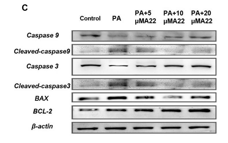
Application: WB Species: human Sample: HepG2

Application: WB Species: Mouse Sample:
Restrictive clause
Affinity Biosciences tests all products strictly. Citations are provided as a resource for additional applications that have not been validated by Affinity Biosciences. Please choose the appropriate format for each application and consult Materials and Methods sections for additional details about the use of any product in these publications.
For Research Use Only.
Not for use in diagnostic or therapeutic procedures. Not for resale. Not for distribution without written consent. Affinity Biosciences will not be held responsible for patent infringement or other violations that may occur with the use of our products. Affinity Biosciences, Affinity Biosciences Logo and all other trademarks are the property of Affinity Biosciences LTD.